| Description | Shipped From Abroad
Delivery & Availability:
Typically 10-21 working days – excluding furniture and heavy/bulky equipment. Please contact us for further information.
| Shipped from abroad
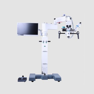
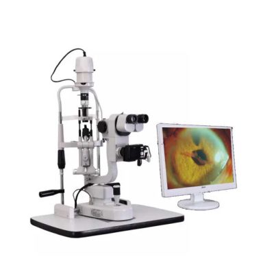
- Digital Slit lamp system,Galilean optical system,Five magnifications.
- Independent image process and management software system
- More than 10 million pixel image resolution
Delivery & Availability:
Typically 14 working days – excluding furniture and heavy/bulky equipment. Please contact us for further information.
| Shipped from abroad
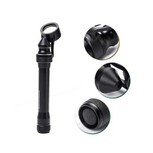
This ultra-portable is an excellent diagnostic instrument for the examination of anterior segment structures and ocular abnormalities.
Delivery & Availability:
Typically 14 working days – excluding furniture and heavy/bulky equipment. Please contact us for further information.
| Shipped from abroad
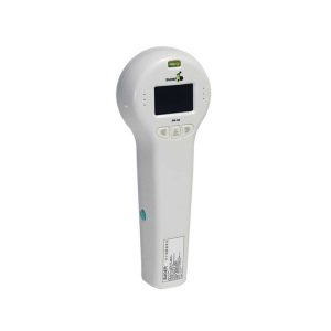
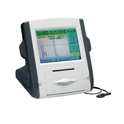
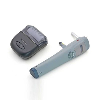
Ultra-small corneal curvature tester, is mainly used to measure the corneal curvature radius and diopter, wireless output print data.
Delivery & Availability:
Typically 14 working days – excluding furniture and heavy/bulky equipment. Please contact us for further information.
| Shipped from abroad
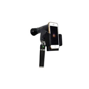
The brand-new Pantoscopic Ophthalmoscope is a portable digital imaging device which makes it possible to view and take pictures of the eyes.
Delivery & Availability:
Typically 14 working days – excluding furniture and heavy/bulky equipment. Please contact us for further information.
| Shipped from abroad
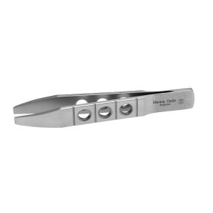
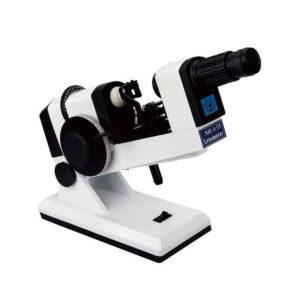
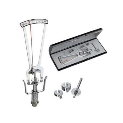
The NJC-4 Lensmeter is used to measure the diopters of spherical lens and cylinder lens, the axis of cylinder lens, the strength and the baseline direction of shuttle lens, it can also stamp the optical center of lens, the axis of cylinder len and the base direction of shuttle lens.
Delivery & Availability:
Typically 14 working days – excluding furniture and heavy/bulky equipment. Please contact us for further information.
|
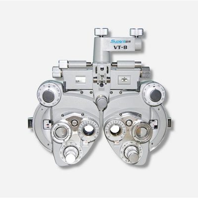
| Content | | Slit Lamp with Workstation Features:
- Digital Slit lamp system, Galilean optical system, Five magnifications
- Independent image process and management software system
- More than 10 million pixel image resolution
Technical Specifications:
| Microscope Type |
Galileo Parallel |
| Digital System |
External Digital Slit Lamp with 10 Mega pixels |
| Magnification Change Way |
Drum Five Magnifications |
| Eyepiece Magnification |
12.5x |
| Total Magnifications |
6x, 10x, 16x, 25x,40x |
| Diopter Adjustment |
-5D ~+5D |
| Slit Width |
0-14MM Continuous |
| Slit Height |
1-14MM Continuous |
| Slit Angle |
0°- 180° Adjustable |
| Slit Inclination Angle |
5°,10°,15°, 20° |
| Light Spot Diameter |
0.2mm, 2mm, 3mm, 5mm, 10mm, 14mm |
| Filter |
Heat Absorption; Grey; Red free; Cobalt Blue |
| Fixation |
Red LED |
| Illumination Bulb |
12V/50W German OSRAM Halogen Tungsten Lamp |
| Features:
- Ultra-portable: This ultra-portable is an excellent diagnostic instrument for the examination of anterior segment structures and ocular abnormalities. Its easy-to-operate optical system produces a high-brightness, continuously adjustable slit image ideal for pediatric and geriatric setting, emergency department screenings, ward rounds, beside examination, post-op evaluations, and mission work;
- LED illumination: the first and the only portable slit lamp in the world applying LED illumination system. The most prominent advantage of our LED illumination system gives the examiner the clearest image without glare;
- Comfortable using experience: The low heat radiation from our LED lamp makes the most comfortable examine experience for the patient;
- Sharpest Slit: With the blade imaging system S150 gives equally sharpest slit as the best classic slit lamp in the world;
- No need to replace illumination lamp: The LED lamp used in S150 portable slit lamp is rated 20,000 hours at full power. Its lifetime is almost 10 times longer than a normal halogen lamp;
- Power-saving: S150 can work for long hours without changing batteries.
Technical Specifications:
- Magnification: 5x
- Range of slit length: 5mm~11mm
- Minimum slit width: 0.2mm
- Working distance: 12~32mm
- Power supply: AA batteries x 2
- Illumination: LED blub(3.3V/1W)
- Battery life: More than 3h(AA alkaline batteries x 2)
- Net weight: 150g(without batteries)
How to Operate:
- Press the Power Switch to turn the slit lamp on/off
- Loosen the Locking Screw
- Twist the Lamp Body while holding the Magnifier Bracket still till the slit is in the required angle
- Push or pull the Lamp Body while holding the Magnifier Bracket still to set the Magnifier into a position comfortable for observation
- Fasten the Locking Screw
- Pull the Lamp Head back and forth for adjusting the width and the length of the slit, as well as the intensity of the light
| Portable Keratometer Features:
Ultra-small corneal curvature tester, is mainly used to measure the corneal curvature radius and diopter, wireless output print data.
Technical Specifications:
| Model |
SW-100 |
| Measuring Range |
6.5mm~9.5mm: ±0.05mm: 0.01mm |
| Precision |
± 0.05mm |
| Resolution of Curvature Radius of Cornea |
0.01mm |
| Single Measuring Time |
0.03s |
| Output |
Wireless Infrared Thermal Printer |
| Can observe the eye directly through the screen. |
| Weight |
0.5Kg(with batteries) |
| Dimension |
240mmx90mmx60mm |
| Power |
500mW+15% |
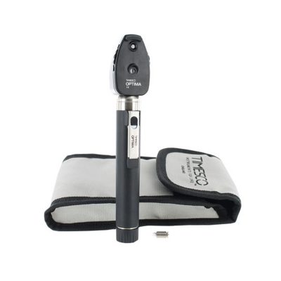
| The brand-new Pantoscopic Ophthalmoscope is a portable digital imaging device which makes it possible to view and take pictures of the eyes. The optical access of the Pantoscopic Ophthalmoscope is aligned to the visual axis of the smartphone camera by the adaptor which allows to you take pictures of the fundus and retinal nerve in high resolution. You could save pictures for each patient or email and print as needed. The Pantoscopic Ophthalmoscope provides a 5X larger view of the fundus compared with the standard ophthalmoscope. It has a wider view field of 230. Without dilating the pupil, the fundus imagines could be captured at any time and places.
Features:
- Rapid capture of fundus images on your cellphone.
- Get eyeground documentation in an easy and low-cost way.
- Send images to your peers across the continum of care instantly.
- Easy carry to capture images at any clinical circumstance and work efficiently.
- Make patient education easier.
- 5X larger view of the fundus vs. standard ophthalmoscopes in an undilated eye.
- Halogen lamp provides bright, white light.
- Apertures and Filters: Looking at the pupil through an add-on corneal viewing lens, the practitioner can detect lens opacities easily.
| The NJC-4 Lensmeter is used to measure the diopters of the spherical lens and cylinder lens, the axis of cylinder lens, the strength and the baseline direction of shuttle lens, it can also stamp the optical center of the lens, the axis of cylinder lens, and the base direction of shuttle lens. This instrument (Both Ac and Dc are permitted Two cells When Dc) has clear readings and graduations as well as high objective precision and reliabilities except that it can be operated easily and conveniently, AU the lenses and the made glasses can be measured by it, therefore, it is a required and ideal measuring instrument for glasses manufactures, glasses stores and ophthalmological hospitals.
Technical Specifications:
- Eyepiece focusing range: ±5D
- The top point diopter measuring range: 0—±20D
- The minimum measuring square value: 0.125D
- The shuttle lens measuring range: 5A
- The shuttle lens square value: 1A
- The cylinder lens axial range: 0—180°
- The cylinder lens axial square value: 5°
- The maximum diameter of the measured lenses l00m/m
- Power supply: AC 110V-6V DC 3V
- Note: This item is AC 110V; The battery is not included!
|


Reviews
There are no reviews yet.