| Description | Shipped From Abroad
Delivery & Availability:
Typically 10-21 working days – excluding furniture and heavy/bulky equipment. Please contact us for further information.
| In Stock
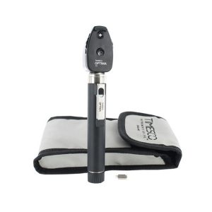
- Durable and light weight
- Supplied in a soft pouch
- Long life bright white Xenon bulb
- Macular beam dioptres lenses from 0 to +20 and 0 to -20
- 3 year guarantee (excludes consumables)
Delivery & Availability:
Typically 5-7 working days – excluding furniture and heavy/bulky equipment. Please contact us for further information.
| Shipped from abroad
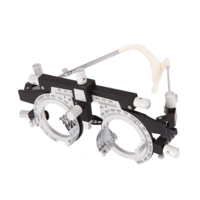
The product can quickly and precisely measure the astigmatism axis and is one of the necessary instruments in optometry inspection.
Delivery & Availability:
Typically 14 working days – excluding furniture and heavy/bulky equipment. Please contact us for further information.
| Shipped from abroad
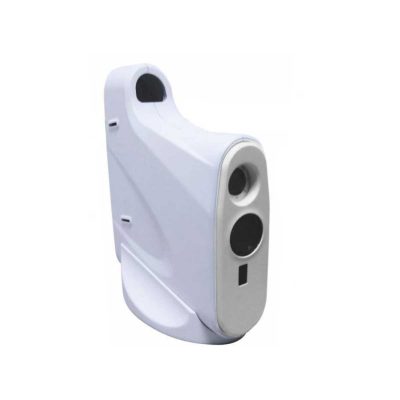
The product is designed on the principle basis of Goldman tonometer. It can be connected with slit lamp(Carl Zeiss type).
Delivery & Availability:
Typically 14 working days – excluding furniture and heavy/bulky equipment. Please contact us for further information.
| Shipped from abroad
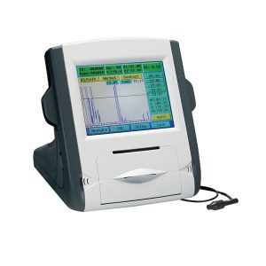
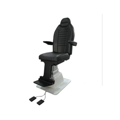
- Large color liquid-crystal screen
- Touch screen input, easy operation
- Curve freezing: Manual/Auto mode, controlled by pedal
- Built-in speed thermal printer
- Enter the name & ID; easy to check archive
Delivery & Availability:
Typically 14 working days – excluding furniture and heavy/bulky equipment. Please contact us for further information.
| Shipped from abroad
- Top Optical System.
- High eyepoint, comfort.
- Five steps drum zoom design, easier to use 6x, 10x, 16x, 25x, 40x magnification.
- High Definition Eyepieces, More Comfortable for Viewing.
Delivery & Availability:
Typically 14 working days – excluding furniture and heavy/bulky equipment. Please contact us for further information.
|
| Content | | Timesco Ophthalmoscope features a head made from lightweight hermetically sealed durable plastic, precision optics and a latex free rubber eyebrow rest. A bright white light from long life standard bulbs provides crystal clear illumination in ophthalmic diagnostic procedures.
- Durable and lightweight
- Supplied in a soft pouch
- Long life bright white Standard bulb
- Macular beam dioptres lenses from 0 to +20 and 0 to -20
- 3-year guarantee (excludes consumables)
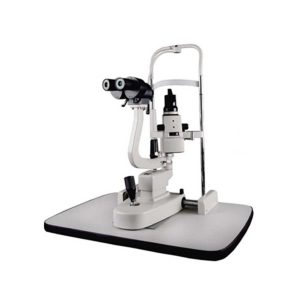
| The product can quickly and precisely measure the astigmatism axis and is one of the necessary instruments in optometry inspection.
Features:
- The filament can be rotated for 360° and move upward and downward. The brightness of the streak can be adjusted.
- Quickly and precisely measure the astigmatism axis.
- The light can be converged, radiated, and paralleled.
- Automatic Power Off function effectively protects the bulb.
- Two-steps adjustable brightness for bulb: S(Bright) and W(Dark).
Technical Specifications:
| Working Distance |
1 m |
| Streak |
3 mm-20 mm |
| Streak Rotation |
360° |
| Illumination Source |
3V/2W, halogen bulb |
| Input Voltage |
AC 220V±10%, 50Hz±1Hz |
| Power Consumption |
4.5 VA |
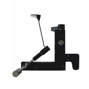
| Applanation Tonometer is designed on the principle basis of Goldman tonometer. It can be connected with slit lamp(Carl Zeiss type).
Features of Applanation Tonometer:
- When used with slit lamp, it can be used to examine the eyes and measure ocular pressure.
- Accurate measurement and total tolerance are less than 0.066KPa(0.55Hg).
- Directly get the ocular pressure and do not need to look up the conversion table.
- The measured ocular pressure is not affected by hardness of eye.
- The affected?ocular volume is just 0.56 cubic millimeter.
- Adjustable measuring pressure ensures the long-term stability and reliability.
Technical Specifications Applanation Tonometer:
- Measuring Range: 0~1064 Kpa
- Light Ring Displacement: 1.53×2=3.06mm
- Diameter of Prism Head: 7mm
- Moving Range of Prism Head: 3mm
| Feature:
- Large color liquid-crystal screen
- Touch screen input, easy operation
- Curve freezing: Manual/Auto mode, controlled by a pedal
- Built-in speed thermal printer
- Enter the name & ID; easy to check the archive
Technical Specifications:
| Model |
SW-1000 Ophthalmic A Scan Biometer |
| A Scan |
| A scan probe |
10MHz import small size probe, built-in luminotron |
| Measuring range |
15mm-40mm |
| Measurement precision |
±0. 05mm; with macula lutea trace function |
| Measurement |
Anterior chamber depth, lens thickness, vitreous body length, total length and average |
| Method of measurement |
immersion and contact |
| Eye mode |
Phakic/ Aphakic/Dense/ various IOL |
| IOL formula |
SRK-II, SRK-T, BINKHORST- Ⅱ, HOLLADAY, HOFFER-Q, HAIGIS |
| Storage |
10 cases, 5 readings each case |
| Output |
A scan waveform and IOL calculation sheet
|
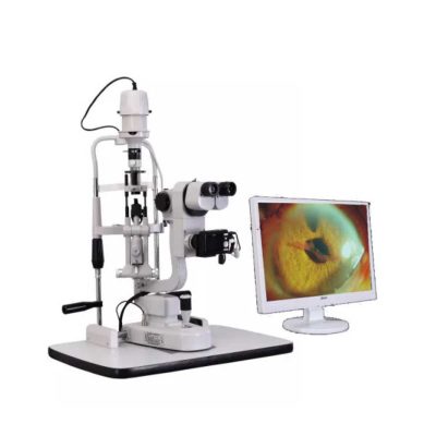
| Slit Lamp Features:
- Top Optical System.
- High eyepoint, comfort.
- Five steps drum zoom design, easier to use 6x, 10x, 16x, 25x, 40x magnification.
- High Definition Eyepieces, More Comfortable for Viewing.
Technical Specifications:
| Microscope Type |
Galileo Parallel |
| Optics |
Super Optic System |
| Magnification Change Way |
Drum Rotation |
| Eyepiece Magnification |
12.5x |
| Total Magnifications |
6x 10x, 16x, 25x, 40x |
| Diopter Adjustment |
-5D ~+5D |
| Slit Width |
0-14MM Continuous |
| Slit Height |
1-14MM Continuous |
| Slit Angle |
0°- 180° Adjustable |
| Light Source |
LED |
| Light Spot Diameter |
0.2mm, 2mm, 3mm, 5mm, 10mm, 14mm |
| Filter |
Heat Absorption; Grey; Redfree; Cobalt Blue |
| Fixation |
Red LED |
| Electrical |
|
| Illumination Bulb |
LED |
| Input Voltage |
110V/220V (±10%) |
| Applanation Tonometer Interface |
Included |
|


Reviews
There are no reviews yet.