| Description | Shipped From Abroad
18mm
Delivery & Availability:
Typically 10-21 working days – excluding furniture and heavy/bulky equipment. Please contact us for further information.
| Shipped from abroad
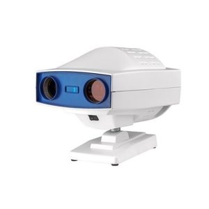
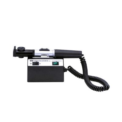
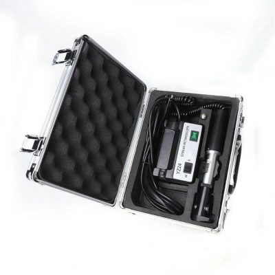
This ultra-portable is an excellent diagnostic instrument for the examination of anterior segment structures and ocular abnormalities.
Delivery & Availability:
Typically 14 working days – excluding furniture and heavy/bulky equipment. Please contact us for further information.
| Ship from abroad
- Easy installation and maintenance: It is quite easy to replace light bulks, and unnecessary to adjust its focus and position.
- Customer Programmable: Icons can be displayed in predefined sequence by programming.
- Display Single Icon: Display single icon by multi-function mask plate.
- Tilted Placement: Perfect image position can be obtained by adjusting the horizontal axis of projector.
- Standards parts: Metal plate with polarized coating (410x 410mm)
Delivery & Availability:
Typically 14 working days – excluding furniture and heavy/bulky equipment. Please contact us for further information.
| Ship from abroad
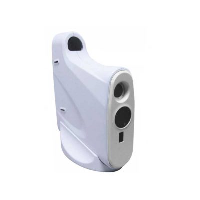
- Equipped with comprehensive measuring functions, it provides SPH, CYL, AXIS and pupil distance optometry
- Durable and easy to operate
- Easily and intuitively read the sphere focal scale value
- High eco-friendly materials
- Design fitting the face curve and no stimulation
Delivery & Availability:
Typically 14 working days – excluding furniture and heavy/bulky equipment. Please contact us for further information.
| Shipped from abroad
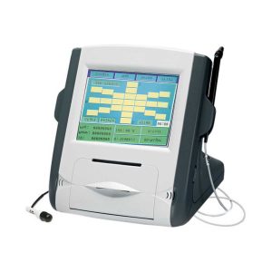
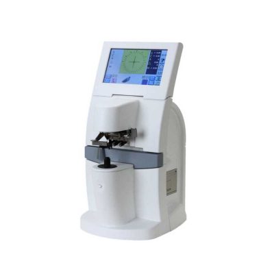
- Large color liquid-crystal screen.
- Touch screen input, easy operation.
- Curve freezing: Manual/Auto mode, controlled by pedal.
- Built-in speed thermal printer.
- Can display ultrasound waveform when measuring.
Delivery & Availability:
Typically 14 working days – excluding furniture and heavy/bulky equipment. Please contact us for further information.
| Shipped from abroad
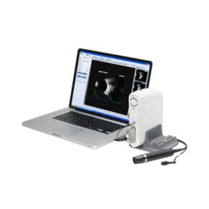
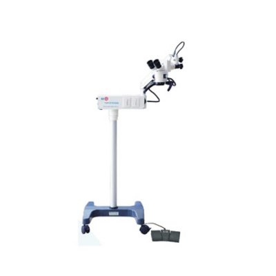
- Software image workstation
- B, B+B, B+A, A modes
- Video review for 100 images
- PDF report output
- Optional 20MHz B Probe: vitreous plus function
Delivery & Availability:
Typically 14 working days – excluding furniture and heavy/bulky equipment. Please contact us for further information.
|
| Content | | Features:
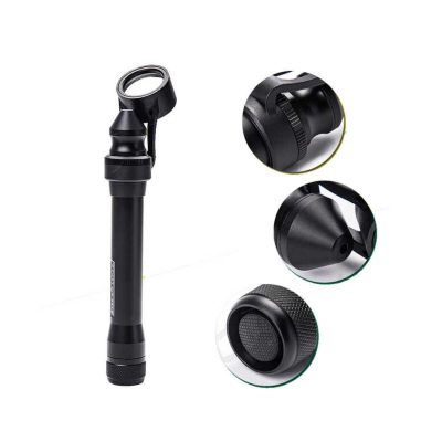
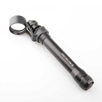
- Ultra-portable: This ultra-portable is an excellent diagnostic instrument for the examination of anterior segment structures and ocular abnormalities. Its easy-to-operate optical system produces a high-brightness, continuously adjustable slit image ideal for pediatric and geriatric setting, emergency department screenings, ward rounds, beside examination, post-op evaluations, and mission work;
- LED illumination: the first and the only portable slit lamp in the world applying LED illumination system. The most prominent advantage of our LED illumination system gives the examiner the clearest image without glare;
- Comfortable using experience: The low heat radiation from our LED lamp makes the most comfortable examine experience for the patient;
- Sharpest Slit: With the blade imaging system S150 gives equally sharpest slit as the best classic slit lamp in the world;
- No need to replace illumination lamp: The LED lamp used in S150 portable slit lamp is rated 20,000 hours at full power. Its lifetime is almost 10 times longer than a normal halogen lamp;
- Power-saving: S150 can work for long hours without changing batteries.
Technical Specifications:
- Magnification: 5x
- Range of slit length: 5mm~11mm
- Minimum slit width: 0.2mm
- Working distance: 12~32mm
- Power supply: AA batteries x 2
- Illumination: LED blub(3.3V/1W)
- Battery life: More than 3h(AA alkaline batteries x 2)
- Net weight: 150g(without batteries)
How to Operate:
- Press the Power Switch to turn the slit lamp on/off
- Loosen the Locking Screw
- Twist the Lamp Body while holding the Magnifier Bracket still till the slit is in the required angle
- Push or pull the Lamp Body while holding the Magnifier Bracket still to set the Magnifier into a position comfortable for observation
- Fasten the Locking Screw
- Pull the Lamp Head back and forth for adjusting the width and the length of the slit, as well as the intensity of the light
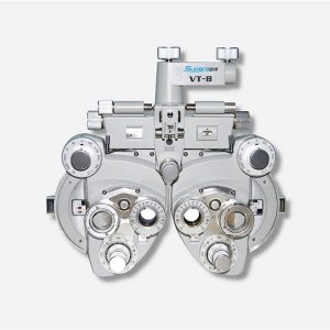
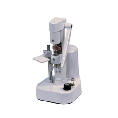
| Digital Chart Projector-Features:
- Easy installation and maintenance: It is quite easy to replace light bulks, and unnecessary to adjust its focus and position.
- Customer Programmable: Icons can be displayed in predefined sequence by programming.
- Display Single Icon: Display single icon by multi-function mask plate.
- Tilted Placement: Perfect image position can be obtained by adjusting the horizontal axis of projector.
- Standards parts: Metal plate with polarized coating (410x 410mm)
Technical Specifications of Digital Chart Projector:
|
Projection Distance |
1.5m~6m |
|
Projection Magnification |
30x (at 5m) |
|
Projection Size |
330x270mm (at 5 m) |
|
Chart Switching Speed |
One chart per 0.03s |
|
Mask Switching Speed |
1 open, 5 horizontal different lines, 8 vertical lines, 21 single letters, 1 red/green |
|
Program |
2 sets programs, each program contains up to 30 steps |
|
Speed of mask conversion |
One mask per 0.03s |
|
Lamp |
LED lamp |
|
Auto-off function |
After 10 minutes idle time |
|
Power Source |
AC 220V, 50Hz or 110V, 60Hz |
|
Power consumption |
40W |
|
Accessories |
Remote control, polarized metal screen, halogen lamp, polarized glasses, fuses (2), batteries (2) |
| Features:
- Equipped with comprehensive measuring functions, it provides SPH, CYL, AXIS and pupil distance optometry
- Durable and easy to operate
- Easily and intuitively read the sphere focal scale value
- High eco-friendly materials
- Design fitting the face curve and no stimulation
- Easy to take and clean
- Free switch between the cross-cylindrical lens and the rotary prism
- When the rotating risk is turning by the sphere, it can make sphere power adjust 3.00D for big scope.
- It is designed expediently and smartly for a particular cross cylinder. Supporting supplementary lens could increase scope of measurement.
Technical Specifications:
|
Sphere |
Range:-19.00~+16.75m-1 Step: 0.25m-1, 3.00m-1 |
|
Cylinder |
Range: 0.00~-6.00m-1(Measuring Range With Accessories0.00~-8.00m-1) Step: 0.25m-1 |
|
Cylinder Axis |
Range: 0~180°, Step:5° |
|
Distance of Optical center (also known as Pupil) |
Range: 50~75mmStep: 1mm |
|
Sight Switch |
Range:∞~380mm (distance of Optical center is64mm) |
|
Front Chin Test |
Range: 0~16mm |
|
Distance (from cornea vertex to the lens surface) |
16mm |
|
Standard Accessories Lens |
two pieces of Auxiliary Cylinder -2.00m-1 and -0.12m-1 respectively |
|
Standard Accessories |
one piece of M2 Hexagon wrench , one piece of a Myopia Standard Card, two piece of Myopia Standard Card , one piece of standard card holder , a dust cover |
|
Auxiliary Lens |
“O”:Open aperture
“R”:Retinoscope lens
“R”:Retinoscope lens
“R”:Retinoscope lens
*Lens of +1.50m-1 ,It is suit for the distance of 67 centimeters
“P”:Polaroid
* it is used for examining the dioptric balance of eyes , Implicit strabismus and stereo vision
“RMV”:Red Vertical maddox
*Be used to examine Implicit strabismus
“RMH”:Red horizontal maddox
*Be used to examine Implicit strabismus
“WMV”:Plane Vertical Maddox
*Be used to examine Implicit strabismus
“WMH”:Plane horizontal maddox
*Be used to examine Implicit strabismus
“RL”:Red lens
*Be used to examine eye function, Blending function and Implicit strabismus
“GL”:Green lens
*Be used to examine eye function, Blending function and Implicit strabismus
“+”:Test mark of optical center adjustment
“+.12”:Dioptric of the Spherical Lens is +0.12m-1
*Be used for the semi-adjustment of sphere lens, 0.25m-1
“PH”:1mmPinhole lens
*Be used to exclude visual non-refractive errors of the tested eye
“6ΔU”:6ΔBottom-up prism
*Be used to examine the rotating prism with the detection of nearly horizontal squint
“10ΔI”:10ΔBottom-up prism
*Be used to examine the rotating prism with the detection of nearly horizontal squint
“±0.50”:Cross-cylindrical lens
*Be used to examine the corrected dioptric of the Presbyopia and spherical lens
“OC”:Black lens
|
|
size |
338(L)×99(W)×292(H)mm |
|
NW |
about 5kg |
| Features:
- Large color liquid-crystal screen.
- Touch screen input, easy operation.
- Curve freezing: Manual/Auto mode, controlled by pedal.
- Built-in speed thermal printer.
- Can display ultrasound waveform when measuring.
- Each group is the average of 20 measurements.
- Switch between IOP measured value and actual value.
- Can input name, ID and operator's name.
Technical Specifications:
| P scan probe |
20MHZ, angle of 45 degrees makes easier operation |
| Resolution |
5um |
| Measuring range |
150um~1500um |
| Display |
SINGLE mode and MAP mode |
| Functions of Ophthalmic AB Scan Machine:
- Software image workstation
- B, B+B, B+A, A modes
- Video review for 100 images
- PDF report output
- Optional 20MHz B Probe: vitreous plus function
Technical Specifications:
| A scan |
1.Probe: 10MHz frequencies, with LED
2.Depth: 40mm
3.Precision: ±0.05mm
4.Eye mode: Phakic / Aphakic / Dense / Various IOL
5.Measurement: Anterior chamber depth, lens thickness, vitreous body length, total length and average
6.IOL Formula: SRK-II, SRK-T, BINKHORST, HOLLADAY, HOFFER-Q, HAIGIS, Stat.
7.Calculation: Average and standard deviation
8.Store: 10 Scanning results for each eye |
| B scan |
1.Probe: 10MHz/20MHz (optional), Magnetic driven, noiseless
2.Scanning Mode: Sector Scanning
3.Resolution: Lateral ≤0.3mm; Vertical≤0.2mm
4.Geometric Location Precision: Lateral≤10%; Vertical≤5%
5.Depth: 60mm
6.Enhance the part of vitreous body and retina
7.Gain of probe:30dB-105dB
8.Scanning Angle : 53°
9.Gray Scale: 256
10.False Color: Multi colors OTC
11.Measure Mode: distances, perimeter and area
12.Movies: 100 images movie review,AVI ZIP JPG format image output
13.Output: PDF format case report, connect to normal printer |
| Others |
1.Display Mode :B, B+B, B+A, A
2.Hint: preset keyword
3.Case Search: Multi-keywords
4.Working Platform: Windows XP, VISTA, WINDOWS7
5.User-defined report template |
|


Reviews
There are no reviews yet.