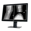

Coronis Fusion 4MP (MDCC‑4430)
$0.00
Shipped From Abroad
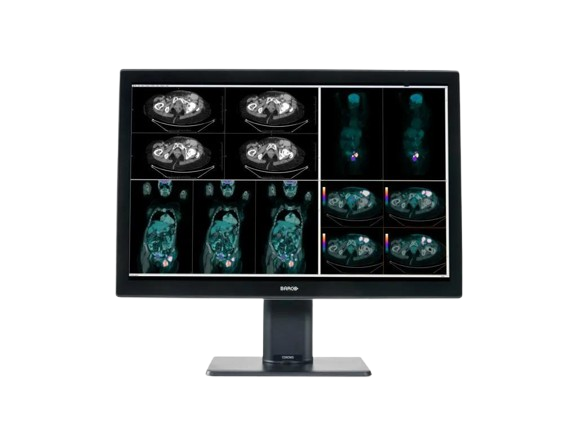
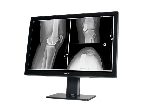
The Barco Coronis Fusion 4MP (MDCC-4430) is a high-fidelity medical display designed for diagnostic radiology. With a 4-megapixel resolution, wide color gamut, and image-enhancement features, it supports mammography, CT, MRI, and multi-modality imaging workflows.
Typically 10-21 working days – excluding furniture and heavy/bulky equipment. Please contact us for further information.
Description
The Barco Coronis Fusion 4MP (MDCC-4430) delivers optimized diagnostic visualization for radiologists by combining high-quality optics with advanced processing. Its native resolution of 2560 × 1600 pixels and large active area (655 × 410 mm) provide generous screen real estate for image review. The display includes uniformity correction and Barco’s image-enhancement technologies to maintain consistency across the display. It supports multi-modality imaging in breast, vascular, and general radiology settings. With efficient design and compatibility for clinical workflows, it balances performance and ergonomics in diagnostic environments.
Key Features
-
Native 4MP resolution (2560 × 1600)
-
Active screen area: 655 × 410 mm
-
Uniformity correction to ensure brightness consistency
-
Image-enhancement tools for clarity in clinical reading
-
Wide gamut/color support (multi-modality capability)
-
Optimized for breast imaging, mammography, CT, and MRI
-
Efficient, lightweight design for diagnostic reading rooms
A brighter diagnosis
Coronis Fusion, Barco’s renowned multi-modality display for radiologists, now comes in a new, energy-efficient, and lightweight design. Bright on so many levels, Coronis Fusion has been designed to help radiologists provide care with confidence.
See more details quickly thanks to the high brightness, high contrast ratio, and best-in-class image quality. What’s more, the wide color gamut in combination with SteadyColor™ calibration technology helps you see even more colors and more details on the 30-inch screen.
Smart workflow
Designed for your comfort and to boost productivity, Coronis Fusion comes with smart image-enhancing features and workflow tools. The display’s wide viewing angle, combined with the SoftGlow™ task and wall light, helps reduce eye strain. Thanks to SpotView, radiologists can further improve detection accuracy as well as reading productivity.
Effortless quality & compliance
Like all of Barco’s medical display systems, Coronis Fusion comes with QAWeb Enterprise, a cloud-based technology for automated calibration, Quality Assurance, and compliance to ensure maximum uptime of the display with no need for human intervention.
A one-stop-shop solution
Barco medical display systems include exclusive display controllers that are validated with the latest workstations and with all major PACS applications. All components are under the full 5-year warranty (including the display backlight) for complete peace of mind.
Ensuring diagnostic confidence with MDR Class IIa
Our radiology displays are MDR-certified as Class IIa. Their product information has been reviewed and cleared by independent medical and technical experts, and is audited yearly. In other words, we ensure diagnostic confidence and peace of mind for our users.
Specifications
| Category | Specification |
|---|---|
| Screen Technology | IPS |
| Active Screen Size (Diagonal) | 772 mm (30.4″) |
| Active Screen Size (H × V) | 655 × 410 mm (25.8″ × 16.1″) |
| Aspect Ratio | 16:10 |
| Resolution | Native 4MP (2560 × 1600 pixels); Configurable to 2 × 2MP+ (1280 × 1600 pixels); 2 × 2MP (1200 × 1600 pixels) |
| Pixel Pitch | 0.256 mm |
| Color Imaging / Gray Imaging | Yes / Yes |
| Bit Depth | 30-bit |
| Viewing Angle (H/V) | 178° |
| Uniformity Correction | Color PPU |
| SteadyColor | Yes (with Barco Display Controller and QAWeb Enterprise 2.2+) |
| Ambient Light Presets / Sensor | Yes / Yes |
| Front Sensor | Yes (I-Guard, Coronis) |
| Maximum Luminance (Typical) | 1050 cd/m² |
| DICOM Calibrated Luminance | 600 cd/m² |
| Contrast Ratio | 2000:1 |
| Response Time | 18 ms |
| Housing Color | Black / White |
| Video Input Signals | 2 × DisplayPort 1.2 |
| Video Output Signals | 1 × DisplayPort (MST) |
| USB Ports | 1 × USB 2.0 upstream; 2 × USB 2.0 downstream; 1 × USB 2.0 downstream with high-power charging |
| Power Rating | 100–240 Vac, 50/60 Hz, 3.6–1.6 A |
| Power Consumption | 75 W (nominal @ 600 cd/m²); <0.5 W (hibernate/standby) |
| Dimensions with Stand (W × H × D) | 714 × 524~624 × 240 mm |
| Dimensions without Stand (W × H × D) | 714 × 478 × 74 mm |
| Dimensions Packaged (W × H × D) | 800 × 650 × 295 mm |
| Net Weight with Stand | MDCC-4430: 17.7 kg; MDCC-4430 NC: 16.3 kg |
| Net Weight without Stand | MDCC-4430: 13.1 kg; MDCC-4430 NC: 11.7 kg |
| Net Weight Packaged | MDCC-4430: 22.3 kg; MDCC-4430 NC: 20.9 kg |
| Tilt / Swivel / Pivot | -5° to +25° / ±30° / N/A |
| Height Adjustment Range | 100 mm |
| Mounting Standard | VESA (100 mm) |
| Screen Protection | MDCC-4430: Protective, anti-reflective glass; MDCC-4430 NC: No glass cover |
| Recommended Modalities | All digital images except digital mammography |
| Certifications | FDA 510(k) K191845, CE0123, CCC, INMETRO, KC, BIS, EAC |
| Safety Standards | IEC/EN/UL/CSA 60950-1, 62368-1, 60601-1, AAMI ES 60601-1 |
| EMI Standards | IEC/EN 60601-1-2, FCC Part 15 Class B, ICES-001 Level B, VCCI |
| Environmental Compliance | China Energy Label, EU RoHS, China RoHS, REACH, Canada Health, WEEE, Packaging Directive |
| Supplied Accessories | User guide, Documentation disc, System sheet, Video cables, Mains cables, USB cable |
| Optional Accessories | Graphics board, Touch pad, QA software (QAWeb) |
| Warranty | 5 years (includes 40,000 hrs backlight warranty) |
| Operating Temperature | 0–35°C (specs: 10–30°C) |
| Storage Temperature | -20–60°C |
| Operating Humidity | 20–85% RH (non-condensing) |
| Storage Humidity | 20–85% RH (non-condensing) |
| Operating Pressure | ≥70 kPa |
| Storage Pressure | 50–106 kPa |
Quick Comparison
| Coronis Fusion 4MP (MDCC‑4430) remove | IBIS Neeo R9 Digital Surgical C-Arm remove | Anke MRI Openmark 5000 Permanent System remove | Single X-Ray Viewing Box remove | DrGem GXR-SD 400mA Floor Mounted Digital X-ray remove | Sonoscape S8 Exp Portable Ultrasound remove | ||||||||||||||||||||||||||||||||||||||||||||||||||||||||||||||||||||||||||||||||||||||||||||||||||||||||||||||||||||||||||||||||||||||||||||||||||||||||||||||||||||||||||||||||||||||||||||||||||||||||||||||||||||||||||||||||||||||||||||||||||||||||||||||||||||||||||||||||||||||||||||||||||||||||||||||||||||||||||||||||||||||||||||||||||||||||||||||||||||||||||||||||||||||||||||||||||||||||||||||||
|---|---|---|---|---|---|---|---|---|---|---|---|---|---|---|---|---|---|---|---|---|---|---|---|---|---|---|---|---|---|---|---|---|---|---|---|---|---|---|---|---|---|---|---|---|---|---|---|---|---|---|---|---|---|---|---|---|---|---|---|---|---|---|---|---|---|---|---|---|---|---|---|---|---|---|---|---|---|---|---|---|---|---|---|---|---|---|---|---|---|---|---|---|---|---|---|---|---|---|---|---|---|---|---|---|---|---|---|---|---|---|---|---|---|---|---|---|---|---|---|---|---|---|---|---|---|---|---|---|---|---|---|---|---|---|---|---|---|---|---|---|---|---|---|---|---|---|---|---|---|---|---|---|---|---|---|---|---|---|---|---|---|---|---|---|---|---|---|---|---|---|---|---|---|---|---|---|---|---|---|---|---|---|---|---|---|---|---|---|---|---|---|---|---|---|---|---|---|---|---|---|---|---|---|---|---|---|---|---|---|---|---|---|---|---|---|---|---|---|---|---|---|---|---|---|---|---|---|---|---|---|---|---|---|---|---|---|---|---|---|---|---|---|---|---|---|---|---|---|---|---|---|---|---|---|---|---|---|---|---|---|---|---|---|---|---|---|---|---|---|---|---|---|---|---|---|---|---|---|---|---|---|---|---|---|---|---|---|---|---|---|---|---|---|---|---|---|---|---|---|---|---|---|---|---|---|---|---|---|---|---|---|---|---|---|---|---|---|---|---|---|---|---|---|---|---|---|---|---|---|---|---|---|---|---|---|---|---|---|---|---|---|---|---|---|---|---|---|---|---|---|---|---|---|---|---|---|---|---|---|---|---|---|---|---|---|---|---|---|---|---|---|---|---|---|---|---|---|---|---|---|---|---|---|---|---|---|---|---|---|---|---|---|---|---|---|---|---|---|---|---|---|---|---|---|---|
| Name | Coronis Fusion 4MP (MDCC‑4430) remove | IBIS Neeo R9 Digital Surgical C-Arm remove | Anke MRI Openmark 5000 Permanent System remove | Single X-Ray Viewing Box remove | DrGem GXR-SD 400mA Floor Mounted Digital X-ray remove | Sonoscape S8 Exp Portable Ultrasound remove | |||||||||||||||||||||||||||||||||||||||||||||||||||||||||||||||||||||||||||||||||||||||||||||||||||||||||||||||||||||||||||||||||||||||||||||||||||||||||||||||||||||||||||||||||||||||||||||||||||||||||||||||||||||||||||||||||||||||||||||||||||||||||||||||||||||||||||||||||||||||||||||||||||||||||||||||||||||||||||||||||||||||||||||||||||||||||||||||||||||||||||||||||||||||||||||||||||||||||||||||
| Image | 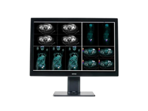 |  |  |  |  |  | |||||||||||||||||||||||||||||||||||||||||||||||||||||||||||||||||||||||||||||||||||||||||||||||||||||||||||||||||||||||||||||||||||||||||||||||||||||||||||||||||||||||||||||||||||||||||||||||||||||||||||||||||||||||||||||||||||||||||||||||||||||||||||||||||||||||||||||||||||||||||||||||||||||||||||||||||||||||||||||||||||||||||||||||||||||||||||||||||||||||||||||||||||||||||||||||||||||||||||||||
| SKU | SF1033560011-1 | SF1033560092-3 | SF1033560084-203 | SF1033560074-5 | SF1033560012-15 | ||||||||||||||||||||||||||||||||||||||||||||||||||||||||||||||||||||||||||||||||||||||||||||||||||||||||||||||||||||||||||||||||||||||||||||||||||||||||||||||||||||||||||||||||||||||||||||||||||||||||||||||||||||||||||||||||||||||||||||||||||||||||||||||||||||||||||||||||||||||||||||||||||||||||||||||||||||||||||||||||||||||||||||||||||||||||||||||||||||||||||||||||||||||||||||||||||||||||||||||||
| Rating | |||||||||||||||||||||||||||||||||||||||||||||||||||||||||||||||||||||||||||||||||||||||||||||||||||||||||||||||||||||||||||||||||||||||||||||||||||||||||||||||||||||||||||||||||||||||||||||||||||||||||||||||||||||||||||||||||||||||||||||||||||||||||||||||||||||||||||||||||||||||||||||||||||||||||||||||||||||||||||||||||||||||||||||||||||||||||||||||||||||||||||||||||||||||||||||||||||||||||||||||||||||
| Price |
|
|
| $95.20 |
| $9,350.00 | |||||||||||||||||||||||||||||||||||||||||||||||||||||||||||||||||||||||||||||||||||||||||||||||||||||||||||||||||||||||||||||||||||||||||||||||||||||||||||||||||||||||||||||||||||||||||||||||||||||||||||||||||||||||||||||||||||||||||||||||||||||||||||||||||||||||||||||||||||||||||||||||||||||||||||||||||||||||||||||||||||||||||||||||||||||||||||||||||||||||||||||||||||||||||||||||||||||||||||||||
| Stock | |||||||||||||||||||||||||||||||||||||||||||||||||||||||||||||||||||||||||||||||||||||||||||||||||||||||||||||||||||||||||||||||||||||||||||||||||||||||||||||||||||||||||||||||||||||||||||||||||||||||||||||||||||||||||||||||||||||||||||||||||||||||||||||||||||||||||||||||||||||||||||||||||||||||||||||||||||||||||||||||||||||||||||||||||||||||||||||||||||||||||||||||||||||||||||||||||||||||||||||||||||||
| Availability | |||||||||||||||||||||||||||||||||||||||||||||||||||||||||||||||||||||||||||||||||||||||||||||||||||||||||||||||||||||||||||||||||||||||||||||||||||||||||||||||||||||||||||||||||||||||||||||||||||||||||||||||||||||||||||||||||||||||||||||||||||||||||||||||||||||||||||||||||||||||||||||||||||||||||||||||||||||||||||||||||||||||||||||||||||||||||||||||||||||||||||||||||||||||||||||||||||||||||||||||||||||
| Add to cart | |||||||||||||||||||||||||||||||||||||||||||||||||||||||||||||||||||||||||||||||||||||||||||||||||||||||||||||||||||||||||||||||||||||||||||||||||||||||||||||||||||||||||||||||||||||||||||||||||||||||||||||||||||||||||||||||||||||||||||||||||||||||||||||||||||||||||||||||||||||||||||||||||||||||||||||||||||||||||||||||||||||||||||||||||||||||||||||||||||||||||||||||||||||||||||||||||||||||||||||||||||||
| Description | Shipped From Abroad
The Barco Coronis Fusion 4MP (MDCC-4430) is a high-fidelity medical display designed for diagnostic radiology. With a 4-megapixel resolution, wide color gamut, and image-enhancement features, it supports mammography, CT, MRI, and multi-modality imaging workflows.
Delivery & Availability:
Typically 10-21 working days – excluding furniture and heavy/bulky equipment. Please contact us for further information.
| Shipped from Abroad Our Neeo “C” arms are easy to place, use and are specifically designed to be used in orthopedics, traumatology, abdominal surgery, urology, cardiology and operating rooms. Delivery & Availability: Typically 21 working days – excluding furniture and heavy/bulky equipment. Please contact us for further information. | Shipped from Abroad
OPENMARK 5000 is 0.51T MRI. It's approved by FDA and has CE mark. It adopts two-pillar magnet design with 280 degree openness and equipped with powerful
RF and gradient system, together with advanced imaging technology, making it as a high-end system which is comparable to high-field MRI.
Delivery & Availability: Typically 90 working days – excluding furniture and heavy/bulky equipment. Please contact us for further information. | In stock
| In Stock The GXR-SD Digital X-ray is a diagnostic digital radiography system that provides reliable high quality digital radiographic images with a reduced dose. The GXR-SD DR systems offer comprehensive digital solutions to all radiography needs, featuring ACQUIDR digital imaging system with stationary or portable digital flat-panel detectors as well as reliable high-frequency x-ray generators that are known worldwide for their excellent performance, lifetime and stability. Patient tables and wall stands are also offered. Delivery & Availability: Typically 21 working days – excluding furniture and heavy/bulky equipment. Please contact us for further information. | Shipped from Abroad With ultra-modern innovative design and the clinically-proven technologies, S8 Exp is portable ultrasound scanner well equipped as a low-physical-effort and enhanced-image-quality ultrasound scanner, which not only provides optimized solutions for versatile applications, but does help to improve the user-experience for both routine and non-traditional challenges. Delivery & Availability: Typically 5-7 working days – excluding furniture and heavy/bulky equipment. Please contact us for further information. | |||||||||||||||||||||||||||||||||||||||||||||||||||||||||||||||||||||||||||||||||||||||||||||||||||||||||||||||||||||||||||||||||||||||||||||||||||||||||||||||||||||||||||||||||||||||||||||||||||||||||||||||||||||||||||||||||||||||||||||||||||||||||||||||||||||||||||||||||||||||||||||||||||||||||||||||||||||||||||||||||||||||||||||||||||||||||||||||||||||||||||||||||||||||||||||||||||||||||||||||
| Content | The Barco Coronis Fusion 4MP (MDCC-4430) delivers optimized diagnostic visualization for radiologists by combining high-quality optics with advanced processing. Its native resolution of 2560 × 1600 pixels and large active area (655 × 410 mm) provide generous screen real estate for image review. The display includes uniformity correction and Barco’s image-enhancement technologies to maintain consistency across the display. It supports multi-modality imaging in breast, vascular, and general radiology settings. With efficient design and compatibility for clinical workflows, it balances performance and ergonomics in diagnostic environments. Key Features
A brighter diagnosisCoronis Fusion, Barco's renowned multi-modality display for radiologists, now comes in a new, energy-efficient, and lightweight design. Bright on so many levels, Coronis Fusion has been designed to help radiologists provide care with confidence. See more details quickly thanks to the high brightness, high contrast ratio, and best-in-class image quality. What's more, the wide color gamut in combination with SteadyColor™ calibration technology helps you see even more colors and more details on the 30-inch screen.Smart workflowDesigned for your comfort and to boost productivity, Coronis Fusion comes with smart image-enhancing features and workflow tools. The display's wide viewing angle, combined with the SoftGlow™ task and wall light, helps reduce eye strain. Thanks to SpotView, radiologists can further improve detection accuracy as well as reading productivity.Effortless quality & complianceLike all of Barco's medical display systems, Coronis Fusion comes with QAWeb Enterprise, a cloud-based technology for automated calibration, Quality Assurance, and compliance to ensure maximum uptime of the display with no need for human intervention.A one-stop-shop solutionBarco medical display systems include exclusive display controllers that are validated with the latest workstations and with all major PACS applications. All components are under the full 5-year warranty (including the display backlight) for complete peace of mind.Ensuring diagnostic confidence with MDR Class IIaOur radiology displays are MDR-certified as Class IIa. Their product information has been reviewed and cleared by independent medical and technical experts, and is audited yearly. In other words, we ensure diagnostic confidence and peace of mind for our users.Specifications
| Our Neeo “C” arms are easy to place, use and are specifically designed to be used in orthopedics, traumatology, abdominal surgery, urology, cardiology and operating rooms.
Using Neeo with the RTP (Real Time Processing) option it is possible to perform vascular, urological and cardiological diagnostics. One of the main functions, digital image subtraction, allows to see, as an example, the passage of contrast liquids in a tissue or in a venous or arterial duct; thanks to the possibility of looping, the acquired video can be reproduced several times to monitor more accurately the passage of the fluid within the area in question. Angiographic measurement is another useful function in the vascular field (QA Quantitative Angiography) that allows the measurement of stenoses. Finally, fluoroscopy allows the correct positioning of stents or expanders.
Neeo is used in various interventional and diagnostic procedures in traumatology and orthopedics wards and operating rooms as well. Thanks to low-dose fluoroscopy, it is possible to use the device for positioning bone or subcutaneous grafts, inserting K-wire (Kirschner wire) for stabilization of bone fragments or for the correct positioning of prostheses. The low dose emitted ensures safe use for both the patient and the surgeon or doctor on the operating field.
On the control panel there is a large touch screen display that allows to adjust the basic functions of the equipment. From this display it is possible to select and adjust the fluoroscopic data for the examination, activate or deactivate the laser pointer, select between pulsed, one shot or standard fluoroscopy, rotate the image and perform all operations on collimator. The four side buttons on the display offer the possibility to move the bow vertically thanks to an extremely silent motor.
Neeo has two 19 “medical grade monitors that can be positioned according to the needs of the medical practitioner. Work monitors and feedback monitors are separated to be managed independently. The possible movements are: rotation, revolution, tilting and possibility of height adjustment.
Features:
Click Here To Download Catalogue | OPENMARK 5000 is 0.51T MRI. It's approved by FDA and has CE mark. It adopts two-pillar magnet design with 280 degree openness and equipped with powerful
RF and gradient system, together with advanced imaging technology, making it as a high-end system which is comparable to high-field MRI.
Features:
Click Here To Download Catalogue |
| DrGem GXR-SD 400mA Floor Mounted Digital X-ray system matches with a radiographic room which perfectly fits your workow and can be easily upgraded to DR system with the help of DR interface and PC interface in GXR generator as well as Bucky suitable to Flat Panel Detector. GXR X-ray system is equipped with a high frequency X-ray generator which consistently produces high quality radiograph in favor of high quality X-ray output with a very small kV ripple and accurate mA and mAs. GXR X-ray system is designed to provide convenience to operator and comfort to patient
Features of DrGem GXR-SD 400mA Floor Mounted Digital X-ray:
Click Here To Download Catalogue | Sonoscape S8 Exp Portable Ultrasound scannerDETAILS Agile and Versatile With ultra-modern innovative design and the clinically-proven technologies, S8 Exp Portable Ultrasound scanner is well equipped as a low-physical-effort and enhanced-image-quality ultrasound scanner, which not only provides optimized solutions for versatile applications but does help to improve the user experience for both routine and non-traditional challenges. Working with S8 Exp, it will trigger your unlimited reverie and endow you with endless charm. Carrying forward the classical design of SonoScape's portable ultrasound products, S8 Exp successfully combines the best ergonomics, attractive design and ease of use. This charismatic identity is also enhanced by a sophisticated color palette—with sedate grey as its interior paint color and pearl white as exterior cover, S8 Exp reveals a style of aristocrat and strong character among SonoScape's ultrasound systems. Workflow The S8 Exp is a portable ultrasound scanner that adapts to your workflow, whether you are in the consulting room, at the bedside, or at a remote location. With easy-to-use new platform designed for sonographers' needs and full connection interfaces for easy connectivity and data sharing, S8 Exp leads to improved user comfort and clinical outcome as well as patient throughput and working efficiency. Powerful Platform Embedded with SonoScape's core imaging technologies such as μ-scan, PHI and Spatial Compound, S8 Exp boasts exceptional 2D image, sensitive spectral, Color and Power Doppler, displaying well-defined anatomy and pathology and facilitating a highly optimized diagnostic user environment for conclusive diagnoses. Besides, S8 Exp offers a comprehensive selection of electronic probes to maximally extend its capabilities to meet a wide range of applications including the abdomen, pediatric, OB/GYN, cardiovascular, musculoskeletal, etc. The advanced probe technologies also effectively enhance the image quality and confidence in reaching clinical diagnoses even in difficult patients.Click Here To Download Catalogue | |||||||||||||||||||||||||||||||||||||||||||||||||||||||||||||||||||||||||||||||||||||||||||||||||||||||||||||||||||||||||||||||||||||||||||||||||||||||||||||||||||||||||||||||||||||||||||||||||||||||||||||||||||||||||||||||||||||||||||||||||||||||||||||||||||||||||||||||||||||||||||||||||||||||||||||||||||||||||||||||||||||||||||||||||||||||||||||||||||||||||||||||||||||||||||||||||||||||||||||||
| Weight | N/A | N/A | N/A | N/A | N/A | N/A | |||||||||||||||||||||||||||||||||||||||||||||||||||||||||||||||||||||||||||||||||||||||||||||||||||||||||||||||||||||||||||||||||||||||||||||||||||||||||||||||||||||||||||||||||||||||||||||||||||||||||||||||||||||||||||||||||||||||||||||||||||||||||||||||||||||||||||||||||||||||||||||||||||||||||||||||||||||||||||||||||||||||||||||||||||||||||||||||||||||||||||||||||||||||||||||||||||||||||||||||
| Dimensions | N/A | N/A | N/A | N/A | N/A | N/A | |||||||||||||||||||||||||||||||||||||||||||||||||||||||||||||||||||||||||||||||||||||||||||||||||||||||||||||||||||||||||||||||||||||||||||||||||||||||||||||||||||||||||||||||||||||||||||||||||||||||||||||||||||||||||||||||||||||||||||||||||||||||||||||||||||||||||||||||||||||||||||||||||||||||||||||||||||||||||||||||||||||||||||||||||||||||||||||||||||||||||||||||||||||||||||||||||||||||||||||||
| Additional information |
|


Reviews
There are no reviews yet.