The Barco Coronis OneLook (MDMC-32133) is a 32MP diagnostic display designed for mammography and advanced radiology. It delivers superior resolution, high luminance, precise grayscale and color imaging, with optimized workflow tools for breast imaging, MRI, CT, and ultrasound.
Delivery & Availability: Typically 10-21 working days – excluding furniture and heavy/bulky equipment. Please contact us for further information.
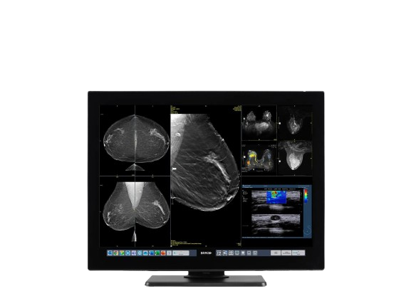
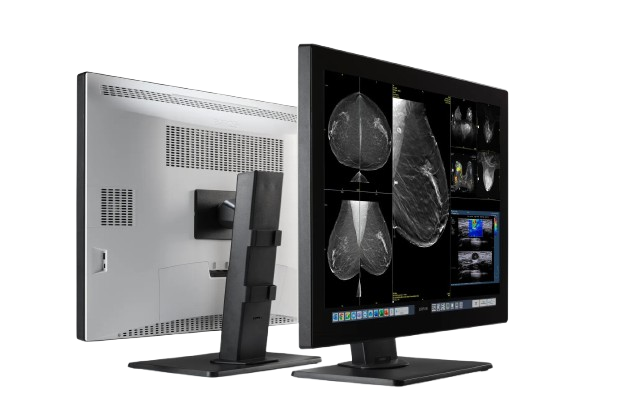
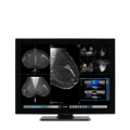
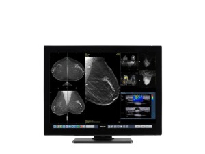
The Coronis OneLook (MDMC-32133) is Barco’s flagship display solution, optimized for mammography and advanced radiology visualization. With its 32 MP panel and high luminance, it allows radiologists to view full-resolution mammograms without zooming or panning. Equipped with Barco’s RapidFrame technology to support crisp motion in 3D cine imaging, it also features an expanded, calibrated color range and modular custom on-screen shortcuts. Integrated with QAWeb for quality assurance and compliance, the display is built to elevate accuracy, productivity, and image fidelity in diagnostic workflows.
Highest resolution ever in breast imaging
Meet the Coronis OneLook® display solution, brought to you by the team behind the world-class Coronis Uniti. From supporting orthopedics to finding tiny breast calcifications: Coronis OneLook is Barco’s paragon, coming after decades of research and development – your perfect radiology assistant, which will serve for years to come.
Coronis OneLook gives you more detail than you’ve ever imagined: it boasts the highest number of image lines and pixels we have ever achieved. All this, combined with crisp, consistent grayscales and colors and new productivity tools, is true to the Barco range.
Key Features
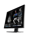
See complete images at a look
Straight from the acquisition machine, every detail is present without you needing to zoom in and pan in steps. An industry first!
Extreme medicalcontrast
Coronis OneLook’s brightness of 1,200 cd/m² and 1300:1 contrast ratio result in a whopping 770 just noticeable differences, which can be boosted up as high as 849 with the I-Luminate brightness booster. The OpticalGlass reduces reflections, so you see your images in all their sharpness.
Excellent Color representation
Coronis OneLook’s color range is 30% wider than that of our renowned Coronis Uniti monitor. It also features our SteadyColor calibration technology, ensuring consistent color representation.
Personalize your workflow
Coronis OneLook has been optimized with our latest insights into radiology workflow and comfort. In addition to our suite of Intuitive Workflow Tools, it includes customizable on-screen touch buttons, so you can instantly invoke functions and launch your go-to applications without any clicks
Crystal-clear moving images
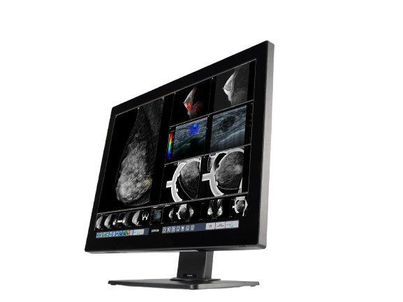
Optimized for 3D cine examinations of CT, ultrasound, and breast MRI, Coronis OneLook is equipped with RapidFrame for crisp and in-focus moving images. This Barco-patented technology can lead to a 10% higher detection of small details in moving images.
Large screen surface
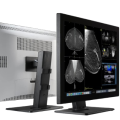
Coronis OneLook’s large 33” screen allows you to layout multiple medical images on the screen at the same time. This format, called Fusion, has been proven to improve productivity by 19%.
Specifications
Display Specifications
Category
Details
Screen Technology
IPS
Active Screen Size (Diagonal)
850 mm (33″)
Active Screen Size (H × V)
708 mm × 472 mm (28″ × 19″)
Aspect Ratio
3:2
Resolution
32MP (6848 × 4656 @ 60 Hz) with touchbar
Active Area
6848 × 4560 pixels @ 60 Hz (without touchbar)
Pixel Pitch
0.103 mm
Color Imaging / Gray Imaging
Yes / Yes
Bit Depth
10-bit per R, G, B (with Barco Display Controller)
Shipped From Abroad
The Barco Coronis OneLook (MDMC-32133) is a 32MP diagnostic display designed for mammography and advanced radiology. It delivers superior resolution, high luminance, precise grayscale and color imaging, with optimized workflow tools for breast imaging, MRI, CT, and ultrasound.
Delivery & Availability:Typically 10-21 working days – excluding furniture and heavy/bulky equipment. Please contact us for further information.
Shipped from Abroad
SuperMark 1.5T is a new generation superconducting MRI system based on years of experience in production and research. It's applicable to whole body scan, such as, nervous system, spine, joint soft tissue, pelvic and abdominal cavity, etc
Delivery & Availability:
Typically 90 working days – excluding furniture and heavy/bulky equipment. Please contact us for further information.
Shipped from Abroad
This Machine gives a possibility to perform computed tomography without any problems and on high quality level. This device is used to conduct exams of internal organs and their functioning. With its help, a physician has a possibility to assess the condition of the human body as a whole.
Delivery & Availability:
Typically 90 working days – excluding furniture and heavy/bulky equipment. Please contact us for further information.
Shipped from Abroad
Easily accomplish more with SonoScape’s new P50 ultrasound system. Incorporating single crystal clarity, automatic corrections and calculation, and user defined flexibility promises a confident diagnostic experience as well as opening new doors of opportunity for ultrasound use.
Delivery & Availability:
Typically 7-14 working days – excluding furniture and heavy/bulky equipment. Please contact us for further information.
Shipped from Abroad
As SonoScape steps forward to add value and efficiency to ultrasound, the latest S22 was designed in a user-friendly platform to address current and future demanding needs. It represents an excellent mix in performance and price.
Delivery & Availability:
Typically 5-7 working days – excluding furniture and heavy/bulky equipment. Please contact us for further information.
Shipped from Abroad
SonoScape has developed a new probe and function for the E1 Exp. With these additions the E1 Exp will bring users a more efficient examination experience with satisfying image quality and a smooth workflow.
Delivery & Availability:
Typically 5-7 working days – excluding furniture and heavy/bulky equipment. Please contact us for further information.
Content
https://youtu.be/qYwbJXpkPgE?si=WGJARl2Us_WguIGy
The Coronis OneLook (MDMC-32133) is Barco’s flagship display solution, optimized for mammography and advanced radiology visualization. With its 32 MP panel and high luminance, it allows radiologists to view full-resolution mammograms without zooming or panning. Equipped with Barco’s RapidFrame technology to support crisp motion in 3D cine imaging, it also features an expanded, calibrated color range and modular custom on-screen shortcuts. Integrated with QAWeb for quality assurance and compliance, the display is built to elevate accuracy, productivity, and image fidelity in diagnostic workflows.
Highest resolution ever in breast imaging
Meet the Coronis OneLook® display solution, brought to you by the team behind the world-class Coronis Uniti. From supporting orthopedics to finding tiny breast calcifications: Coronis OneLook is Barco’s paragon, coming after decades of research and development – your perfect radiology assistant, which will serve for years to come.
Coronis OneLook gives you more detail than you’ve ever imagined: it boasts the highest number of image lines and pixels we have ever achieved. All this, combined with crisp, consistent grayscales and colors and new productivity tools, is true to the Barco range.
Key Features
See complete images at a look
Straight from the acquisition machine, every detail is present without you needing to zoom in and pan in steps. An industry first!
Extreme medicalcontrast
Coronis OneLook’s brightness of 1,200 cd/m² and 1300:1 contrast ratio result in a whopping 770 just noticeable differences, which can be boosted up as high as 849 with the I-Luminate brightness booster. The OpticalGlass reduces reflections, so you see your images in all their sharpness.
Excellent Color representation
Coronis OneLook’s color range is 30% wider than that of our renowned Coronis Uniti monitor. It also features our SteadyColor calibration technology, ensuring consistent color representation.
Personalize your workflow
Coronis OneLook has been optimized with our latest insights into radiology workflow and comfort. In addition to our suite of Intuitive Workflow Tools, it includes customizable on-screen touch buttons, so you can instantly invoke functions and launch your go-to applications without any clicks
Crystal-clear moving images
Optimized for 3D cine examinations of CT, ultrasound, and breast MRI, Coronis OneLook is equipped with RapidFrame for crisp and in-focus moving images. This Barco-patented technology can lead to a 10% higher detection of small details in moving images.
Large screen surface
Coronis OneLook’s large 33” screen allows you to layout multiple medical images on the screen at the same time. This format, called Fusion, has been proven to improve productivity by 19%.
Specifications
Display Specifications
Category
Details
Screen Technology
IPS
Active Screen Size (Diagonal)
850 mm (33")
Active Screen Size (H × V)
708 mm × 472 mm (28" × 19")
Aspect Ratio
3:2
Resolution
32MP (6848 × 4656 @ 60 Hz) with touchbar
Active Area
6848 × 4560 pixels @ 60 Hz (without touchbar)
Pixel Pitch
0.103 mm
Color Imaging / Gray Imaging
Yes / Yes
Bit Depth
10-bit per R, G, B (with Barco Display Controller)
SuperMark 1.5T is a new generation superconducting MRI system based on years of experience in production and research. It's applicable to whole body scan, such as, nervous system, spine, joint soft tissue, pelvic and abdominal cavity, etc. SuperMark 1.5T provides not only conventional pulse sequences and clinical diagnosis functions, but also provides advanced functional applications, for instance, 3D angiography and water imaging. It adopts brand new ANKE APEX operating system which ensures easy operation and fast diagnosis.
Technical Advantages:
Reliable short cavity superconducting magnet system with zero liquid helium
consumption
New generation fully digitalized and extensible multichannel spectrometer
Powerful high efficiency and high fidelity gradient system; Multi-channel PA RF
receiving coil with intelligent identification
English operating system and high extensible computer system
High resolution conventional clinical images; Practical advanced functional
imaging
Superconducting MRI System:
Highly open and humanization design -> Streamlined design
Rich sequences and technology satisfy clinical needs -> Efficient service
Low Investment:
High cost performance superconducting MRI system
Zero liquid helium consumption, low running and maintenance cost
Core technology by independent R & D supports full upgrade
This Machine gives a possibility to perform computed tomography without any problems and on high quality level. This device is used to conduct exams of internal organs and their functioning. With its help, a physician has a possibility to assess the condition of the human body as a whole.
Features:
It is easy to use;
Convenience;
Multi functionality;
Obtained images are of high definition;
High-definition 3D images of the area under study;
The procedure is pain-free;
The data is processed fast;
The image can be stored in the computer memory;
The diagnostics does not take a lot of time;
Acceptable radiation dose.
Technical Specifications:
No.
Technical Features
Descriptions
1
Gantry
1.01
Gantry type
Low voltage slip-ring
1.02
Gantry driven type
Strap-driven
1.03
Patient opening
70cm
1.04
Gantry tilt mode
Digital gantry tilt
1.05
Digital tilt capability
±50°
1.06
Detector type
OptiWave rare-earth ceramic detector
1.07
Numbers of detector rows
16
1.08
Width of Z-axle detector
20mm
1.09
Detector columns of channels per row
848
1.10
Numbers of detector columns
13568
1.11
Data-transfer type
RF, optical fiber communication
1.12
Distance of focus-ISO-center
53cm
1.13
Distance of focus-detector
94cm
1.14
3D laser orientation
Provided
1.15
13" integrated display panel
Provided
1.16
Adose automatic exposure control (mA
Modulation)
Provided
1.17
Auto-voice manager
Breath Graphical Display
Hold Message (Record/Playback)
Breath Message (Record/Playback)
1.18
AccuSaving energy conservation management
Provided
2
HVPS and X-ray tube
2.01
Maximum continuous output of HVgenerator
42kW
2.02
Tube kV selections
70kV, 80kV, 100 kV, 120 kV, 140 kV
2.03
Tube mA range
10~350mA
2.04
Tube anode heat capacity
3.5MHU
2.05
Max. anode cooling rate
735kHU/min
2.06
Type of cooling
Oil cooling + Air cooling
2.07
Tube focus
Large: 1.2mm×1.4mm
Small: 0.7mm×0.8mm
2.08
Collimator width selection
4-level election
2.09
Focus spot tracking technology
Provided
3
Patient table
3.01
Maximum horizontal-movable range
1850mm
3.02
Table horizontal-scannablerange
1800mm
3.03
Table horizontal-position repeatability
±0.25mm
3.04
Minimum height above floor
430mm
3.05
Maximum vertical-movable range
500mm
3.06
Maximum speed of vertical movement
35mm
3.07
Maximum speed of horizontal movement
150mm/s
3.08
Maximum patient weight
205kg
3.09
Foot pedal of patient table control
Provided
4
Computer
4.01
CPU
3.5GHz
4.02
Memory
32GB
4.03
Storage of hard-disk
1TB×2
4.04
Monitor
24’’ LCD Monitor
4.05
Resolution of monitor
1920×1200
4.06
Image-data external storage type
CD/DVD/USB
4.07
Time of image reconstruction (512×512)
33.3ms/image
4.08
Speed of image reconstruction (512×12)
30fps
4.09
DICOM 3.0 interface
Provided
4.10
Printer DICOM 3.0 interface
Provided
4.11
Auto filming
Provided
4.12
Worklist function
Provided
5
Scan parameters
5.01
Shortest 360 degree rotation time
0.75s
5.02
Allowed rotation times
0.75s, 1.0s, 1.5s, 2.0s, 3.0s, 4.0s
5.03
Maximum slice numbers per rotation
32
5.04
Minimum slice thickness of scan
1.25mm
5.05
Minimum slice thickness of reconstruction
0.625mm
5.06
Maximum slice thickness of scan
20mm
5.07
Nominal reconstruction slice thickness
0.625mm, 1.25mm, 2.5mm, 5.0mm, 7.5mm,
10mm, 20mm
5.08
Speed of image reconstruction (512×512)
30 frames/s
5.09
Scan FOV
50cm
5.10
Image reconstruction matrix
512×512, 1024×1024 (Optional)
5.11
Image reconstruction matrix
512×512, 1024×1024 (Optional)
5.12
Image display matrix
512×512, 1024×1024 (Optional)
5.13
Maximum continuous scan duration
120s
5.14
Maximum continuous scan length
180cm
5.15
Direction of TOPO
Front-back, Left-right
5.16
Max. length of TOPO
180cm
5.17
Range of pitch
0.5~1.5
5.18
Scan mode
Scout scan
Axial scan
Helical scan
Cine scan
6
Image Quality
6.01
High contrast resolution
21lp/cm@0%MTF
6.02
Low contrast resolution
2.0mm@0.30%
6.03
Isotropic imaging resolution
0.24mm
6.04
Range of CT numbers
-32767~32768
6.05
Image noise
≤0.29@28mGy
7
Advanced application
7.01
Multi-Planar Reconstruction (MPR)
Provided
7.02
Curve Multi-Planar Reconstruction (CPR)
Provided
7.03
Surface Shaded Display (SSD)
Provided
7.04
Volume Rendering (VR)
Provided
7.05
Maximum Intensity Projection (MIP)
Provided
7.06
Minimum Intensity Projection (MinIP)
Provided
7.07
Virtual Endoscopy (VE)
Provided
7.08
CT angiography (CTA)
Provided
7.09
Tissue segmentation
Provided
7.10
One click bone remove
Provided
7.11
One click patient table remove
Provided
7.12
Bolus-tracking Technology
Provided
7.13
Spiral auto start
Provided
7.14
Cine display
Provided
7.15
AbastTM bone artifact suppression technology
Provided
7.16
AmastTM metal artifact suppression technology
Provided
7.17
Admir3D all-domain iterative reconstruction
Provided
7.18
Low-dose pediatric scan technology
Provided
7.19
Low-dose lung scan technology
Provided
7.20
AccuHead grey-white matter enhanced
technology
Provided
7.21
AccuOrgan lung high resolution scan technology
Provided
7.22
AccuOrgan inner-ear high resolution scan
technology
Provided
7.23
AccuOrgan body high resolution scan technology
Provided
7.24
AccuOrgan bone high resolution scan technology
Provided
7.25
AccuMatter dual-energy with Admir3D for new
application
DETAILSPowerful Compact Precision
Taking into consideration the evolving expectations and needs for ultrasound, the P50 is a slim and unobtrusive trolley system that is comfortable in tight, congested spaces with little room to work in. Providing everything you need for a comfortable examination in a small space for both you and your patient.
Single Crystal Transducer
Wideband single crystal probes greatly improve the signal ratio, acquire stunning images and provide superior sensitivity and resolution for both the near and far-fields.
μ-Scan+
The new generation μ-Scan imaging technologies give you better image quality by reducing noise, improving signal strength and improving visualization.
Dynamic Color
Dynamic colour improves upon already existing colour Doppler technologies for clear capture of colour flow and detail visualization of even tiny veins with lower velocities.
Solution for Radiology
P50, is a leading-edge ultrasound system that can meet the demands of any clinical setting. You can experience a superior performance in multi-dimensional imaging for a full range of clinical applications – abdominal, breast and cardiovascular.
C-xlasto Imaging
By understanding that tissue stiffness varies depending on the type of tissue, we can use C-xlasto Imaging to easily find abnormalities and tumours within soft tissue. The differences in tissue responses are detected and visualized in real-time by the elastography algorithms through different representations, which can be particularly helpful in analyzing breast, thyroid and musculoskeletal structures. Predominately used only in linear probes, SonoScape’s new transvaginal and bi-plane probe for gynaecology and urology are breaking the mould and expanding elastography applications.
Real-time Color Panoramic
With the combination of colour flow and real-time panoramic, visualizing the blood flow of an entire vein or artery is now an easy task. Accomplished in real-time for the convenience of the sonographers, any mistakes can also be easily backtracked and corrected without interrupting the scan.
Contrast Imaging
Contrast Imaging on P50 makes full use of the infra harmonic signal and second harmonic signal to improve the image resolution and deep penetration. What’s more, the Dynamic Acoustic Control technology effectively controls the acoustic pressure for the contrast agent, decreasing the required agent dose and assures uniform image quality, guaranteeing longer contrast agent duration and better lesion perfusion of delayed phase observation.
Solution for OB/GYN
P50 has superior image quality, automated measurement tools, and a variety of volume technologies to provide ideal solutions for clinical examinations such as pregnancy examinations, and gynecologic disease diagnosis. With a new 4D transvaginal probe, P50 helps you to see and detect fetal abnormalities and significantly improves your diagnostic confidence during your examinations.
S-Live Silhouette
A unique transparent 3D anatomical image of the fetus for improved initial anatomical review. By using this new application, the system can create completely different fetal images from conventional ultrasound images, which can depict the fetal's intracorporeal anatomical structure.
Pelvic Floor 4D
Working in conjunction with SonoScape’s latest transvaginal probes, trans-perineal 4D pelvic floor ultrasound provides a useful clinical assessment of the impact of vaginal delivery on the female anterior compartment. Allowing doctors to judge whether the pelvic organs prolapsed or not, the extent of prolapse, and determining whether the pelvic muscles tore correctly.
S-Guide
S-Guide gives the user an extensive list of example obstetric ultrasound images as reference guides and a convenient checklist system to keep track of their progress during their obstetrics examination.
Auto Face
Automatically removes masking layers in front of the fetus’s face for a clearer vision of the fetus’s face.
AVC Follicle
AVC Follicle automatically identifies how many follicles are present and calculates their individual volumes.
Solution for Cardiology
P50 provides clear 2D clinical images and Doppler sensitivity to assess critical cardiac performance. Compatible with SonoScape’s single crystal probes, the P50 can provide images with better resolution and penetration in Cardiac diagnosis.
Tissue Doppler Imaging
Tissue Doppler Imaging allows clinical doctors to quantitatively evaluate local myocardial movements and functions, facilitating them with the ability to analyze and compare the motions of the different parts of the patient’s heart.
Stress Echo
Stress echocardiography is the combination of 2D echocardiography with physical, pharmacological or electrical stress of the patient. It also then provides users with report management tools such as configurable template editor, multiple loops to select one for storage, wall motion scoring, stress echo report, etc
Auto IMT
Auto IMT is used when determining the level of vascular sclerosis present in the patient by automatically tracing and calculating the thickness of the carotid vessels. What distinguishes the P50 is that it provides an instant and accurate Mean and Max index at the touch of a single button.
Auto EF
Automated 2D Cardiac Quantification is a fully intelligent trace function for endocardium with 19 easily-adjustable points providing rapid access to proven 2D EF and volumes.
DETAILS
As SonoScape steps forward to add value and efficiency to ultrasound, the latest S22 was designed in a user-friendly platform to address current and future demanding needs. It represents an excellent mix in performance and price.
S22, is a shared service ultrasound system with a slim and elegant package that has combined mobility with utility to fit in specific clinical situations including emergency department, ICU, operating room and so on. Furthermore, its ergonomic design, easy operating and flexible data management will give you a memorable experience.
SPECIFICATION
• Large high-resolution widescreen LED
• Sensitive touch screen
• Four transducer sockets plus one socket for pencil probe
• A comprehensive selection of probes: linear, Convex, Micro-convex, Volumetric, Endocavity, Bi-plane, Phased Array, TEE, Intraoperative, Pencil
• Premium application technology: 4D, μ-scan speckle reduction, compound imaging, Pulse Inversion Harmonic Imaging, Color M-Mode, Steer M-Mode, PDI, TDI, Real-time Panoramic Imaging, Trapezoid Imaging, Auto-IMT…
• Full patient database and image management solutions: DICOM 3.0, AVI/JPG, USB 2.0, HDD, DVD, PDF report
• Multi-Language Input Keyboard
• Built-in battery
DETAILSEfficient Diagnosis
μ-Scan, Speckle Reduction & Edge Enhancement
Spatial Compound Imaging
PIH - Pure Inversion Harmonic
Wide Scan - Enlarged Image Area
Tissue-Specific Imaging
SR Flow
Ergonomic Designs
Up to 2 Transducer Ports
Light Weight and Compact
15.6 inch Anti-flickering HD LED Screen
Tilting Monitor Angle Adjustment
Backlit Keyboard and Intelligent Panel
Long-lasting Battery for 90 mins
Ease of Use
Quick Boot Up
Auto-Brightness Adjustment
Auto Image Optimization
Auto IMT
Auto Trace
Equipped Accessories
Wi-Fi and Bluetooth Available
DICOM
500GB Hard Disk
Height Adjustable Trolley
Durable, Carry-on Site Suitcase


Reviews
There are no reviews yet.