| Description | Shipped From Abroad
Delivery & Availability:
Typically 10-21 working days – excluding furniture and heavy/bulky equipment. Please contact us for further information.
| Shipped from abroad
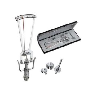
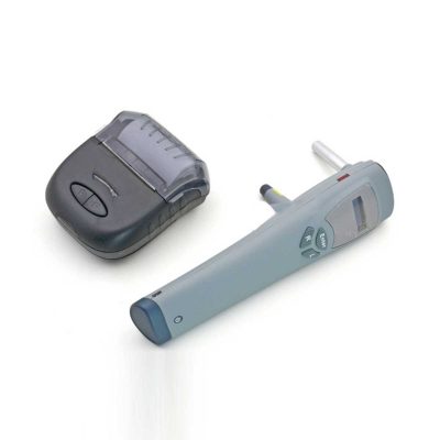
This product is used to measure the intraocular pressure (IOP) by measuring the depth produced on the surface of the cornea by a load of a known weight. Each division on the scale corresponds to 1/20mm corneal depth.
Delivery & Availability:
Typically 14 working days – excluding furniture and heavy/bulky equipment. Please contact us for further information.
| Shipped from abroad
- Top Optical System.
- High eyepoint, comfort.
- Five steps drum zoom design, easier to use 6x, 10x, 16x, 25x, 40x magnification.
- High Definition Eyepieces, More Comfortable for Viewing.
Delivery & Availability:
Typically 14 working days – excluding furniture and heavy/bulky equipment. Please contact us for further information.
| Ship from abroad
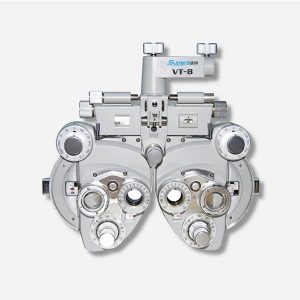
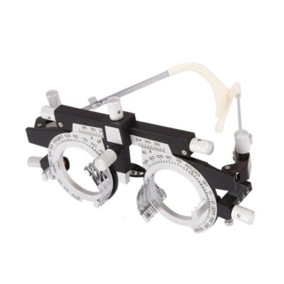
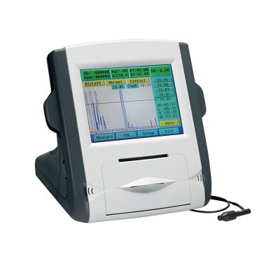
- Equipped with comprehensive measuring functions, it provides SPH, CYL, AXIS and pupil distance optometry
- Durable and easy to operate
- Easily and intuitively read the sphere focal scale value
- High eco-friendly materials
- Design fitting the face curve and no stimulation
Delivery & Availability:
Typically 14 working days – excluding furniture and heavy/bulky equipment. Please contact us for further information.
| Shipped from abroad
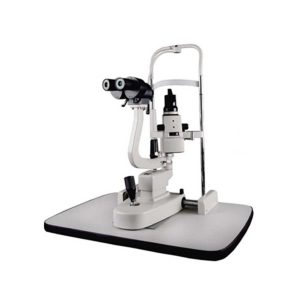
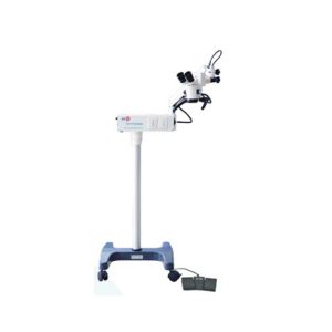
YZ20P5 Operation Microscope is a simple binocular coaxial microscope for a single man. The Operation Microscope is small, light, and convenient. It has high agility and can meet the requirements for general ophthalmology operation. The microscope fits mobile medical treatment.
Delivery & Availability:
Typically 14 working days – excluding furniture and heavy/bulky equipment. Please contact us for further information.
| Shipped from abroad
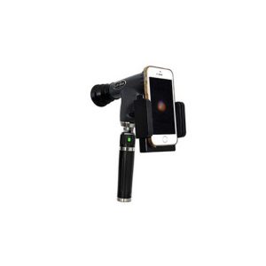
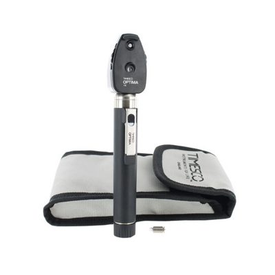
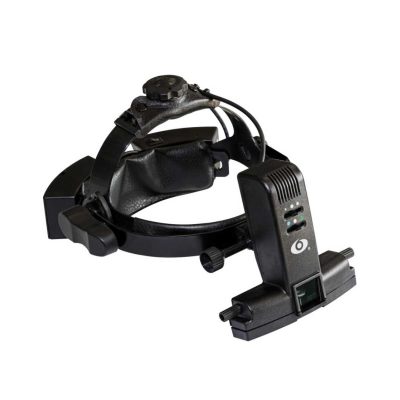
The brand-new Pantoscopic Ophthalmoscope is a portable digital imaging device which makes it possible to view and take pictures of the eyes.
Delivery & Availability:
Typically 14 working days – excluding furniture and heavy/bulky equipment. Please contact us for further information.
|
| Content | | This product is used to measure the intraocular pressure (IOP) by measuring the depth produced on the surface of the cornea by a load of a known weight. Each division on the scale corresponds to 1/20mm corneal depth.
Features:
- Schiotz type
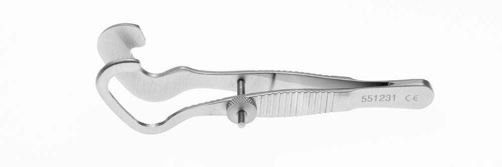
- All the parts that contact the cornea are made of stainless steel and are anti-acid, which does no harm to issues
- The instrument is safe, reliable, accurate, and convenient to carry
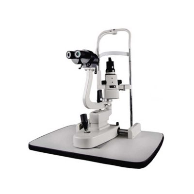
| Slit Lamp Features:
- Top Optical System.
- High eyepoint, comfort.
- Five steps drum zoom design, easier to use 6x, 10x, 16x, 25x, 40x magnification.
- High Definition Eyepieces, More Comfortable for Viewing.
Technical Specifications:
| Microscope Type |
Galileo Parallel |
| Optics |
Super Optic System |
| Magnification Change Way |
Drum Rotation |
| Eyepiece Magnification |
12.5x |
| Total Magnifications |
6x 10x, 16x, 25x, 40x |
| Diopter Adjustment |
-5D ~+5D |
| Slit Width |
0-14MM Continuous |
| Slit Height |
1-14MM Continuous |
| Slit Angle |
0°- 180° Adjustable |
| Light Source |
LED |
| Light Spot Diameter |
0.2mm, 2mm, 3mm, 5mm, 10mm, 14mm |
| Filter |
Heat Absorption; Grey; Redfree; Cobalt Blue |
| Fixation |
Red LED |
| Electrical |
|
| Illumination Bulb |
LED |
| Input Voltage |
110V/220V (±10%) |
| Applanation Tonometer Interface |
Included |
| Features:
- Equipped with comprehensive measuring functions, it provides SPH, CYL, AXIS and pupil distance optometry
- Durable and easy to operate
- Easily and intuitively read the sphere focal scale value
- High eco-friendly materials
- Design fitting the face curve and no stimulation
- Easy to take and clean
- Free switch between the cross-cylindrical lens and the rotary prism
- When the rotating risk is turning by the sphere, it can make sphere power adjust 3.00D for big scope.
- It is designed expediently and smartly for a particular cross cylinder. Supporting supplementary lens could increase scope of measurement.
Technical Specifications:
|
Sphere |
Range:-19.00~+16.75m-1 Step: 0.25m-1, 3.00m-1 |
|
Cylinder |
Range: 0.00~-6.00m-1(Measuring Range With Accessories0.00~-8.00m-1) Step: 0.25m-1 |
|
Cylinder Axis |
Range: 0~180°, Step:5° |
|
Distance of Optical center (also known as Pupil) |
Range: 50~75mmStep: 1mm |
|
Sight Switch |
Range:∞~380mm (distance of Optical center is64mm) |
|
Front Chin Test |
Range: 0~16mm |
|
Distance (from cornea vertex to the lens surface) |
16mm |
|
Standard Accessories Lens |
two pieces of Auxiliary Cylinder -2.00m-1 and -0.12m-1 respectively |
|
Standard Accessories |
one piece of M2 Hexagon wrench , one piece of a Myopia Standard Card, two piece of Myopia Standard Card , one piece of standard card holder , a dust cover |
|
Auxiliary Lens |
“O”:Open aperture
“R”:Retinoscope lens
“R”:Retinoscope lens
“R”:Retinoscope lens
*Lens of +1.50m-1 ,It is suit for the distance of 67 centimeters
“P”:Polaroid
* it is used for examining the dioptric balance of eyes , Implicit strabismus and stereo vision
“RMV”:Red Vertical maddox
*Be used to examine Implicit strabismus
“RMH”:Red horizontal maddox
*Be used to examine Implicit strabismus
“WMV”:Plane Vertical Maddox
*Be used to examine Implicit strabismus
“WMH”:Plane horizontal maddox
*Be used to examine Implicit strabismus
“RL”:Red lens
*Be used to examine eye function, Blending function and Implicit strabismus
“GL”:Green lens
*Be used to examine eye function, Blending function and Implicit strabismus
“+”:Test mark of optical center adjustment
“+.12”:Dioptric of the Spherical Lens is +0.12m-1
*Be used for the semi-adjustment of sphere lens, 0.25m-1
“PH”:1mmPinhole lens
*Be used to exclude visual non-refractive errors of the tested eye
“6ΔU”:6ΔBottom-up prism
*Be used to examine the rotating prism with the detection of nearly horizontal squint
“10ΔI”:10ΔBottom-up prism
*Be used to examine the rotating prism with the detection of nearly horizontal squint
“±0.50”:Cross-cylindrical lens
*Be used to examine the corrected dioptric of the Presbyopia and spherical lens
“OC”:Black lens
|
|
size |
338(L)×99(W)×292(H)mm |
|
NW |
about 5kg |
| Operation Microscope(YZ20P5) is a simple binocular coaxial microscope for a single man. The Microscope is small, light, and convenient. It has high agility and can meet the requirements for general ophthalmology operation. The microscope fits mobile medical treatment.
Features of Operation Microscope:
- Multi-layer coating technology is used on optical lenses to enhance the transmission rate and prevent mildew.
- Foot-controlled focus, three-step magnification change, good depth of field of view, and good binocular fusion to meet the need of cataract surgery.
- Using apochromatic technology to make different wavelengths of the light focus near the focal point behind the lens, so as to make the operator's vision more clearly.
- The machine weighs only 41Kg to be light and compact and is especially applicable for mobile medical.
- Optional desktop components to make the machine more portable and can also be customized according to special requirements to meet the needs in ophthalmology, ENT and other surgery.
- Optional F250/F300/F400 lens.
Technical Specifications of Operation Microscope:
|
Eyepiece Magnification
|
12.5×
|
|
Focal Length of Objective Lens
|
F=200
|
|
Working Distance
|
190 mm
|
|
Magnification for Main Microscope
|
5.3×, 8×, 16×
|
|
Diameter of Field
|
37 mm, 25 mm, 16.7 mm
|
|
Diopter Adjustment
|
±5D
|
|
Pupillary Distance
|
50 mm ~ 70 mm
|
|
Maximum Resolution
|
100 LP/mm
|
|
Light Source
|
12V/100W halogen lamp for medical use
|
|
Illumination Type
|
6° coaxial illumination of the cold light source
|
|
Coaxial Illuminance
|
≥30,000 Lx
|
|
Reaching Radius of Arm
|
870 mm
|
|
Adjustable Vertical Range
|
700 mm~1100 mm
|
| Fine Focusing Range |
30 mm |
|
Input Voltage
|
AC 220V/110V±10%, 50Hz/60Hz
|
|
Power Consumption
|
120 VA
|
|
Fuse
|
AC 250V T4.0A, AC 125V T8.0A
|
|
Electrical Safety Standard
|
IEC 60601-1, Class I
|
|
Packing Volume
|
0.2 m3,1 carton
|
|
Total Weight
|
41 kg
|
| The brand-new Pantoscopic Ophthalmoscope is a portable digital imaging device which makes it possible to view and take pictures of the eyes. The optical access of the Pantoscopic Ophthalmoscope is aligned to the visual axis of the smartphone camera by the adaptor which allows to you take pictures of the fundus and retinal nerve in high resolution. You could save pictures for each patient or email and print as needed. The Pantoscopic Ophthalmoscope provides a 5X larger view of the fundus compared with the standard ophthalmoscope. It has a wider view field of 230. Without dilating the pupil, the fundus imagines could be captured at any time and places.
Features:
- Rapid capture of fundus images on your cellphone.
- Get eyeground documentation in an easy and low-cost way.
- Send images to your peers across the continum of care instantly.
- Easy carry to capture images at any clinical circumstance and work efficiently.
- Make patient education easier.
- 5X larger view of the fundus vs. standard ophthalmoscopes in an undilated eye.
- Halogen lamp provides bright, white light.
- Apertures and Filters: Looking at the pupil through an add-on corneal viewing lens, the practitioner can detect lens opacities easily.
|


Reviews
There are no reviews yet.