| Description | Shipped From Abroad
Delivery & Availability:
Typically 10-21 working days – excluding furniture and heavy/bulky equipment. Please contact us for further information.
| Shipped from abroad
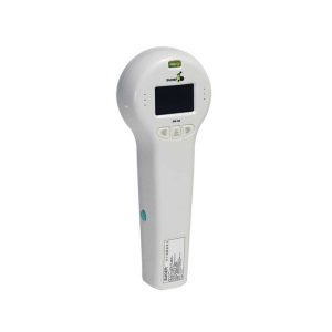
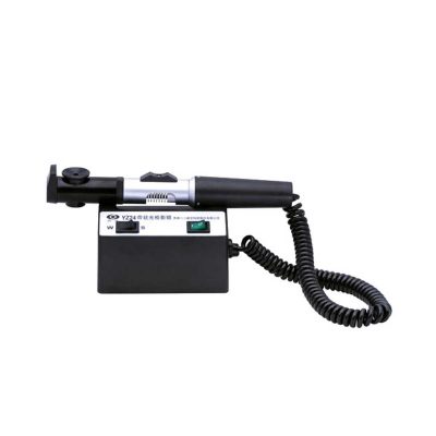
Ultra-small corneal curvature tester, is mainly used to measure the corneal curvature radius and diopter, wireless output print data.
Delivery & Availability:
Typically 14 working days – excluding furniture and heavy/bulky equipment. Please contact us for further information.
| Ship from abroad
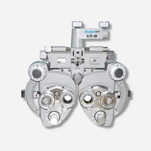
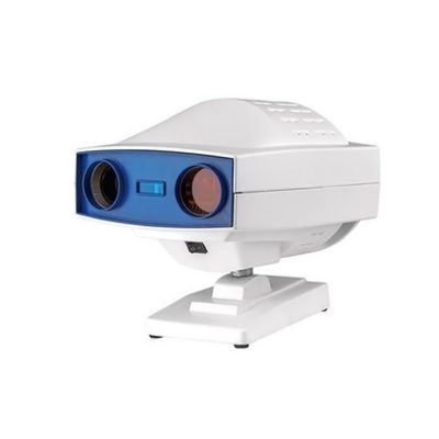
- Equipped with comprehensive measuring functions, it provides SPH, CYL, AXIS and pupil distance optometry
- Durable and easy to operate
- Easily and intuitively read the sphere focal scale value
- High eco-friendly materials
- Design fitting the face curve and no stimulation
Delivery & Availability:
Typically 14 working days – excluding furniture and heavy/bulky equipment. Please contact us for further information.
| In Stock
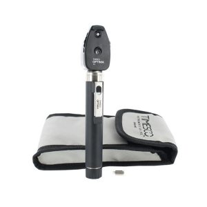
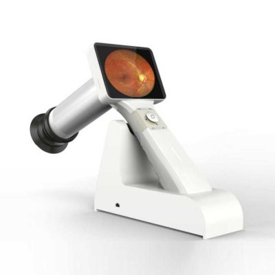
- Durable and light weight
- Supplied in a soft pouch
- Long life bright white Xenon bulb
- Macular beam dioptres lenses from 0 to +20 and 0 to -20
- 3 year guarantee (excludes consumables)
Delivery & Availability:
Typically 5-7 working days – excluding furniture and heavy/bulky equipment. Please contact us for further information.
| Shipped from abroad
- Light rack with integral casting, CNC machining, the measuring system is more stable, good consistency.
- Color LCD display, 5.7 inches man-machine interface is more comfortable.
- PD automatic measurement function, automatically PD value.
Delivery & Availability:
Typically 14 working days – excluding furniture and heavy/bulky equipment. Please contact us for further information.
| Shipped from abroad
This ultra-portable is an excellent diagnostic instrument for the examination of anterior segment structures and ocular abnormalities.
Delivery & Availability:
Typically 14 working days – excluding furniture and heavy/bulky equipment. Please contact us for further information.
|
| Content | | Portable Keratometer Features:
Ultra-small corneal curvature tester, is mainly used to measure the corneal curvature radius and diopter, wireless output print data.
Technical Specifications:
| Model |
SW-100 |
| Measuring Range |
6.5mm~9.5mm: ±0.05mm: 0.01mm |
| Precision |
± 0.05mm |
| Resolution of Curvature Radius of Cornea |
0.01mm |
| Single Measuring Time |
0.03s |
| Output |
Wireless Infrared Thermal Printer |
| Can observe the eye directly through the screen. |
| Weight |
0.5Kg(with batteries) |
| Dimension |
240mmx90mmx60mm |
| Power |
500mW+15% |
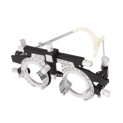
| Features:
- Equipped with comprehensive measuring functions, it provides SPH, CYL, AXIS and pupil distance optometry
- Durable and easy to operate
- Easily and intuitively read the sphere focal scale value
- High eco-friendly materials
- Design fitting the face curve and no stimulation
- Easy to take and clean
- Free switch between the cross-cylindrical lens and the rotary prism
- When the rotating risk is turning by the sphere, it can make sphere power adjust 3.00D for big scope.
- It is designed expediently and smartly for a particular cross cylinder. Supporting supplementary lens could increase scope of measurement.
Technical Specifications:
|
Sphere |
Range:-19.00~+16.75m-1 Step: 0.25m-1, 3.00m-1 |
|
Cylinder |
Range: 0.00~-6.00m-1(Measuring Range With Accessories0.00~-8.00m-1) Step: 0.25m-1 |
|
Cylinder Axis |
Range: 0~180°, Step:5° |
|
Distance of Optical center (also known as Pupil) |
Range: 50~75mmStep: 1mm |
|
Sight Switch |
Range:∞~380mm (distance of Optical center is64mm) |
|
Front Chin Test |
Range: 0~16mm |
|
Distance (from cornea vertex to the lens surface) |
16mm |
|
Standard Accessories Lens |
two pieces of Auxiliary Cylinder -2.00m-1 and -0.12m-1 respectively |
|
Standard Accessories |
one piece of M2 Hexagon wrench , one piece of a Myopia Standard Card, two piece of Myopia Standard Card , one piece of standard card holder , a dust cover |
|
Auxiliary Lens |
“O”:Open aperture
“R”:Retinoscope lens
“R”:Retinoscope lens
“R”:Retinoscope lens
*Lens of +1.50m-1 ,It is suit for the distance of 67 centimeters
“P”:Polaroid
* it is used for examining the dioptric balance of eyes , Implicit strabismus and stereo vision
“RMV”:Red Vertical maddox
*Be used to examine Implicit strabismus
“RMH”:Red horizontal maddox
*Be used to examine Implicit strabismus
“WMV”:Plane Vertical Maddox
*Be used to examine Implicit strabismus
“WMH”:Plane horizontal maddox
*Be used to examine Implicit strabismus
“RL”:Red lens
*Be used to examine eye function, Blending function and Implicit strabismus
“GL”:Green lens
*Be used to examine eye function, Blending function and Implicit strabismus
“+”:Test mark of optical center adjustment
“+.12”:Dioptric of the Spherical Lens is +0.12m-1
*Be used for the semi-adjustment of sphere lens, 0.25m-1
“PH”:1mmPinhole lens
*Be used to exclude visual non-refractive errors of the tested eye
“6ΔU”:6ΔBottom-up prism
*Be used to examine the rotating prism with the detection of nearly horizontal squint
“10ΔI”:10ΔBottom-up prism
*Be used to examine the rotating prism with the detection of nearly horizontal squint
“±0.50”:Cross-cylindrical lens
*Be used to examine the corrected dioptric of the Presbyopia and spherical lens
“OC”:Black lens
|
|
size |
338(L)×99(W)×292(H)mm |
|
NW |
about 5kg |
| Timesco Ophthalmoscope features a head made from lightweight hermetically sealed durable plastic, precision optics and a latex free rubber eyebrow rest. A bright white light from long life standard bulbs provides crystal clear illumination in ophthalmic diagnostic procedures.
- Durable and lightweight
- Supplied in a soft pouch
- Long life bright white Standard bulb
- Macular beam dioptres lenses from 0 to +20 and 0 to -20
- 3-year guarantee (excludes consumables)
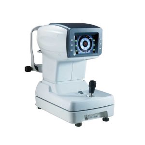
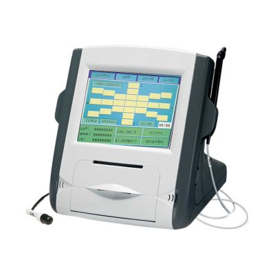
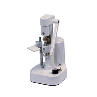
| Features:
- Using the ARM processor and the latest domestic image processing, the system is fast and the image is clear.
- Japan’s mature optical path system, humanized automatic mist measurement process, to reduce the error caused by adjustment, more precise measurements.
- Light rack with integral casting, CNC machining, the measuring system is more stable, good consistency.
- Color LCD display, 5.7 inches man-machine interface is more comfortable.
- PD automatic measurement function, automatically PD value.
- One key lock function, quickly locking mobile platforms.
- Innovative designs, according to the structure of human body engineering, to bring customers good aesthetic feeling and comfortable.
- Data transmission with CV7200, improved efficiency of online 5x magnification options, respond to different viewing needs.
Technical Specifications:
Measurement Range
Vertex distance.......................................0.0,12.0,13.75,15mm
Sphere.......................................-20D~+20D(0.12/0.25D step)
Cylinder...........................................0~±10D(0.12/0.25D step)
Axis...............................................................1~180°(1° step)
PD distance............................................................30~85mm
PD..........................................................................10~86mm
Minimum pupil diameter..............................................Ø2.0mm
Target................................................automatic fogging target
Hardware specifications
Monitor.............................................................5.7" color LCD
Printer......................................................Fast thermal printer
Power saving function....................OFF, 5/15 mins (selectable)
Power supply................................AC110~220V, 60/50Hz, 50W
Dimension/weight..............................288x500x480mm(14kgs) | Features:
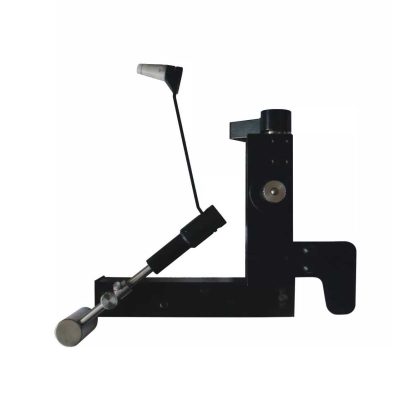
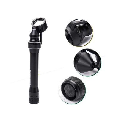
- Ultra-portable: This ultra-portable is an excellent diagnostic instrument for the examination of anterior segment structures and ocular abnormalities. Its easy-to-operate optical system produces a high-brightness, continuously adjustable slit image ideal for pediatric and geriatric setting, emergency department screenings, ward rounds, beside examination, post-op evaluations, and mission work;
- LED illumination: the first and the only portable slit lamp in the world applying LED illumination system. The most prominent advantage of our LED illumination system gives the examiner the clearest image without glare;
- Comfortable using experience: The low heat radiation from our LED lamp makes the most comfortable examine experience for the patient;
- Sharpest Slit: With the blade imaging system S150 gives equally sharpest slit as the best classic slit lamp in the world;
- No need to replace illumination lamp: The LED lamp used in S150 portable slit lamp is rated 20,000 hours at full power. Its lifetime is almost 10 times longer than a normal halogen lamp;
- Power-saving: S150 can work for long hours without changing batteries.
Technical Specifications:
- Magnification: 5x
- Range of slit length: 5mm~11mm
- Minimum slit width: 0.2mm
- Working distance: 12~32mm
- Power supply: AA batteries x 2
- Illumination: LED blub(3.3V/1W)
- Battery life: More than 3h(AA alkaline batteries x 2)
- Net weight: 150g(without batteries)
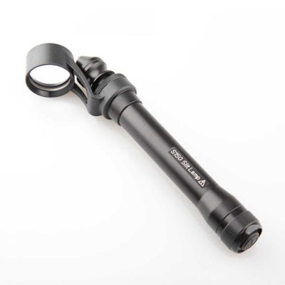
How to Operate:
- Press the Power Switch to turn the slit lamp on/off
- Loosen the Locking Screw
- Twist the Lamp Body while holding the Magnifier Bracket still till the slit is in the required angle
- Push or pull the Lamp Body while holding the Magnifier Bracket still to set the Magnifier into a position comfortable for observation
- Fasten the Locking Screw
- Pull the Lamp Head back and forth for adjusting the width and the length of the slit, as well as the intensity of the light
|


Reviews
There are no reviews yet.