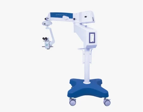
FOCUS ADV PLUS MICROSCOPE
$0.00
Shipped From Abroad
Typically 10-21 working days – excluding furniture and heavy/bulky equipment. Please contact us for further information.
Description
Description
Electromagnetic Brake
Through the electromagnetic brakes, any and all positioning of the microscope can be done with total precision and comfort. Guaranteeing less effort, ease and time optimization.
LCD screen
The touch screen LCD display is used to view and activate all equipment settings. With its touch screen, all controls can be performed with a simple touch.
Pedal control
The automation of functions, with their control through a multitasking pedal, allows the Ophthalmologist and Otorhinolaryngologist complete freedom of their hands during the surgical procedure.
Quick Comparison
| FOCUS ADV PLUS MICROSCOPE remove | Auto Refractometer remove | Ear Irrigation and acumen removal remove | Operation Microscope remove | Ophthalmic AB Scan Machine remove | Slit Lamp remove | |||||||||||||||||||||||||||||||||||||||||||||||||||||||||||||||||||||||||||||||||
|---|---|---|---|---|---|---|---|---|---|---|---|---|---|---|---|---|---|---|---|---|---|---|---|---|---|---|---|---|---|---|---|---|---|---|---|---|---|---|---|---|---|---|---|---|---|---|---|---|---|---|---|---|---|---|---|---|---|---|---|---|---|---|---|---|---|---|---|---|---|---|---|---|---|---|---|---|---|---|---|---|---|---|---|---|---|---|
| Name | FOCUS ADV PLUS MICROSCOPE remove | Auto Refractometer remove | Ear Irrigation and acumen removal remove | Operation Microscope remove | Ophthalmic AB Scan Machine remove | Slit Lamp remove | ||||||||||||||||||||||||||||||||||||||||||||||||||||||||||||||||||||||||||||||||
| Image |  |  |  | 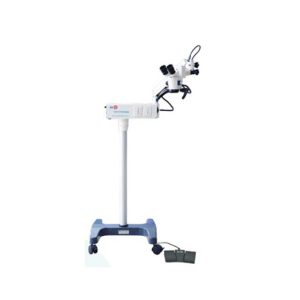 | 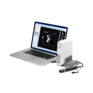 | 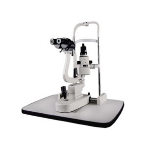 | ||||||||||||||||||||||||||||||||||||||||||||||||||||||||||||||||||||||||||||||||
| SKU | SF103356013091-7 | SF1033560107-14 | SF103356013012 | SF1033560107-24 | SF1033560107-8 | SF1033560107-15 | ||||||||||||||||||||||||||||||||||||||||||||||||||||||||||||||||||||||||||||||||
| Rating | ||||||||||||||||||||||||||||||||||||||||||||||||||||||||||||||||||||||||||||||||||||||
| Price |
| $2,035.00 |
| $2,695.00 | $4,895.00 | $1,375.00 | ||||||||||||||||||||||||||||||||||||||||||||||||||||||||||||||||||||||||||||||||
| Stock | ||||||||||||||||||||||||||||||||||||||||||||||||||||||||||||||||||||||||||||||||||||||
| Availability | ||||||||||||||||||||||||||||||||||||||||||||||||||||||||||||||||||||||||||||||||||||||
| Add to cart | ||||||||||||||||||||||||||||||||||||||||||||||||||||||||||||||||||||||||||||||||||||||
| Description | Shipped From Abroad
FOCUS ADVANCED PLUS microscope, a line of equipment specially designed in detail, to meet the most demanding standards of Ophthalmological and ENT surgical procedures. Microsurgeries currently require cutting-edge technology with the ability to perform extremely complex procedures in an agile and simple way. Through the Electromagnetic Brake, activated by a button, any and all positioning of the microscope can be done with complete accuracy, comfort and lightness. This ensures less effort and ease during all surgical procedures. For optimizing surgical processes, the NEVRO microscope is the most efficient and advanced.
Delivery & Availability:
Typically 10-21 working days – excluding furniture and heavy/bulky equipment. Please contact us for further information. | Shipped from abroad
| In Stock
Features:
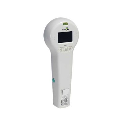
●Professional
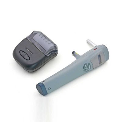
Same ear wax removal tool as those used by doctors, you can easily eliminate ear wax buildup at home, really save your money and time on medical visiting. Safe and Environmentally Friendly.
●Quick & Easy
This ear wax removal kit is a quick, effective treatment for excess ear wax buildup. Fill the bottle with solution, Twist on the disposable tip, Use the trigger handle to spray solution into the ear canal. So Easy.
Delivery & Availability:
Typically 7-14 working days – excluding furniture and heavy/bulky equipment. Please contact us for further information.
| Shipped from abroad
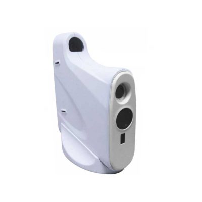
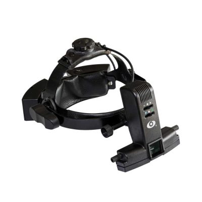
YZ20P5 Operation Microscope is a simple binocular coaxial microscope for a single man. The Operation Microscope is small, light, and convenient. It has high agility and can meet the requirements for general ophthalmology operation. The microscope fits mobile medical treatment.
| Shipped from abroad
| Shipped from abroad
| ||||||||||||||||||||||||||||||||||||||||||||||||||||||||||||||||||||||||||||||||
| Content | Description
Electromagnetic Brake
Through the electromagnetic brakes, any and all positioning of the microscope can be done with total precision and comfort. Guaranteeing less effort, ease and time optimization.
LCD screen
The touch screen LCD display is used to view and activate all equipment settings. With its touch screen, all controls can be performed with a simple touch.
Pedal control
The automation of functions, with their control through a multitasking pedal, allows the Ophthalmologist and Otorhinolaryngologist complete freedom of their hands during the surgical procedure.
| Features:
| Features: ●Professional Same ear wax removal tool as those used by doctors, you can easily eliminate ear wax buildup at home, really save your money and time on medical visiting. Safe and Environmentally Friendly. ●Quick & Easy This ear wax removal kit is a quick, effective treatment for excess ear wax buildup. Fill the bottle with solution, Twist on the disposable tip, Use the trigger handle to spray solution into the ear canal. So Easy. ●Standard Capacity of the ear cleaner solution bottle is 10.6Oz, it has the most suitable size to hold in hand. Working at condition 32-122℉(0-50℃). Recommend to fill 1/5 of the bottle with OTC hydrogen peroxide, and 4/5 with very warm water. ●Complete Ear Washer System Our earwax removal kit comes with 1× Ear Washer Bottle, 1× Wash Basin, 1× Rubber Bulb, 1× Short Injection Head, 1× Long Hose Injection Head, 5× Disposable Tip, 1× User Manual. | Operation Microscope(YZ20P5) is a simple binocular coaxial microscope for a single man. The Microscope is small, light, and convenient. It has high agility and can meet the requirements for general ophthalmology operation. The microscope fits mobile medical treatment.
Features of Operation Microscope:
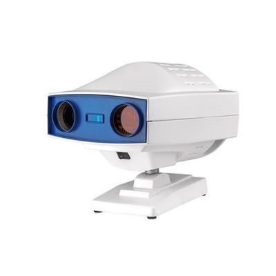
Click Here To Download Catalogue | Functions of Ophthalmic AB Scan Machine:
| Slit Lamp Features:
| ||||||||||||||||||||||||||||||||||||||||||||||||||||||||||||||||||||||||||||||||
| Weight | N/A | N/A | N/A | N/A | N/A | N/A | ||||||||||||||||||||||||||||||||||||||||||||||||||||||||||||||||||||||||||||||||
| Dimensions | N/A | N/A | N/A | N/A | N/A | N/A | ||||||||||||||||||||||||||||||||||||||||||||||||||||||||||||||||||||||||||||||||
| Additional information |


Reviews
There are no reviews yet.