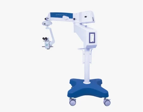
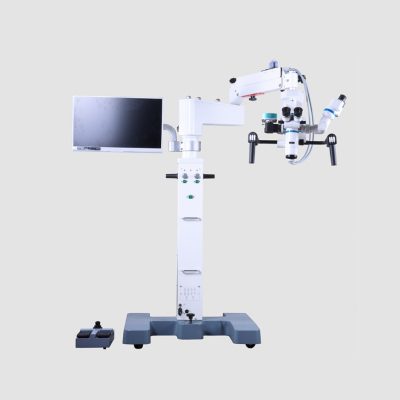
FOCUS ADV PLUS MICROSCOPE
$0.00
Shipped From Abroad
FOCUS ADVANCED PLUS microscope, a line of equipment specially designed in detail, to meet the most demanding standards of Ophthalmological and ENT surgical procedures. Microsurgeries currently require cutting-edge technology with the ability to perform extremely complex procedures in an agile and simple way. Through the Electromagnetic Brake, activated by a button, any and all positioning of the microscope can be done with complete accuracy, comfort and lightness. This ensures less effort and ease during all surgical procedures. For optimizing surgical processes, the NEVRO microscope is the most efficient and advanced.
Delivery & Availability:
Typically 10-21 working days – excluding furniture and heavy/bulky equipment. Please contact us for further information.
Typically 10-21 working days – excluding furniture and heavy/bulky equipment. Please contact us for further information.
Description
Description
Electromagnetic Brake
Through the electromagnetic brakes, any and all positioning of the microscope can be done with total precision and comfort. Guaranteeing less effort, ease and time optimization.
LCD screen
The touch screen LCD display is used to view and activate all equipment settings. With its touch screen, all controls can be performed with a simple touch.
Pedal control
The automation of functions, with their control through a multitasking pedal, allows the Ophthalmologist and Otorhinolaryngologist complete freedom of their hands during the surgical procedure.
Quick Comparison
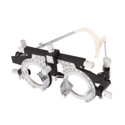
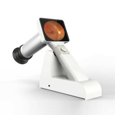
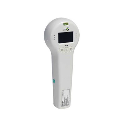
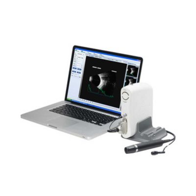
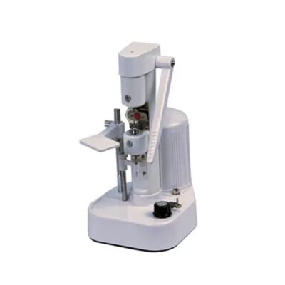
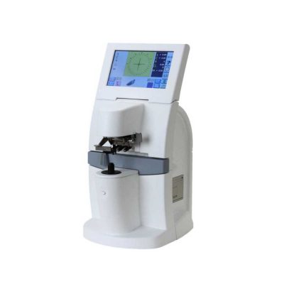
| FOCUS ADV PLUS MICROSCOPE remove | Auto Refractometer remove | Portable Rebound Tonometer remove | Timesco Ophthalmoscope remove | Binocular Indirect Ophthalmoscope remove | Applanation Tonometer remove | ||||||||||||||||||||||||||||||||||||||||||
|---|---|---|---|---|---|---|---|---|---|---|---|---|---|---|---|---|---|---|---|---|---|---|---|---|---|---|---|---|---|---|---|---|---|---|---|---|---|---|---|---|---|---|---|---|---|---|---|
| Name | FOCUS ADV PLUS MICROSCOPE remove | Auto Refractometer remove | Portable Rebound Tonometer remove | Timesco Ophthalmoscope remove | Binocular Indirect Ophthalmoscope remove | Applanation Tonometer remove | |||||||||||||||||||||||||||||||||||||||||
| Image |  |  |  |  |  |  | |||||||||||||||||||||||||||||||||||||||||
| SKU | SF103356013091-7 | SF1033560107-14 | SF1033560107-17 | SF1033560084-282 | SF1033560107-4 | SF1033560107-1 | |||||||||||||||||||||||||||||||||||||||||
| Rating | |||||||||||||||||||||||||||||||||||||||||||||||
| Price |
| $2,035.00 | $1,815.00 | $140.00 | $880.00 |
| |||||||||||||||||||||||||||||||||||||||||
| Stock | |||||||||||||||||||||||||||||||||||||||||||||||
| Availability | |||||||||||||||||||||||||||||||||||||||||||||||
| Add to cart | |||||||||||||||||||||||||||||||||||||||||||||||
| Description | Shipped From Abroad
FOCUS ADVANCED PLUS microscope, a line of equipment specially designed in detail, to meet the most demanding standards of Ophthalmological and ENT surgical procedures. Microsurgeries currently require cutting-edge technology with the ability to perform extremely complex procedures in an agile and simple way. Through the Electromagnetic Brake, activated by a button, any and all positioning of the microscope can be done with complete accuracy, comfort and lightness. This ensures less effort and ease during all surgical procedures. For optimizing surgical processes, the NEVRO microscope is the most efficient and advanced.
Delivery & Availability:
Typically 10-21 working days – excluding furniture and heavy/bulky equipment. Please contact us for further information. | Shipped from abroad
| Shipped from abroad
Tonometer SW-500 with vertical and horizontal two working modes, wireless output print data.
| In Stock
| Shipped from abroad
Super lightweight design, reduce fatigue, operation is very convenient.
| Shipped from abroad
The product is designed on the principle basis of Goldman tonometer. It can be connected with slit lamp(Carl Zeiss type).
| |||||||||||||||||||||||||||||||||||||||||
| Content | Description
Electromagnetic Brake
Through the electromagnetic brakes, any and all positioning of the microscope can be done with total precision and comfort. Guaranteeing less effort, ease and time optimization.
LCD screen
The touch screen LCD display is used to view and activate all equipment settings. With its touch screen, all controls can be performed with a simple touch.
Pedal control
The automation of functions, with their control through a multitasking pedal, allows the Ophthalmologist and Otorhinolaryngologist complete freedom of their hands during the surgical procedure.
| Features:
| Tonometer SW-500 with vertical and horizontal two working modes, wireless output print data. The equipment is used to measure intraocular pressure, using the principle of: the probe hits the surfaces of different hardness at a certain speed, has a different reaction when the probe rebounds. Be of advantages of high accuracy, portable, without anesthesia, without the cross-infection, etc.
Features:
| Timesco Ophthalmoscope features a head made from lightweight hermetically sealed durable plastic, precision optics and a latex free rubber eyebrow rest. A bright white light from long life standard bulbs provides crystal clear illumination in ophthalmic diagnostic procedures.
Click Here To Download Catalogue | Ophthalmoscope Features:
| Applanation Tonometer is designed on the principle basis of Goldman tonometer. It can be connected with slit lamp(Carl Zeiss type).
Features of Applanation Tonometer:
| |||||||||||||||||||||||||||||||||||||||||
| Weight | N/A | N/A | N/A | N/A | N/A | N/A | |||||||||||||||||||||||||||||||||||||||||
| Dimensions | N/A | N/A | N/A | N/A | N/A | N/A | |||||||||||||||||||||||||||||||||||||||||
| Additional information | |||||||||||||||||||||||||||||||||||||||||||||||


Reviews
There are no reviews yet.