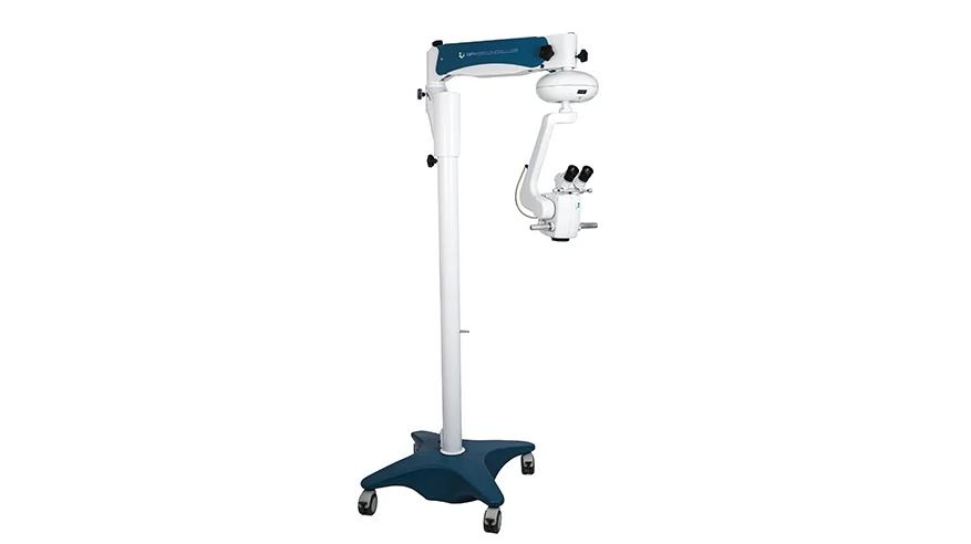
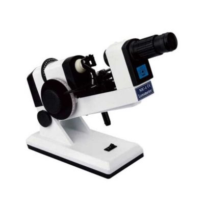
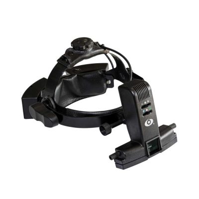
FOCUS ADVANCED MICROSCOPE
$0.00
Shipped From Abroad
The FOCUS series is a line of equipment specially designed in detail to meet the most demanding standards of anterior and posterior segment surgical procedures.
The optics developed for FOCUS line microscopes enable external transmission of LED light, generating excellent clarity and image depth of the observed field.
Ideal for Cataract and Retina procedures.
The optics developed for FOCUS ADVANCED line microscopes enable external transmission of LED light (oblique), generating excellent clarity and image depth of the observed field. The equipment has an independent coaxial lighting system, activated via a multifunction pedal, ensuring total image clarity and excellent red reflection quality, providing total safety and visibility during cataract procedures.
Having functions such as zoom, microfocusing, intensity control activated via pedal and being compatible with the best non-contact lens adapters for vitreectomy on the market, it enables all retinal procedures with total quality.
Typically 10-21 working days – excluding furniture and heavy/bulky equipment. Please contact us for further information.
Description
Description
Versatility in multiple configuration possibilities
Focus Microscopes stand out in the market for presenting multidisciplinary characteristics and high optical quality. Additionally, they allow you to add options and accessories according to the need for use, including binoculars, double iris, image inverter, among others.
The Focus* line of microscopes has a pedal-operated motorized microfocusing and zooming system, aiming for greater precision and comfort for the user during surgical procedures.
Pedal controlled XY positioner
The DFVasconcellos XY Positioner allows precise alignment between the user’s vision and the surgical field. This becomes an increasingly necessary resource as microscopy techniques become more widespread.
Quick Comparison
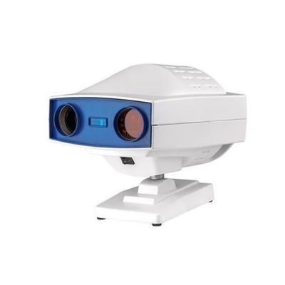
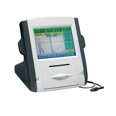
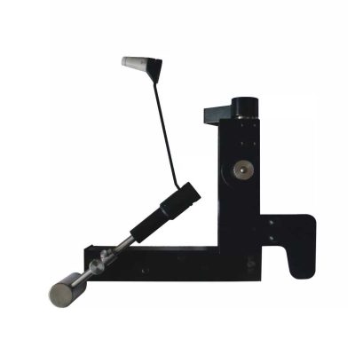
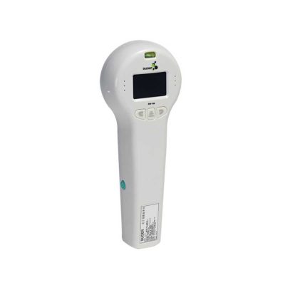
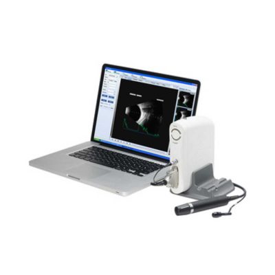
| FOCUS ADVANCED MICROSCOPE remove | Automatic Computer Goldmann (Visual Field Analyzer) remove | Pantoscopic Ophthalmoscope remove | Ophthalmic AB Scan Machine remove | ENT/Neurosurgery Operating Microscope remove | Slit Lamp with Workstation remove | |||||||||||||||||||||||||||||||||||||||||||||||||||||||||||||||||
|---|---|---|---|---|---|---|---|---|---|---|---|---|---|---|---|---|---|---|---|---|---|---|---|---|---|---|---|---|---|---|---|---|---|---|---|---|---|---|---|---|---|---|---|---|---|---|---|---|---|---|---|---|---|---|---|---|---|---|---|---|---|---|---|---|---|---|---|---|---|---|
| Name | FOCUS ADVANCED MICROSCOPE remove | Automatic Computer Goldmann (Visual Field Analyzer) remove | Pantoscopic Ophthalmoscope remove | Ophthalmic AB Scan Machine remove | ENT/Neurosurgery Operating Microscope remove | Slit Lamp with Workstation remove | ||||||||||||||||||||||||||||||||||||||||||||||||||||||||||||||||
| Image |  | 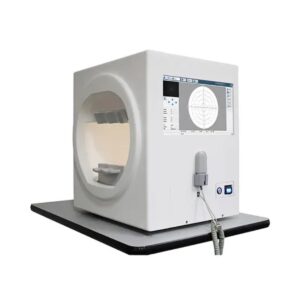 | 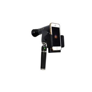 | 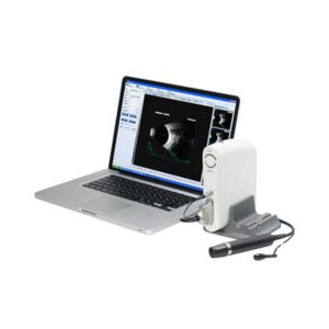 | 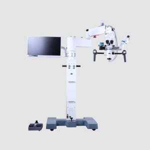 | 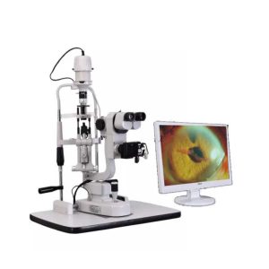 | ||||||||||||||||||||||||||||||||||||||||||||||||||||||||||||||||
| SKU | SF103356013091-8 | SF103356013013 | SF1033560107-3 | SF1033560107-8 | SF1033560109-1 | SF1033560107-7 | ||||||||||||||||||||||||||||||||||||||||||||||||||||||||||||||||
| Rating | ||||||||||||||||||||||||||||||||||||||||||||||||||||||||||||||||||||||
| Price |
| $3,850.00 |
| $4,895.00 |
| $3,740.00 | ||||||||||||||||||||||||||||||||||||||||||||||||||||||||||||||||
| Stock | ||||||||||||||||||||||||||||||||||||||||||||||||||||||||||||||||||||||
| Availability | ||||||||||||||||||||||||||||||||||||||||||||||||||||||||||||||||||||||
| Add to cart | ||||||||||||||||||||||||||||||||||||||||||||||||||||||||||||||||||||||
| Description | Shipped From Abroad
The FOCUS series is a line of equipment specially designed in detail to meet the most demanding standards of anterior and posterior segment surgical procedures.
The optics developed for FOCUS line microscopes enable external transmission of LED light, generating excellent clarity and image depth of the observed field.
Ideal for Cataract and Retina procedures.
The optics developed for FOCUS ADVANCED line microscopes enable external transmission of LED light (oblique), generating excellent clarity and image depth of the observed field. The equipment has an independent coaxial lighting system, activated via a multifunction pedal, ensuring total image clarity and excellent red reflection quality, providing total safety and visibility during cataract procedures.
Having functions such as zoom, microfocusing, intensity control activated via pedal and being compatible with the best non-contact lens adapters for vitreectomy on the market, it enables all retinal procedures with total quality.
Delivery & Availability:
Typically 10-21 working days – excluding furniture and heavy/bulky equipment. Please contact us for further information. | In Stock
Features:
The Bio-1000 automated perimeter absorbs the advantages of international advanced perimetry devices. It comprises the highly integrated computer, optics, machinery and electronics systems.
Delivery & Availability:
Typically 7-14 working days – excluding furniture and heavy/bulky equipment. Please contact us for further information.
| Shipped from abroad
The brand-new Pantoscopic Ophthalmoscope is a portable digital imaging device which makes it possible to view and take pictures of the eyes.
| Shipped from abroad
| Shipped from abroad
Corder Microscope has Fluid, Responsive and Accurate.Fluid. Responsive. Accurate. These were a few of the principles guiding every phase in the design of the Corder Microscope. With the choicest mechanical machined components, the Corder Microscope has the grace and agility to adjust to every desired position on command. Well designed Apochromatic optics treated with Corder's Mcoatings produce true-to life sharp images with high depth, definition and contrast. | Shipped from abroad
| ||||||||||||||||||||||||||||||||||||||||||||||||||||||||||||||||
| Content | Description
Versatility in multiple configuration possibilities
Focus Microscopes stand out in the market for presenting multidisciplinary characteristics and high optical quality. Additionally, they allow you to add options and accessories according to the need for use, including binoculars, double iris, image inverter, among others.
The Focus* line of microscopes has a pedal-operated motorized microfocusing and zooming system, aiming for greater precision and comfort for the user during surgical procedures.
Pedal controlled XY positioner
The DFVasconcellos XY Positioner allows precise alignment between the user's vision and the surgical field. This becomes an increasingly necessary resource as microscopy techniques become more widespread.
| The Bio-1000 automated perimeter absorbs the advantages of international advanced perimetry devices. It comprises the highly integrated computer, optics, machinery and electronics systems. Incorporated with the advanced configuration, comprehensive software inspection categories, and strictly in accordance with international Goldman standard, it provide scientific means for glaucoma, fundus disease, visual pathway injury and neurological diseases.
Feature:
* Comprehensive real-time monitoring,Heiji-krakau physiological blind spot monitoring,gaze tracking/head position tracking,automatic measurement of pupil diameter, reduce the impact of pupil effect on visual field detection.
* Personalized design,accurate clinical analysis,accurate and repid examination strategy.
* Under international Goldman standard,providing a variety of classic test procedures and report analysis.
Technical Specification:
Click Here To Download Catalogue | The brand-new Pantoscopic Ophthalmoscope is a portable digital imaging device which makes it possible to view and take pictures of the eyes. The optical access of the Pantoscopic Ophthalmoscope is aligned to the visual axis of the smartphone camera by the adaptor which allows to you take pictures of the fundus and retinal nerve in high resolution. You could save pictures for each patient or email and print as needed. The Pantoscopic Ophthalmoscope provides a 5X larger view of the fundus compared with the standard ophthalmoscope. It has a wider view field of 230. Without dilating the pupil, the fundus imagines could be captured at any time and places.
Features:
| Functions of Ophthalmic AB Scan Machine:
| Features:Corder Microscope has Fluid, Responsive and Accurate.Fluid. Responsive. Accurate. These were a few of the principles guiding every phase in the design of the Corder Microscope. With the choicest mechanical machined components, the Corder Microscope has the grace and agility to adjust to every desired position on command. Well designed Apochromatic optics treated with Corder's Mcoatings produce true-to life sharp images with high depth, definition and contrast. More comfortable operation Tiltable binocular tubes available, which can incline more than 60° depending on the posture and physique of the operating surgeon. Movable range: 30° (straight) to 90° (inclined) Corder microscope configured with XYZ motorized movement operated through a comfortable foot /Handle control, a veryeffective co-axial illumnation and 50W halogen light source makes it ideal for Neuro surgeries.Doctor-patient communication is easierTo address digital documentation needs, a host of digital SLR, video camera, and CCD adapters are made available with the ProLine in addition to Corder's proprietary iVu multi-functional imaging solution. 1080P full hd image quality, efficient image management during the operation. Integrate your digital workflow to facilitate case management and facilitate more intuitive patient communication. Technical Permeants: Magnification: motorized zoom system, 1:6 zoom ratio, magnification 3x~16x Focusing range: 50mm Binocular tube: 30°~90° tiltable tube ,(0° ~200° optional) Eyepiece: 12.5x / 10x Objective lens: F 300mm(175mm, 250mm, 350mm optional) pupil distance: 55mm~75mm diopter adjustment: +6D ~ -6D Field of view: Φ74~Φ12mm X-Y translator: Motorized by foot switch or handle controller, ±30mm Assistant tube: 360° Rotating assistant tube Reset functions: YES Illumination System: Coaxial illumination Light source: Halogen lamp Light intensity adjustment: Continuous brightness adjustment 0-100000lux Fiber optic illumination: Dual fiber Field of illumination: Φ50mm Filter: Red free filter, small spot Accessories CCD Camera system: Beam splitter, CCD adapter, CCD, Display XENON LAMP: 150000lux Integrated Video Adapter: SONY / CANON CameraClick Here To Download Catalogue | Slit Lamp with Workstation Features:
| ||||||||||||||||||||||||||||||||||||||||||||||||||||||||||||||||
| Weight | N/A | N/A | N/A | N/A | N/A | N/A | ||||||||||||||||||||||||||||||||||||||||||||||||||||||||||||||||
| Dimensions | N/A | N/A | N/A | N/A | N/A | N/A | ||||||||||||||||||||||||||||||||||||||||||||||||||||||||||||||||
| Additional information |


Reviews
There are no reviews yet.