FOCUS STANDARD MICROSCOPE
$0.00
Shipped From Abroad
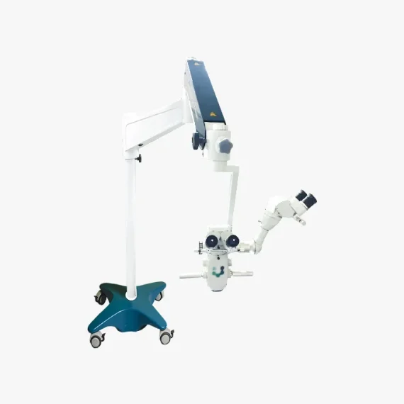
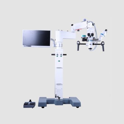
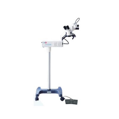
The FOCUS series is a line of equipment specially designed in detail to meet the most demanding standards of anterior and posterior segment surgical procedures.The optics developed for FOCUS line microscopes enable external transmission of LED light, generating excellent clarity and image depth of the observed field.
Ideal for Cataract procedures.
Delivery & Availability:
Typically 10-21 working days – excluding furniture and heavy/bulky equipment. Please contact us for further information.
Typically 10-21 working days – excluding furniture and heavy/bulky equipment. Please contact us for further information.
Description
Description
Versatility in multiple configuration possibilities
Focus Microscopes stand out in the market for presenting multidisciplinary characteristics and high optical quality. Additionally, they allow you to add options and accessories according to the need for use, including binoculars, double iris, image inverter, among others.
The Focus* line of microscopes has a pedal-operated motorized microfocusing and zooming system, aiming for greater precision and comfort for the user during surgical procedures.
Quick Comparison
| FOCUS STANDARD MICROSCOPE remove | Tonometer remove | Handheld Digital Auto-refractometer remove | Manual lensmeter remove | Binocular Indirect Ophthalmoscope remove | Auto Refractometer remove | |||||||||||||||||||||||||||||||||||||||||||||
|---|---|---|---|---|---|---|---|---|---|---|---|---|---|---|---|---|---|---|---|---|---|---|---|---|---|---|---|---|---|---|---|---|---|---|---|---|---|---|---|---|---|---|---|---|---|---|---|---|---|---|
| Name | FOCUS STANDARD MICROSCOPE remove | Tonometer remove | Handheld Digital Auto-refractometer remove | Manual lensmeter remove | Binocular Indirect Ophthalmoscope remove | Auto Refractometer remove | ||||||||||||||||||||||||||||||||||||||||||||
| Image |  | 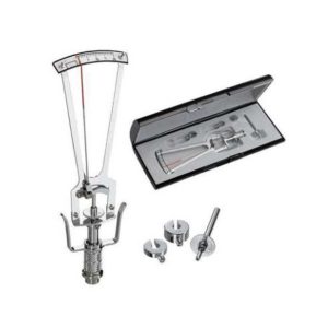 | 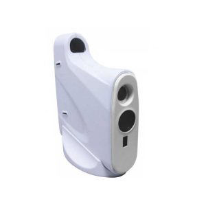 | 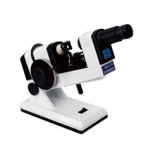 | 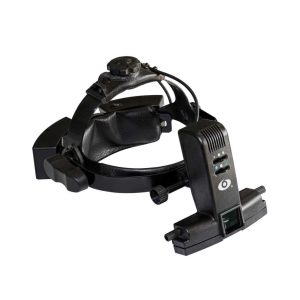 | 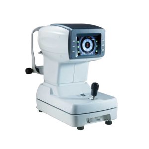 | ||||||||||||||||||||||||||||||||||||||||||||
| SKU | SF103356013091-9 | SF1033560107-9 | SF1033560107-2 | SF1033560107-22 | SF1033560107-4 | SF1033560107-14 | ||||||||||||||||||||||||||||||||||||||||||||
| Rating | ||||||||||||||||||||||||||||||||||||||||||||||||||
| Price |
| $220.00 |
|
| $880.00 | $2,035.00 | ||||||||||||||||||||||||||||||||||||||||||||
| Stock | ||||||||||||||||||||||||||||||||||||||||||||||||||
| Availability | ||||||||||||||||||||||||||||||||||||||||||||||||||
| Add to cart | ||||||||||||||||||||||||||||||||||||||||||||||||||
| Description | Shipped From Abroad
The FOCUS series is a line of equipment specially designed in detail to meet the most demanding standards of anterior and posterior segment surgical procedures.The optics developed for FOCUS line microscopes enable external transmission of LED light, generating excellent clarity and image depth of the observed field.
Ideal for Cataract procedures.
Delivery & Availability:
Typically 10-21 working days – excluding furniture and heavy/bulky equipment. Please contact us for further information.
| Shipped from abroad
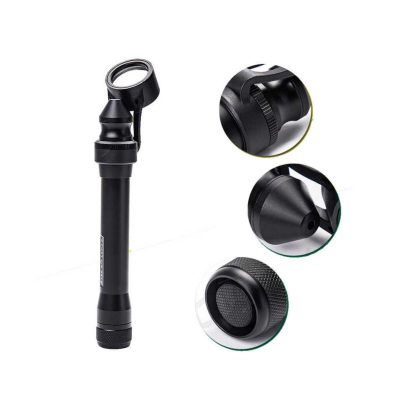
This product is used to measure the intraocular pressure (IOP) by measuring the depth produced on the surface of the cornea by a load of a known weight. Each division on the scale corresponds to 1/20mm corneal depth.
| Shipped from abroad
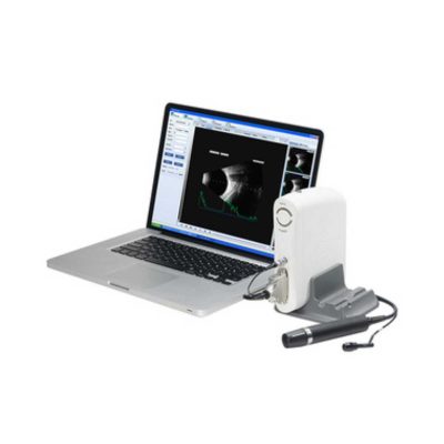
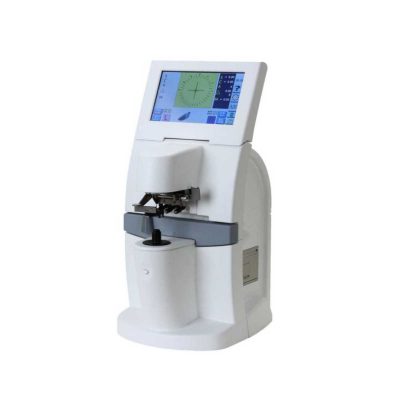
AutoSight 900 is a portable vision screener for patients at any age. Its working principle is the refraction of light.
| Shipped from abroad
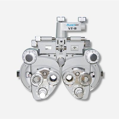
The NJC-4 Lensmeter is used to measure the diopters of spherical lens and cylinder lens, the axis of cylinder lens, the strength and the baseline direction of shuttle lens, it can also stamp the optical center of lens, the axis of cylinder len and the base direction of shuttle lens.
| Shipped from abroad
Super lightweight design, reduce fatigue, operation is very convenient.
| Shipped from abroad
| ||||||||||||||||||||||||||||||||||||||||||||
| Content | Description
Versatility in multiple configuration possibilities
Focus Microscopes stand out in the market for presenting multidisciplinary characteristics and high optical quality. Additionally, they allow you to add options and accessories according to the need for use, including binoculars, double iris, image inverter, among others.
The Focus* line of microscopes has a pedal-operated motorized microfocusing and zooming system, aiming for greater precision and comfort for the user during surgical procedures.
| This product is used to measure the intraocular pressure (IOP) by measuring the depth produced on the surface of the cornea by a load of a known weight. Each division on the scale corresponds to 1/20mm corneal depth.
Features:
| Handheld Digital Auto-refractometer(AutoSight 900) is a portable vision screener for patients at any age. Its working principle is the refraction of light. Optical rays are focused on a sensor after passing through the eye's refractive system. The spherical power, cylindrical power, and axis of both eyes can be obtained by digital signal processing.
Features of Handheld Digital Auto-refractometer:
| The NJC-4 Lensmeter is used to measure the diopters of the spherical lens and cylinder lens, the axis of cylinder lens, the strength and the baseline direction of shuttle lens, it can also stamp the optical center of the lens, the axis of cylinder lens, and the base direction of shuttle lens. This instrument (Both Ac and Dc are permitted Two cells When Dc) has clear readings and graduations as well as high objective precision and reliabilities except that it can be operated easily and conveniently, AU the lenses and the made glasses can be measured by it, therefore, it is a required and ideal measuring instrument for glasses manufactures, glasses stores and ophthalmological hospitals.
Technical Specifications:
| Ophthalmoscope Features:
| Features:
| ||||||||||||||||||||||||||||||||||||||||||||
| Weight | N/A | N/A | N/A | N/A | N/A | N/A | ||||||||||||||||||||||||||||||||||||||||||||
| Dimensions | N/A | N/A | N/A | N/A | N/A | N/A | ||||||||||||||||||||||||||||||||||||||||||||
| Additional information | ||||||||||||||||||||||||||||||||||||||||||||||||||


Reviews
There are no reviews yet.