G-scan Brio: New Weight-Bearing MRI system
$0.00
Shipped From Abroad
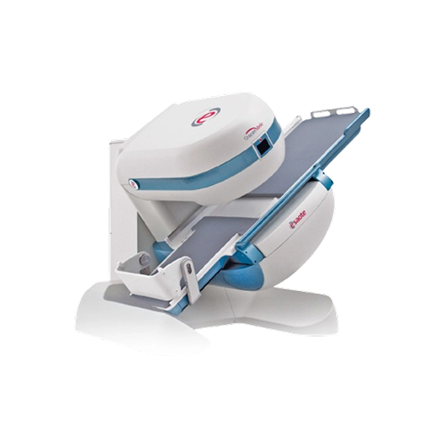
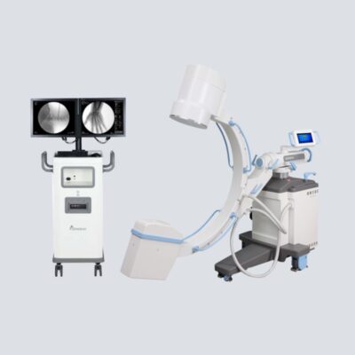
The G-scan Brio Weight-Bearing MRI system is a revolutionary MRI approach for all musculoskeletal applications, which allows you to increase your diagnostic accuracy and confidence. The open and tilting design is a new and innovative way of doing MRI in which the position of the patient becomes an integral part of the outcome of the examination.
Typically 10-21 working days – excluding furniture and heavy/bulky equipment. Please contact us for further information.
Description
The key to confidence
- Minimum space of installation, plug-and-play system.
- Designed for the spine and joints.
- Possibility to combine weight-bearing exam with dynamic imaging.
- Low power consumption.
Adds weight to your diagnosis
Many symptoms and pathologies occur or are emphasised when the patient is in the weight-bearing position. Conventional MRI may not demonstrate the pathology related to particular symptoms, whereas the G-scan Brio gives you a new point of view so you can accurately diagnose MSK pathologies that occur in the weight-bearing position. With the G-scan Brio you can gain a more complete understanding of the spine and joint under examination. The forces of gravity generate bio-mechanical changes in the human anatomy, so MR imaging in the natural standing position allows you to obtain extra details that would not normally be seen.
Imaging & Visualization
The G-scan Brio system’s primary innovation lies in its ability to change the patient’s position during the scan, effectively adding the force of gravity to the diagnostic process.
-
Weight-Bearing (Tilting) Design: The open and tilting design allows the patient’s position (from supine to upright/standing) to become an integral part of the examination outcome. This means the system can scan patients in the natural standing position, which reveals bio-mechanical changes and additional details in the spine and joints that are often missed in a conventional lying-down (supine) scan. * Dynamic Imaging: It offers the possibility to combine the weight-bearing exam with dynamic imaging to gain a more complete understanding of the pathology under functional stress.
-
Open and Quiet: The open-design magnet provides a non-claustrophobic experience for patients and is notably quiet, making it suitable for all patient types, including pediatric patients.
-
Efficiency and Sustainability: Similar to other Esaote open systems, it is a one-room MRI system requiring minimal installation space (approx. 23 m²) and features low power consumption (<3 kW).
Clinical Applications
The G-scan Brio focuses entirely on enhancing the diagnosis of conditions that are affected by posture and gravity.
-
Musculoskeletal (MSK) Applications: It is a revolutionary MRI approach for all musculoskeletal applications, particularly for conditions of the spine and joints (like the knee, hip, and ankle) where symptoms occur or are emphasised in the weight-bearing position.
-
Diagnostic Confidence: By visualising gravity-induced changes, the system provides a new point of view to accurately diagnose pathologies (e.g., instability, joint shifting) that may not be apparent in conventional scans.
Coils (Probe Types)
The G-scan Brio supports 12 dedicated multi-purpose coils tailored for precise imaging of the spine and joints.
| Anatomical Area | Specific Coil Types |
| Spine | Cervical spine coil (Linear & Large DPA), Lumbar spine coils (4-channels, medium & large sizes), Extra-Large Lumbar Spine coil (2-channels, optional) |
| Joints/Extremities | Knee coil DPA, Shoulder coil DPA, Shoulder coil linear, Shoulder coil (3-channels, optional), Ankle-Foot coil DPA, Hand-Wrist coil DPA |
| Head/Other | 4-Channel Head Coil (optional), Flex coil, Flex coil linear (optional), TMJ bilateral coil (2-channels, optional) |
Quick Comparison
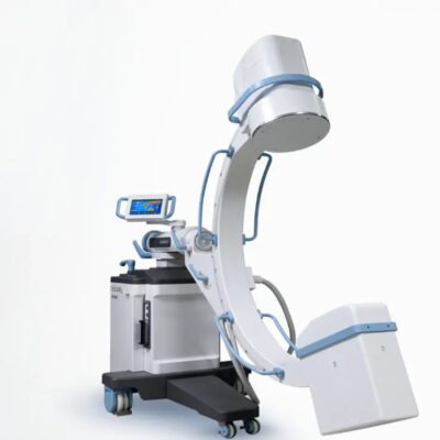
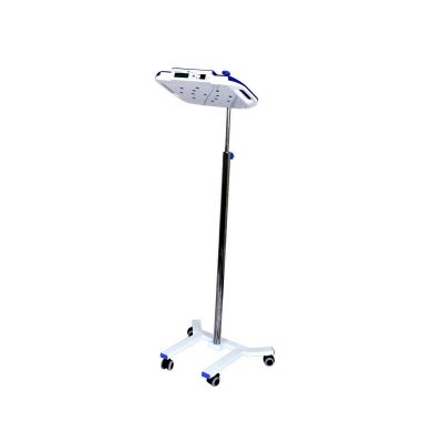
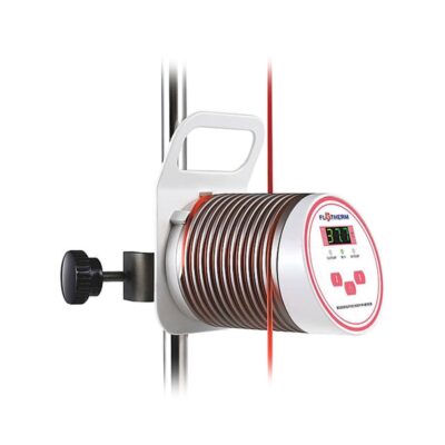
| G-scan Brio: New Weight-Bearing MRI system remove | Keewell FT-1800 Blood & Infusion Warmer remove | DrGem GXR-SD 400mA Floor Mounted Digital X-ray remove | DRGEM DR System remove | DrGem Ceiling Analogue X-ray Machine remove | Biopsy Needle remove | |||||||||
|---|---|---|---|---|---|---|---|---|---|---|---|---|---|---|
| Name | G-scan Brio: New Weight-Bearing MRI system remove | Keewell FT-1800 Blood & Infusion Warmer remove | DrGem GXR-SD 400mA Floor Mounted Digital X-ray remove | DRGEM DR System remove | DrGem Ceiling Analogue X-ray Machine remove | Biopsy Needle remove | ||||||||
| Image | 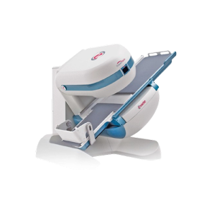 | 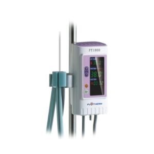 |  | 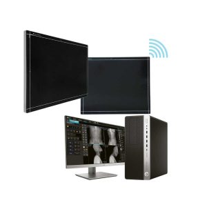 |  | 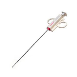 | ||||||||
| SKU | SF1033560076 | SF1033560074-5 | SF1033560074-8 | SF1033560074-7 | SF1033560084-133 | |||||||||
| Rating | ||||||||||||||
| Price |
| $781.00 |
|
|
|
| ||||||||
| Stock | ||||||||||||||
| Availability | ||||||||||||||
| Add to cart | ||||||||||||||
| Description | Shipped From Abroad
The G-scan Brio Weight-Bearing MRI system is a revolutionary MRI approach for all musculoskeletal applications, which allows you to increase your diagnostic accuracy and confidence. The open and tilting design is a new and innovative way of doing MRI in which the position of the patient becomes an integral part of the outcome of the examination.
Delivery & Availability:
Typically 10-21 working days – excluding furniture and heavy/bulky equipment. Please contact us for further information.
| Shipped From Abroad
Safety system:Permanent running self-tests, 24 hours continuous operating Double independent over-heating protections and automatic cut off Visual and acoustic alarm for high temperature / low temperature/sensor faultAdvance Dry Heating Technology:Quick Warming up and effective heatingHeating up to the patient:IV tube is completely wrapped in, no heat lossUser friendly interface:Big LED screen showing set temp., actual temp., heating time and fault situation | In Stock The GXR-SD Digital X-ray is a diagnostic digital radiography system that provides reliable high quality digital radiographic images with a reduced dose. The GXR-SD DR systems offer comprehensive digital solutions to all radiography needs, featuring ACQUIDR digital imaging system with stationary or portable digital flat-panel detectors as well as reliable high-frequency x-ray generators that are known worldwide for their excellent performance, lifetime and stability. Patient tables and wall stands are also offered. Delivery & Availability: Typically 21 working days – excluding furniture and heavy/bulky equipment. Please contact us for further information. | Ship from abroad ACQUIDR is the digital imaging system composed of a Flat Panel Detector(FPD) and an imaging workstation with software. The digital FPD and full-feature imaging software with excellent digital image processing, designed for DRGEM X-ray machine. Delivery & Availability: Typically 21 working days – excluding furniture and heavy/bulky equipment. Please contact us for further information. | Shipped from abroad The DrGem Ceiling Analogue X-ray Machine is a diagnostic radiography system that provides reliable high quality radiographic images with a reduced dose. The reliable high-frequency x-ray generators that are known worldwide for their excellent performance, lifetime and stability. Patient tables and wall stands are also offered. Delivery & Availability: Typically 21 working days – excluding furniture and heavy/bulky equipment. Please contact us for further information. | In stock
| ||||||||
| Content |
https://vimeo.com/905936917?fl=pl&fe=sh
The key to confidence23m2 ONE-ROOM MRI SYSTEM
12x DEDICATED MULTI-PURPOSE COILS
2x DEDICATED SEQUENCES
<3 kW SUSTAINABLE MRI
LAW OF GRAVITY
Adds weight to your diagnosis
Many symptoms and pathologies occur or are emphasised when the patient is in the weight-bearing position. Conventional MRI may not demonstrate the pathology related to particular symptoms, whereas the G-scan Brio gives you a new point of view so you can accurately diagnose MSK pathologies that occur in the weight-bearing position. With the G-scan Brio you can gain a more complete understanding of the spine and joint under examination. The forces of gravity generate bio-mechanical changes in the human anatomy, so MR imaging in the natural standing position allows you to obtain extra details that would not normally be seen.
Imaging & VisualizationThe G-scan Brio system's primary innovation lies in its ability to change the patient's position during the scan, effectively adding the force of gravity to the diagnostic process.
Clinical ApplicationsThe G-scan Brio focuses entirely on enhancing the diagnosis of conditions that are affected by posture and gravity.
Coils (Probe Types)The G-scan Brio supports 12 dedicated multi-purpose coils tailored for precise imaging of the spine and joints.
| Features:Safety system:Permanent running self-tests, 24 hours continuous operating Double independent over-heating protections and automatic cut off Visual and acoustic alarm for high temperature / low temperature/sensor faultAdvance Dry Heating Technology:Quick Warming up and effective heatingHeating up to the patient:IV tube is completely wrapped in, no heat lossUser friendly interface:Big LED screen showing set temp., actual temp., heating time and fault situation Easy and quick to set up, ready to use within minutes.Open system:Accepts standard IV tube, no special disposables needed The most economical warming solution without extra consumable costs TECHNICAL SPECIFICATIONS Model: FT1800 Temperature setting: 33˚C - 41˚C Power supply: a.c.100~240V/50~60Hz Power consumption: Max. 120VA Type of protection against electric shock: Class I Degree of protection against electric shock: BF Applied part; Defibrillation-protected Degree of protection against ingress of liquids: IPX2 Temperature accuracy: ±1.0˚C Overheat protection: 42˚C/43˚C Low temperature alarm 32˚C Warming up time: From 20˚C to 36˚C approx. 2 min. Operating mode: Continuous Dimension (W*D*H): 85×65×175mm Net Weight: 1.2kg Warming profile: - length 1400mm - compatible IV tube 3.5-5.0mm O.D.Click Here To Download Catalogue | DrGem GXR-SD 400mA Floor Mounted Digital X-ray system matches with a radiographic room which perfectly fits your workow and can be easily upgraded to DR system with the help of DR interface and PC interface in GXR generator as well as Bucky suitable to Flat Panel Detector. GXR X-ray system is equipped with a high frequency X-ray generator which consistently produces high quality radiograph in favor of high quality X-ray output with a very small kV ripple and accurate mA and mAs. GXR X-ray system is designed to provide convenience to operator and comfort to patient
Features of DrGem GXR-SD 400mA Floor Mounted Digital X-ray:
Click Here To Download Catalogue | DRGEM ACQUIDR (DRGEM DR System) is the digital imaging system composed of a Flat Panel Detector(FPD) and an imaging workstation with software. The digital FPD and full-feature imaging software with excellent digital image processing will meet all your needs in the diagnostic digital radiographic field.
Features of DRGEM DR System:
| DrGem Ceiling Analogue X-ray Machine is a diagnostic radiography system X-ray Machine that provides reliable high quality radiographic images with a reduced dose. The reliable high-frequency x-ray generators that are known worldwide for their excellent performance, lifetime and stability. Patient tables and wall stands are also offered.
Features of DrGem Ceiling Analogue X-ray Machine
Click Here To Download Catalogue | Bone marrow biopsy needle core can be used to biopsy various organ and be equipped with various needles for a variety of soft tissue biopsies, such as liver, kidney, mammary glands, spleen, lungs or lymph nodes. Small and light weight designing, for easy handling.Two available puncture depths, 10mm(location"1") and 18mm(location"2"), provide convenient clinical choice.The external needle could be took down, then equipped with the core needle portable for convenient orientation and multiple sampling.
| ||||||||
| Weight | N/A | N/A | N/A | N/A | N/A | N/A | ||||||||
| Dimensions | N/A | N/A | N/A | N/A | N/A | N/A | ||||||||
| Additional information |


Reviews
There are no reviews yet.