DrGem Ceiling Analogue X-ray Machine
$0.00
Shipped from abroad
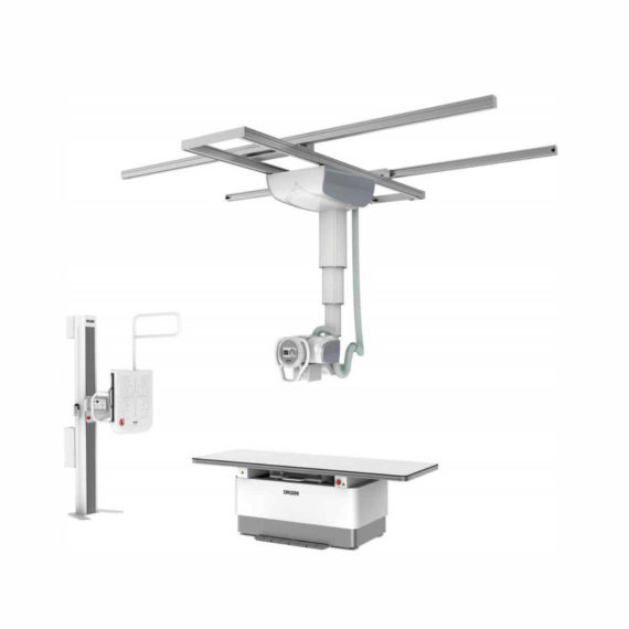
The DrGem Ceiling Analogue X-ray Machine is a diagnostic radiography system that provides reliable high quality radiographic images with a reduced dose. The reliable high-frequency x-ray generators that are known worldwide for their excellent performance, lifetime and stability. Patient tables and wall stands are also offered.
Delivery & Availability:
Typically 21 working days – excluding furniture and heavy/bulky equipment. Please contact us for further information.

Description
DrGem Ceiling Analogue X-ray Machine is a diagnostic radiography system X-ray Machine that provides reliable high quality radiographic images with a reduced dose. The reliable high-frequency x-ray generators that are known worldwide for their excellent performance, lifetime and stability. Patient tables and wall stands are also offered.
Features of DrGem Ceiling Analogue X-ray Machine
- TS-CSA-A (Vertical movement, 1.6m stroke, rail length 3x4meter) including HV cable 15m
- WBS-TA: Vertical movement
- V Stroke:1,450mm in Uprigh Bucky Position,
- 1,526mm in Horizontal Bucky position.
- PBT-4 is a 4 way Floating Tabletop with. A large tabletop with extended travel enables all radiography studies with minimal patient movement. Fully fat tabletop without a frame on the edge makes cleanliness and odors free
Technical Specifications of DrGem Ceiling Analogue X-ray Machine
- Power Rating – 32KW
- Generator – GXR-32S
- Rotor – Dual Speed Starter(DSS)
- Input Power – 400/480VAC, Three phase
- Line Frequency – 50/60Hz
- X-ray tube – DXT-12M, (0.6/1.2mm, 300kHU)
- Tube Voltage – 40 to 150kV, 1kV Step
- Tube Current – 10 to 640mA
- Output – 640mA@81kV, 500mA@104kV, 400mA@130kV, 320mA@150kV
- Time Range – 1ms to 10s
- mAs Range – 0.1 to 800mAs
- Reproducibility – Coecient of Variation : kV < 0.005, Time < 0.005,mAs < 0.01
- Accuracy – kV < ±(1%+1kV), mA < ±(3%+1mA), Time <±(1%+0.5ms), mAs < ±(3%+0.1mAs)
- Linearity – Coecient of Linearity < 0.01 : CL = (X1-X2)/(X1+X2), where X is mR/mAs
- Mechanical Parts:
-TS-CSA-A (Vertical movement, 1.6m, stroke rail length 3x4meter) including HV cable 15m
– PBT-4: 4 way Floating Tabletop with Elevating Feature (66cm).
– WBS-TA: a. Vertical movement
- V Stroke:1,450mm in Upright Bucky
- Position, 1,526mm in Horizontal Bucky position.
– HVC-15: 15M HV cable
– Auto Collimator
Click Here To Download Catalogue
Review(1)
Quick Comparison
| Settings | DrGem Ceiling Analogue X-ray Machine remove | ASPEL AsCARD Green B/W ECG Machine remove | Sonoscape S11 Ultrasound Machine remove | Topaz Digital X-ray Machine remove | DRGEM DR System remove | Sonoscape P20 Ultrasound Machine remove |
|---|---|---|---|---|---|---|
| Name | DrGem Ceiling Analogue X-ray Machine remove | ASPEL AsCARD Green B/W ECG Machine remove | Sonoscape S11 Ultrasound Machine remove | Topaz Digital X-ray Machine remove | DRGEM DR System remove | Sonoscape P20 Ultrasound Machine remove |
| Image |  |  |  |  |  |  |
| SKU | SF1033560074-7 | SF1033560075-8 | SF1033560012-1 | SF1033560074-1 | SF1033560074-8 | SF1033560012-9 |
| Rating | ||||||
| Price |
|
| $6,380.00 |
|
|
|
| Stock | ||||||
| Availability | ||||||
| Add to cart | ||||||
| Description | Shipped from abroad The DrGem Ceiling Analogue X-ray Machine is a diagnostic radiography system that provides reliable high quality radiographic images with a reduced dose. The reliable high-frequency x-ray generators that are known worldwide for their excellent performance, lifetime and stability. Patient tables and wall stands are also offered. Delivery & Availability: Typically 21 working days – excluding furniture and heavy/bulky equipment. Please contact us for further information. | Shipped from Abroad AsCARD Green electrocardiograph is a 1- and 3-channel ECG unit which enables to make electrocardiogram in full 12 leads. Intended for ECG examinations of adult and paediatric patients aimed at identification of cardiological abnormalities, myocardial ischaemia or infarction. The device is intended for use in healthcare facilities by duly trained personnel. ECG examination may be recorded in manual or automatic mode with the ability to perform the analysis and interpretation. Delivery & Availability: Typically 10 working days – excluding furniture and heavy/bulky equipment. Please contact us for further information. | In Stock A Value Choice beyond Your Expectation. SonoScape’s trolley color Doppler system S11 redefines price and performance with practical design. The S11 will go beyond your expectations but not your budget. Delivery & Availability: Typically 2 working days – excluding furniture and heavy/bulky equipment. Please contact us for further information. | In Stock DRGEM’s TOPAZ X-ray machine is a state-of-the-art mobile digital radiography system, designed with maximum comfort for patients and users in mind. From its user-friendly software to smooth movements, TOPAZ is made to improve your workflow and provide you with high-quality images. Delivery & Availability: Typically 21 working days – excluding furniture and heavy/bulky equipment. Please contact us for further information. | Ship from abroad ACQUIDR is the digital imaging system composed of a Flat Panel Detector(FPD) and an imaging workstation with software. The digital FPD and full-feature imaging software with excellent digital image processing, designed for DRGEM X-ray machine. Delivery & Availability: Typically 21 working days – excluding furniture and heavy/bulky equipment. Please contact us for further information. | Shipped from Abroad Incorporating innovative technologies, P20’s user-friendly design with a simple operation panel, intuitive user interface and a variety of intelligent auxiliary scanning tools, will significantly improve your daily examination experience. Besides general imaging applications, P20 has entitled with diagnostic 4D technology which has an extraordinary performance in obstetrics and gynecology applications. Delivery & Availability: Typically 5-7 working days – excluding furniture and heavy/bulky equipment. Please contact us for further information. |
| Content | DrGem Ceiling Analogue X-ray Machine is a diagnostic radiography system X-ray Machine that provides reliable high quality radiographic images with a reduced dose. The reliable high-frequency x-ray generators that are known worldwide for their excellent performance, lifetime and stability. Patient tables and wall stands are also offered.
Features of DrGem Ceiling Analogue X-ray Machine
Click Here To Download Catalogue | AsCARD Green electrocardiograph is a 1- and 3-channel ECG unit which enables to make electrocardiogram in full 12 leads. Intended for ECG examinations of adult and paediatric patients aimed at identification of cardiological abnormalities, myocardial ischaemia or infarction. The device is intended for use in healthcare facilities by duly trained personnel. ECG examination may be recorded in manual or automatic mode with the ability to perform the analysis and interpretation.
Electrocardiograph is based on advanced microprocessor technology. It is equipped with a thermal printer with high-resolution head and graphical LCD display. A hightech membrane keyboard makes the AsCARD Green device operation intuitive, and its menu navigation exceptionally easy. This light-weight, small-footprint and battery powered cause that device can be easily transported to any location. With plastic casing and foil covered keyboard, the device is neat and easy to clean.
Technical Specifications:
Click Here To Download Catalogue | DETAILS
SonoScape’s trolley colour Doppler system S11 redefines price and performance with practical design. The S11 will go beyond your expectations but not your budget. As an easy-to-use ultrasound system, the S11 is integrated with a new software platform, especially optimized for a smooth workflow and convenient operation. The system speeds up the exam process and makes file management easier.
SPECIFICATION
- 15-inch high definition LCD monitor with articulating arm
- Compact and agile trolley design
- 3 active transducer sockets available for a wide range of applications
- Duplex, Color Doppler, DPI, PW Doppler, tissue harmonic imaging, μ-scan speckle reduction imaging, compound imaging, trapezoidal imaging
- Customized settings based on your own working style
- Full patient database and image management solutions
Click Here To Download Catalogue | TOPAZ X-ray machine is among the high end X-ray machine manufactured by DRGEM, a digital X-ray system that provides quality images with little or no effort.
It begins with Advanced Technology
Integrating high technology and over a decade of experience in conventional and digital radiography systems, DRGEM’s TOPAZ X-ray machine is a state-of-the-art mobile digital radiography system, designed with maximum comfort for patients and users. From its user-friendly software to smooth movements, TOPAZ X-ray machine is made to improve your workflow and provide you with high-quality images.
Full Featured Imaging Software & Excellent Digital Image Processing
With a high-performance, built-in touchscreen, TOPAZ X-ray machine offers a user-friendly interface and powerful software for easy operation and increased workflow. The anatomical view-based digital image processing, automatically optimizes and enhances the quality of the image. it also comes with automatic image storage and print with DICOM 3.0 networking capability. additionally, the system offers increasing exam throughput while decreasing examination time.
Click Here To Download Catalogue | DRGEM ACQUIDR (DRGEM DR System) is the digital imaging system composed of a Flat Panel Detector(FPD) and an imaging workstation with software. The digital FPD and full-feature imaging software with excellent digital image processing will meet all your needs in the diagnostic digital radiographic field.
Features of DRGEM DR System:
| DETAILS
Upgraded Images with More Clarity
SonoScape never stops making progress in improving the image quality of its ultrasound products to enhance the confidence of diagnosis for doctors. With extraordinary images provided by P20, the anatomy structures are clearer than ever.
C-Xlasto Imaging
With C-xlasto Imaging, P20 enables comprehensive quantitative elastic analysis. Meanwhile, C-xlasto on P20 is supported by linear, convex and transvaginal probes, to ensure good reproducibility and highly consistent quantitative elastic results.
S-Live
S-Live allows for detailed visualization of subtle anatomical features, thereby enabling intuitive diagnosis with real-time 3D images and enriching patient communication.
Pelvic Floor 4D
Transperineal 4D pelvic floor ultrasound can provide useful clinical values in assessing the vaginal delivery impact on the female anterior compartment, judging whether the pelvic organs are prolapsed or not and the extent, determining if the pelvic muscles were torn accurately.
Anatomic M Mode
Anatomic M Mode helps you observe the myocardial motion at different phases by freely placing sample lines. It accurately measures the myocardial thickness and the heart size of even difficult patients and supports the myocardial function and LV wall-motion assessment.
Tissue Doppler Imaging
P20 is endowed with Tissue Doppler Imaging which provides velocities and other clinical information on myocardial functions, facilitating clinical doctors with the ability to analyze and compare the motions of different parts of the patient's heart.
Click Here To Download Catalogue |
| Weight | N/A | N/A | N/A | N/A | N/A | N/A |
| Dimensions | N/A | N/A | N/A | N/A | N/A | N/A |
| Additional information |


vorbelutrioperbir
Only wanna input that you have a very nice website , I enjoy the design it really stands out.