Genoray Full Field Digital Mammography DMX-600
$0.00
Shipped from Abroad
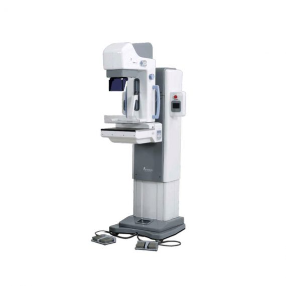
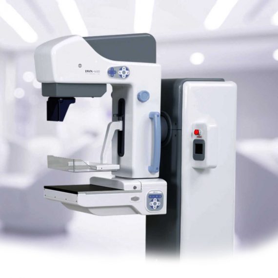
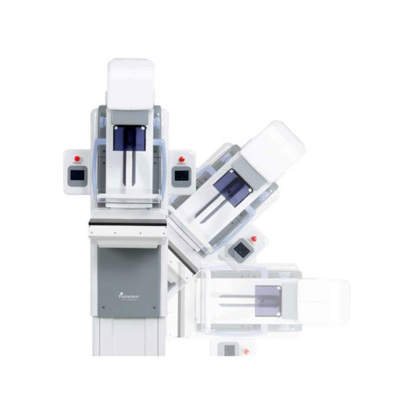
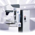
The DMX-600 is a full-field digital mammography system with smart C-arm rotation and fully motorized movements for fast and accurate positioning and efficient examinations. It features crystalline Silicon CMOS active pixel detector for higher contrast and higher resolution images and dual target X-ray tube for dose reduction.
Delivery & Availability:
Typically 21 working days – excluding furniture and heavy/bulky equipment. Please contact us for further information.
Description
The DMX-600 is a full-field digital mammography system with smart C-arm rotation and fully motorized movements for fast and accurate positioning and efficient examinations. It features crystalline Silicon CMOS active pixel detector for higher contrast and higher resolution images and dual target X-ray tube for dose reduction.
Features:
Excellent & High resolution Image quality:
– Innovative and accurate technology of Crystalline Silicon CMOS active pixel detector with higher contrast, higher resolution, brilliant images and economic maintenance cost.
– High performance of dual target X-ray tube for dose reduction.
– High output power of HF Generator with stability and efficiency.
– Stable hardware with the best reproducibility.
Large F.O.V. (Field Of View) by Multi-format technology:
– Optimally suited for screening and diagnosis with the financial advantages.
– Patent Application No. : 10-2011-0139676
– Available large F.O.V. to 23x23cm for almost patient.
User friendly Versatile functions:
– Smart C-arm rotation and fully motorized movements for fast & accurate positioning and efficiency examination by ASP & ASE (CC, RMLO, LMLO)
– Smart Automatic collimation with easy operation.
– Smart compression System by Microprocessor and Intelligent AEC control.
– Ergonomic design for quick and easy set-up.
Smart Software user alike:
– Customized and dedicated acquisition workstation.
– Perfect PACS accessibility with full DICOM capability.
– Extremely fast saving and transferring images for quick access and optimized workflow.
Click Here To Download Catalogue
Quick Comparison
| Genoray Full Field Digital Mammography DMX-600 remove | DrGem Floor Mounted Analogue X-ray remove | DRGEM DR System remove | DrGem GXR-SD 400mA Floor Mounted Digital X-ray remove | ASPEL AsCARD Green B/W ECG Machine remove | ASPEL AsPEKT 712 Holter Monitor and Software remove | |
|---|---|---|---|---|---|---|
| Name | Genoray Full Field Digital Mammography DMX-600 remove | DrGem Floor Mounted Analogue X-ray remove | DRGEM DR System remove | DrGem GXR-SD 400mA Floor Mounted Digital X-ray remove | ASPEL AsCARD Green B/W ECG Machine remove | ASPEL AsPEKT 712 Holter Monitor and Software remove |
| Image |  |  |  |  |  |  |
| SKU | SF1033560097-7 | SF1033560074-6 | SF1033560074-8 | SF1033560074-5 | SF1033560075-8 | SF1033560075-4 |
| Rating | ||||||
| Price |
|
|
|
|
| $1,991.00 |
| Stock | ||||||
| Availability | ||||||
| Add to cart | ||||||
| Description | Shipped from Abroad
The DMX-600 is a full-field digital mammography system with smart C-arm rotation and fully motorized movements for fast and accurate positioning and efficient examinations. It features crystalline Silicon CMOS active pixel detector for higher contrast and higher resolution images and dual target X-ray tube for dose reduction.
| In Stock GXR Analogue X-ray system matches with a radiographic room which perfectly fits your workow and can be easily upgraded to DR system with the help of DR interface and PC interface in GXR generator as well as Bucky suitable to Flat Panel Detector. GXR X-ray system is equipped with a high frequency X-ray generator which consistently produces high quality radiograph in favor of high quality X-ray output with a very small kV ripple and accurate mA and mAs. GXR X-ray system is designed to provide convenience to operator and comfort to patient. Delivery & Availability: Typically 21 working days – excluding furniture and heavy/bulky equipment. Please contact us for further information. | Ship from abroad ACQUIDR is the digital imaging system composed of a Flat Panel Detector(FPD) and an imaging workstation with software. The digital FPD and full-feature imaging software with excellent digital image processing, designed for DRGEM X-ray machine. Delivery & Availability: Typically 21 working days – excluding furniture and heavy/bulky equipment. Please contact us for further information. | In Stock The GXR-SD Digital X-ray is a diagnostic digital radiography system that provides reliable high quality digital radiographic images with a reduced dose. The GXR-SD DR systems offer comprehensive digital solutions to all radiography needs, featuring ACQUIDR digital imaging system with stationary or portable digital flat-panel detectors as well as reliable high-frequency x-ray generators that are known worldwide for their excellent performance, lifetime and stability. Patient tables and wall stands are also offered. Delivery & Availability: Typically 21 working days – excluding furniture and heavy/bulky equipment. Please contact us for further information. | Shipped from Abroad AsCARD Green electrocardiograph is a 1- and 3-channel ECG unit which enables to make electrocardiogram in full 12 leads. Intended for ECG examinations of adult and paediatric patients aimed at identification of cardiological abnormalities, myocardial ischaemia or infarction. The device is intended for use in healthcare facilities by duly trained personnel. ECG examination may be recorded in manual or automatic mode with the ability to perform the analysis and interpretation. Delivery & Availability: Typically 10 working days – excluding furniture and heavy/bulky equipment. Please contact us for further information. | Shipped from Abroad The Holta Monitor allows quick analysis of ECG examination and detection, reviewing and editing capability in the qualitative assessment of VE, VT, Single SVE, PSVT, Pauses, Irregular Rhythm, VT, IVR, Brady - and Tachycardia, Couplets, ST-segment elevation and depression, Maximum, Minimum and averaged Heart Rates, artifacts Delivery & Availability: Typically 10 working days – excluding furniture and heavy/bulky equipment. Please contact us for further information. |
| Content | The DMX-600 is a full-field digital mammography system with smart C-arm rotation and fully motorized movements for fast and accurate positioning and efficient examinations. It features crystalline Silicon CMOS active pixel detector for higher contrast and higher resolution images and dual target X-ray tube for dose reduction.
Features:
Excellent & High resolution Image quality:
- Innovative and accurate technology of Crystalline Silicon CMOS active pixel detector with higher contrast, higher resolution, brilliant images and economic maintenance cost.
- High performance of dual target X-ray tube for dose reduction.
- High output power of HF Generator with stability and efficiency.
- Stable hardware with the best reproducibility.
Large F.O.V. (Field Of View) by Multi-format technology:
- Optimally suited for screening and diagnosis with the financial advantages.
- Patent Application No. : 10-2011-0139676
- Available large F.O.V. to 23x23cm for almost patient.
User friendly Versatile functions:
- Smart C-arm rotation and fully motorized movements for fast & accurate positioning and efficiency examination by ASP & ASE (CC, RMLO, LMLO)
- Smart Automatic collimation with easy operation.
- Smart compression System by Microprocessor and Intelligent AEC control.
- Ergonomic design for quick and easy set-up.
Smart Software user alike:
- Customized and dedicated acquisition workstation.
- Perfect PACS accessibility with full DICOM capability.
- Extremely fast saving and transferring images for quick access and optimized workflow.
Click Here To Download Catalogue | DrGem GXR Floor Mounted Analogue X-ray system matches with a radiographic room which perfectly fits your workflow and can be easily upgraded to DR system with the help of DR interface and PC interface in GXR generator as well as Bucky suitable to Flat Panel Detector. GXR (Analogue X-ray)system is equipped with a high frequency X-ray generator which consistently produces high quality radiograph in favor of high quality X-ray output with a very small kV ripple and accurate mA and mAs. GXR (Analogue X-ray) system is designed to provide convenience to operator and comfort to patient.
Features of DrGem GXR Floor Mounted Analogue X-ray:
Click Here To Download Catalogue | DRGEM ACQUIDR (DRGEM DR System) is the digital imaging system composed of a Flat Panel Detector(FPD) and an imaging workstation with software. The digital FPD and full-feature imaging software with excellent digital image processing will meet all your needs in the diagnostic digital radiographic field.
Features of DRGEM DR System:
| DrGem GXR-SD 400mA Floor Mounted Digital X-ray system matches with a radiographic room which perfectly fits your workow and can be easily upgraded to DR system with the help of DR interface and PC interface in GXR generator as well as Bucky suitable to Flat Panel Detector. GXR X-ray system is equipped with a high frequency X-ray generator which consistently produces high quality radiograph in favor of high quality X-ray output with a very small kV ripple and accurate mA and mAs. GXR X-ray system is designed to provide convenience to operator and comfort to patient
Features of DrGem GXR-SD 400mA Floor Mounted Digital X-ray:
Click Here To Download Catalogue | AsCARD Green electrocardiograph is a 1- and 3-channel ECG unit which enables to make electrocardiogram in full 12 leads. Intended for ECG examinations of adult and paediatric patients aimed at identification of cardiological abnormalities, myocardial ischaemia or infarction. The device is intended for use in healthcare facilities by duly trained personnel. ECG examination may be recorded in manual or automatic mode with the ability to perform the analysis and interpretation.
Electrocardiograph is based on advanced microprocessor technology. It is equipped with a thermal printer with high-resolution head and graphical LCD display. A hightech membrane keyboard makes the AsCARD Green device operation intuitive, and its menu navigation exceptionally easy. This light-weight, small-footprint and battery powered cause that device can be easily transported to any location. With plastic casing and foil covered keyboard, the device is neat and easy to clean.
Technical Specifications:
Click Here To Download Catalogue | The Holter Monitor allows quick analysis of ECG examination (arrhythmias and ST segment).
Technical specifications:
HolCARD 24W Software:
Click Here To Download Catalogue |
| Weight | N/A | N/A | N/A | N/A | N/A | N/A |
| Dimensions | N/A | N/A | N/A | N/A | N/A | N/A |
| Additional information |


Reviews
There are no reviews yet.