
HydroSoft-T Water Softening System
$0.00
Shipped From Abroad
With HydroSoft-T time controlled fully automatic water softening systems; The calcium and magnesium ions in the water are removed by the ion exchange method.
Delivery & Availability:
Typically 10-21 working days – excluding furniture and heavy/bulky equipment. Please contact us for further information.
Typically 10-21 working days – excluding furniture and heavy/bulky equipment. Please contact us for further information.
Description
Time Controlled Water Softening Systems
Time controlled fully automatic water softening systems; These are systems that remove Calcium (Ca) and Magnesium (Mg) ions in water that cause hardness.
Softening systems automatically perform regeneration processes on the desired day of the week as a time period without any intervention at the desired time. The resin that reaches saturation regenerates with salt water in a time-controlled manner and is cleaned from calcium and magnesium ions.
Standard Features
Design Criteria
Quick Comparison
| HydroSoft-T Water Softening System remove | 3B Scientific Pregnancy Models Series, 8 Individual Embryo and Fetus Models - 3B Smart Anatomy remove | 3B Scientific Wearable Breast Self Examination Model - 3B Smart Anatomy remove | 3B Scientific Classic Human Skull Model painted, 3 part - 3B Smart Anatomy remove | 3B Scientific Classic Flexible Human Spine Model - 3B Smart Anatomy remove | 3B Scientific Human Brain Model with Arteries on Base of Head, 8 part - 3B Smart Anatomy remove | |
|---|---|---|---|---|---|---|
| Name | HydroSoft-T Water Softening System remove | 3B Scientific Pregnancy Models Series, 8 Individual Embryo and Fetus Models - 3B Smart Anatomy remove | 3B Scientific Wearable Breast Self Examination Model - 3B Smart Anatomy remove | 3B Scientific Classic Human Skull Model painted, 3 part - 3B Smart Anatomy remove | 3B Scientific Classic Flexible Human Spine Model - 3B Smart Anatomy remove | 3B Scientific Human Brain Model with Arteries on Base of Head, 8 part - 3B Smart Anatomy remove |
| Image | 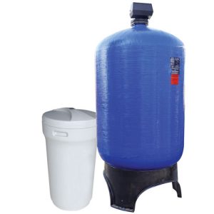 | 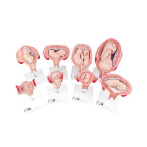 | 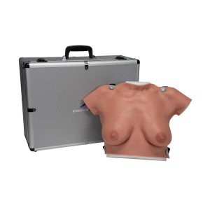 |  | 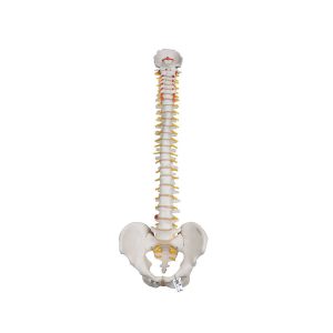 | 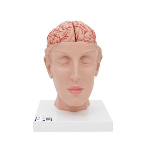 |
| SKU | SF1033560130179-6 | SF1033560099-24 | SF1033560099-21 | SF1033560099-2 | SF1033560099-1 | SF1033560099-15 |
| Rating | ||||||
| Price |
|
|
|
|
|
|
| Stock | ||||||
| Availability | ||||||
| Add to cart | ||||||
| Description | Shipped From Abroad
With HydroSoft-T time controlled fully automatic water softening systems; The calcium and magnesium ions in the water are removed by the ion exchange method.
Delivery & Availability:
Typically 10-21 working days – excluding furniture and heavy/bulky equipment. Please contact us for further information.
| Ship from abroad
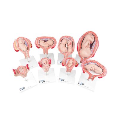
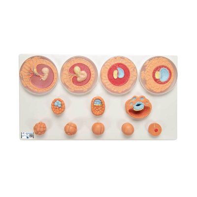
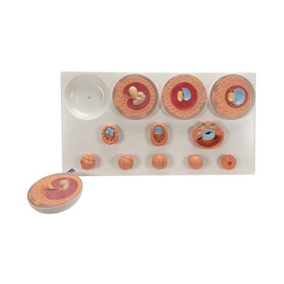
Our most popular series includes 8 models to show the complete stages of development. All models are mounted separately on a stand.
| Ship from abroad
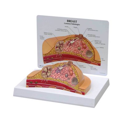
Demonstrate realistic self-examination with our natural casting of a female upper body with medium sized breasts. It can easily be worn, in order to better train and practice breast self-examination.
| Ship from abroad
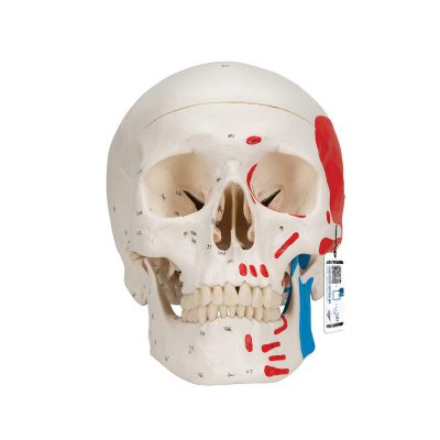
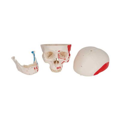
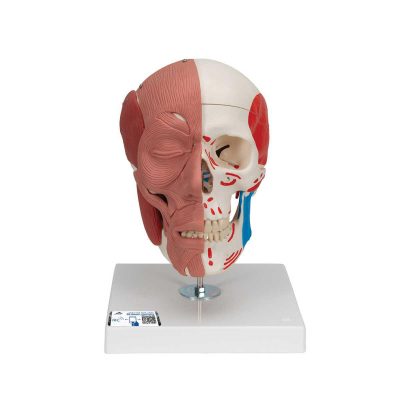
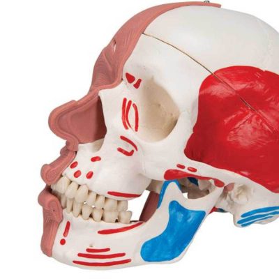
This replica of the human skull is a fantastic tool for teaching and learning the anatomy of the skull. The muscle origins (red) and insertions (blue) are shown in color on the left side of the skull. Cranial bones and structures are numbered on the right side. Jaw hinges with spring to simulate real movement. This skull model identifies over 140 anatomical details.
| Ship from abroad
This spine model for patient and student education is also the most affordable spine model. This spine is fully flexible and designed for hands-on demonstrations.
| Ship from abroad
This C25 deluxe brain comes with opened head to allow detailed study of the brain's position in the skull. The human head is horizontally divided above the skull base. This medially divided deluxe brain model shows the brain arteries in detail.
|
| Content | Time Controlled Water Softening SystemsTime controlled fully automatic water softening systems; These are systems that remove Calcium (Ca) and Magnesium (Mg) ions in water that cause hardness. Softening systems automatically perform regeneration processes on the desired day of the week as a time period without any intervention at the desired time. The resin that reaches saturation regenerates with salt water in a time-controlled manner and is cleaned from calcium and magnesium ions.Standard FeaturesDesign Criteria | Our most popular series includes 8 models to show the complete stages of development. All models are mounted separately on a stand.
| Demonstrate realistic self-examination with our natural casting of a female upper body with medium sized breasts. It can easily be worn, in order to better train and practice breast self-examination.
• Made of new 3B SKINlike™ high-quality silicone
• Displays the skin in finest detail
• Very realistic to the touch and dermatologically tested
• Breast examination is possible in both upright or lying positions
• Benign and malignant tumors in different stages of development hone self-examination skills
- 2 benign tumors
- 4 malignant tumors
- 2 typical anomalies
• Includes “Female Breast” chart
• Supplied with talcum powder, harness, stand and aluminium carrying case | This replica of the human skull is a fantastic tool for teaching and learning the anatomy of the skull. The muscle origins (red) and insertions (blue) are shown in color on the left side of the skull. Cranial bones and structures are numbered on the right side. Jaw hinges with spring to simulate real movement. This skull model identifies over 140 anatomical details.
This high quality skull is a great anatomy teaching tool! | This spine model for patient and student education is also the most affordable spine model. This spine is fully flexible and designed for hands-on demonstrations. The spine contains these features:
This high quality spine is a great tool for teaching/learning the anatomy of the human vertebral column. | This C25 deluxe brain comes with opened head to allow detailed study of the brain's position in the skull. The human head is horizontally divided above the skull base. This medially divided deluxe brain model shows the brain arteries in detail. Both halves of this brain model can be disassembled into:
The classic brain is a great tool for education on the human nervous system and anatomy of the brain. The brain in a head base is delivered on a base. |
| Weight | N/A | N/A | N/A | N/A | N/A | N/A |
| Dimensions | N/A | N/A | N/A | N/A | N/A | N/A |
| Additional information |

Reviews
There are no reviews yet.