
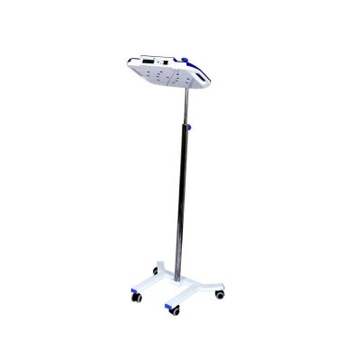
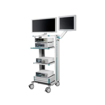
I-Genius-The Intraoperative MRI System
$0.00
Shipped From Abroad
The intraoperative MRI system for neurosurgery
I-Genius is a game-changing intraoperative MRI system that simplifies workflow by providing real-time MR imaging capabilities and delivering valuable information to assist neurosurgeons in making critical decisions during surgical resections.
Typically 10-21 working days – excluding furniture and heavy/bulky equipment. Please contact us for further information.
Description
Next-generation intraoperative MRI
- 1-ROOM
Everything in one room: no more patient transfers or workflow interruptions.
- 1-SURGICAL TABLE
To meet the neurosurgery operating standards.
- 1.2m SAFE ZONE
Thanks to its smart design, the operator can quickly switch from the surgery to the imaging modality.
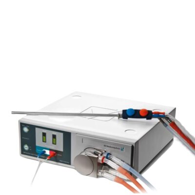
- 3-ADVANCED TOOL
Tools to support the surgeon during the surgery: dedicated cranial fixation system, 55” UHD monitor available in the surgical room for real-time imaging display, customized neuronavigation system (BrainNav by MedCom)*.
MR IMAGING DIRECTLY IN THE OPERATING ROOM
A new era of neurosurgery has begun
Every year, more than 500.000* new cases of primary malignant brain tumors are diagnosed worldwide. Gliomas can be difficult to differentiate from surrounding tissue, making it challenging to estimate residual tumor. In gliomas, incomplete resection may lead to recurrence. Intraoperative MRI with I-Genius transforms the practice of brain tumor surgery by allowing MR imaging directly in the operating room and supporting surgical decisions. To guarantee a seamless and safe workflow within the operating room, Esaote has developed a dedicated MRI solution with an integrated intraoperative patient bed.
Confident vision for smarter surgery
In the operating room, every detail counts. Advanced intraoperative imaging provides neurosurgeons with the clarity and confidence required to make important decisions in critical moments. All protocols are designed with optimized sequences, ensuring imaging tailored to surgical needs. I-Genius redefines intraoperative MRI, breaking down barriers and democratizing access to innovation worldwide. By combining efficiency, flexibility, and sustainability, it makes state-of-the-art MRI accessible to more hospitals, more surgeons, and ultimately more patients.
Imaging & Visualization
The I-Genius system is designed to provide imaging directly in the operating room to support real-time surgical decisions.
-
Real-Time MR Imaging: The core feature is providing real-time MR imaging capabilities directly in the operating room (OR), eliminating the need for patient transfer and workflow interruptions.
-
Optimized Protocols: The protocols are designed with optimized sequences specifically tailored to surgical needs, ensuring image clarity and confidence for neurosurgeons in critical moments.
-
Display: It includes a 55-inch UHD monitor available in the surgical room for real-time image display.
-
Workflow Integration: The system facilitates a safe and seamless workflow by integrating a dedicated MR solution with an integrated intraoperative patient bed.
Clinical Applications
While the I-Genius is a specialized neurosurgical tool, the broader Esaote MR product line (listed on the same page) covers a wide range of applications, including:
| System Focus | Primary Applications | I-Genius Specific Role |
| I-Genius | Intraoperative Neurosurgery | Assisting neurosurgeons in critical decisions during surgical resections, particularly for gliomas and brain tumors, by estimating residual tumor and guiding the resection extent. |
| General Esaote MR (e.g., G-scan, S-scan, O-scan) | Musculoskeletal (MSK) | Orthopedics, Sports Medicine, and Rheumatology (some models feature Weight-bearing imaging). |
| General Esaote MR | Whole Body | General radiology and full-body examinations (on applicable open systems like Magnifico Open). |
| General Esaote MR | Neuroimaging | Standard diagnostic neuroimaging and advanced applications. |
Coils (Probe Types)
In Magnetic Resonance Imaging (MRI), coils are used as transducers to send and receive radiofrequency signals, similar to how probes are used in ultrasound. The I-Genius system features dedicated coils for neurosurgical applications:
-
Headrest Coil: The primary coil for stabilizing and imaging the patient’s head.
-
Large Head Coil: A dedicated coil for comprehensive brain imaging.
-
Flex Coil: An optional flexible coil for specific or smaller area imaging needs.
The system also supports advanced tools like a dedicated cranial fixation system and a customized neuronavigation system (BrainNav by MedCom) to support the surgeon during the procedure.
Quick Comparison
| I-Genius-The Intraoperative MRI System remove | IBIS Neeo R9 Digital Surgical C-Arm remove | Instrument Tray remove | DrGem GXR-SD 400mA Floor Mounted Digital X-ray remove | Operating Light Four (4) Halogen bulbs remove | AVI Bilipod LED Phototherapy Unit remove | ||||||||||||||||||||||||||||||||||||||||
|---|---|---|---|---|---|---|---|---|---|---|---|---|---|---|---|---|---|---|---|---|---|---|---|---|---|---|---|---|---|---|---|---|---|---|---|---|---|---|---|---|---|---|---|---|---|
| Name | I-Genius-The Intraoperative MRI System remove | IBIS Neeo R9 Digital Surgical C-Arm remove | Instrument Tray remove | DrGem GXR-SD 400mA Floor Mounted Digital X-ray remove | Operating Light Four (4) Halogen bulbs remove | AVI Bilipod LED Phototherapy Unit remove | |||||||||||||||||||||||||||||||||||||||
| Image | 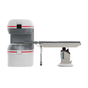 |  |  |  |  |  | |||||||||||||||||||||||||||||||||||||||
| SKU | SF1033560011-1 | SF1033560084-199 | SF1033560074-5 | SF1033560084-170 | SF1033560094-4 | ||||||||||||||||||||||||||||||||||||||||
| Rating | |||||||||||||||||||||||||||||||||||||||||||||
| Price |
|
| $6.90 |
|
| $505.00 | |||||||||||||||||||||||||||||||||||||||
| Stock | |||||||||||||||||||||||||||||||||||||||||||||
| Availability | |||||||||||||||||||||||||||||||||||||||||||||
| Add to cart | |||||||||||||||||||||||||||||||||||||||||||||
| Description | Shipped From Abroad
The intraoperative MRI system for neurosurgery
I-Genius is a game-changing intraoperative MRI system that simplifies workflow by providing real-time MR imaging capabilities and delivering valuable information to assist neurosurgeons in making critical decisions during surgical resections. Delivery & Availability:
Typically 10-21 working days – excluding furniture and heavy/bulky equipment. Please contact us for further information.
| Shipped from Abroad Our Neeo “C” arms are easy to place, use and are specifically designed to be used in orthopedics, traumatology, abdominal surgery, urology, cardiology and operating rooms. Delivery & Availability: Typically 21 working days – excluding furniture and heavy/bulky equipment. Please contact us for further information. | In stock
| In Stock The GXR-SD Digital X-ray is a diagnostic digital radiography system that provides reliable high quality digital radiographic images with a reduced dose. The GXR-SD DR systems offer comprehensive digital solutions to all radiography needs, featuring ACQUIDR digital imaging system with stationary or portable digital flat-panel detectors as well as reliable high-frequency x-ray generators that are known worldwide for their excellent performance, lifetime and stability. Patient tables and wall stands are also offered. Delivery & Availability: Typically 21 working days – excluding furniture and heavy/bulky equipment. Please contact us for further information. | In stock
| Shipped from Abroad
| |||||||||||||||||||||||||||||||||||||||
| Content | https://vimeo.com/1124201105?fl=pl&fe=sh
Next-generation intraoperative MRI
Everything in one room: no more patient transfers or workflow interruptions.
To meet the neurosurgery operating standards.
Thanks to its smart design, the operator can quickly switch from the surgery to the imaging modality.
Tools to support the surgeon during the surgery: dedicated cranial fixation system, 55” UHD monitor available in the surgical room for real-time imaging display, customized neuronavigation system (BrainNav by MedCom)*.
MR IMAGING DIRECTLY IN THE OPERATING ROOM A new era of neurosurgery has begunEvery year, more than 500.000* new cases of primary malignant brain tumors are diagnosed worldwide. Gliomas can be difficult to differentiate from surrounding tissue, making it challenging to estimate residual tumor. In gliomas, incomplete resection may lead to recurrence. Intraoperative MRI with I-Genius transforms the practice of brain tumor surgery by allowing MR imaging directly in the operating room and supporting surgical decisions. To guarantee a seamless and safe workflow within the operating room, Esaote has developed a dedicated MRI solution with an integrated intraoperative patient bed. DESIGNED FOR THE OPERATING ROOM
Confident vision for smarter surgeryIn the operating room, every detail counts. Advanced intraoperative imaging provides neurosurgeons with the clarity and confidence required to make important decisions in critical moments. All protocols are designed with optimized sequences, ensuring imaging tailored to surgical needs. I-Genius redefines intraoperative MRI, breaking down barriers and democratizing access to innovation worldwide. By combining efficiency, flexibility, and sustainability, it makes state-of-the-art MRI accessible to more hospitals, more surgeons, and ultimately more patients. Imaging & VisualizationThe I-Genius system is designed to provide imaging directly in the operating room to support real-time surgical decisions.
Clinical ApplicationsWhile the I-Genius is a specialized neurosurgical tool, the broader Esaote MR product line (listed on the same page) covers a wide range of applications, including:
Coils (Probe Types)In Magnetic Resonance Imaging (MRI), coils are used as transducers to send and receive radiofrequency signals, similar to how probes are used in ultrasound. The I-Genius system features dedicated coils for neurosurgical applications:
The system also supports advanced tools like a dedicated cranial fixation system and a customized neuronavigation system (BrainNav by MedCom) to support the surgeon during the procedure. | Our Neeo “C” arms are easy to place, use and are specifically designed to be used in orthopedics, traumatology, abdominal surgery, urology, cardiology and operating rooms.
Using Neeo with the RTP (Real Time Processing) option it is possible to perform vascular, urological and cardiological diagnostics. One of the main functions, digital image subtraction, allows to see, as an example, the passage of contrast liquids in a tissue or in a venous or arterial duct; thanks to the possibility of looping, the acquired video can be reproduced several times to monitor more accurately the passage of the fluid within the area in question. Angiographic measurement is another useful function in the vascular field (QA Quantitative Angiography) that allows the measurement of stenoses. Finally, fluoroscopy allows the correct positioning of stents or expanders.
Neeo is used in various interventional and diagnostic procedures in traumatology and orthopedics wards and operating rooms as well. Thanks to low-dose fluoroscopy, it is possible to use the device for positioning bone or subcutaneous grafts, inserting K-wire (Kirschner wire) for stabilization of bone fragments or for the correct positioning of prostheses. The low dose emitted ensures safe use for both the patient and the surgeon or doctor on the operating field.
On the control panel there is a large touch screen display that allows to adjust the basic functions of the equipment. From this display it is possible to select and adjust the fluoroscopic data for the examination, activate or deactivate the laser pointer, select between pulsed, one shot or standard fluoroscopy, rotate the image and perform all operations on collimator. The four side buttons on the display offer the possibility to move the bow vertically thanks to an extremely silent motor.
Neeo has two 19 “medical grade monitors that can be positioned according to the needs of the medical practitioner. Work monitors and feedback monitors are separated to be managed independently. The possible movements are: rotation, revolution, tilting and possibility of height adjustment.
Features:
Click Here To Download Catalogue |
| DrGem GXR-SD 400mA Floor Mounted Digital X-ray system matches with a radiographic room which perfectly fits your workow and can be easily upgraded to DR system with the help of DR interface and PC interface in GXR generator as well as Bucky suitable to Flat Panel Detector. GXR X-ray system is equipped with a high frequency X-ray generator which consistently produces high quality radiograph in favor of high quality X-ray output with a very small kV ripple and accurate mA and mAs. GXR X-ray system is designed to provide convenience to operator and comfort to patient
Features of DrGem GXR-SD 400mA Floor Mounted Digital X-ray:
Click Here To Download Catalogue |
| Features:
Click Here To Download Catalogue | |||||||||||||||||||||||||||||||||||||||
| Weight | N/A | N/A | N/A | N/A | N/A | N/A | |||||||||||||||||||||||||||||||||||||||
| Dimensions | N/A | N/A | N/A | N/A | N/A | N/A | |||||||||||||||||||||||||||||||||||||||
| Additional information |


Reviews
There are no reviews yet.