


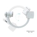
IBIS Neeo R9 Digital Surgical C-Arm
$0.00
Shipped from Abroad
Our Neeo “C” arms are easy to place, use and are specifically designed to be used in orthopedics, traumatology, abdominal surgery, urology, cardiology and operating rooms.
Delivery & Availability:
Typically 21 working days – excluding furniture and heavy/bulky equipment. Please contact us for further information.
Description
Our Neeo “C” arms are easy to place, use and are specifically designed to be used in orthopedics, traumatology, abdominal surgery, urology, cardiology and operating rooms.
Using Neeo with the RTP (Real Time Processing) option it is possible to perform vascular, urological and cardiological diagnostics. One of the main functions, digital image subtraction, allows to see, as an example, the passage of contrast liquids in a tissue or in a venous or arterial duct; thanks to the possibility of looping, the acquired video can be reproduced several times to monitor more accurately the passage of the fluid within the area in question. Angiographic measurement is another useful function in the vascular field (QA Quantitative Angiography) that allows the measurement of stenoses. Finally, fluoroscopy allows the correct positioning of stents or expanders.
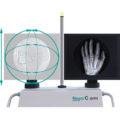
Neeo is used in various interventional and diagnostic procedures in traumatology and orthopedics wards and operating rooms as well. Thanks to low-dose fluoroscopy, it is possible to use the device for positioning bone or subcutaneous grafts, inserting K-wire (Kirschner wire) for stabilization of bone fragments or for the correct positioning of prostheses. The low dose emitted ensures safe use for both the patient and the surgeon or doctor on the operating field.
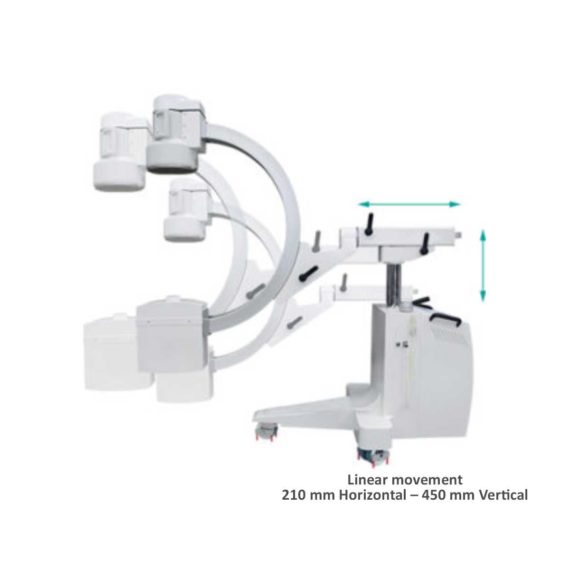
On the control panel there is a large touch screen display that allows to adjust the basic functions of the equipment. From this display it is possible to select and adjust the fluoroscopic data for the examination, activate or deactivate the laser pointer, select between pulsed, one shot or standard fluoroscopy, rotate the image and perform all operations on collimator. The four side buttons on the display offer the possibility to move the bow vertically thanks to an extremely silent motor.
Neeo has two 19 “medical grade monitors that can be positioned according to the needs of the medical practitioner. Work monitors and feedback monitors are separated to be managed independently. The possible movements are: rotation, revolution, tilting and possibility of height adjustment.
Features:
- Continuous fluoroscopy
- Pulsed fluorography
- One Shot fluoroscopy
- Low dose fluoroscopy
- Radiography with cassette (up to 5KW)
- 110,000 images of memory (Standard)
- 19 ″ Medical Flat Monitor (X2)
- Touch control panel
- Image Processing – 1K X 1K, 16 bit
- DAP-Dose Area Meter
- DSA Road Mapping
- Laser Marker
Technical Specification:
- Power: 5KW
- kV Range (kV): 40 – 120
- mAs Range (mAs): 1 – 250
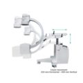
- SID (mm): 1000
- Free Space (mm): 730
DICOM Feature:
- RTP-SW DSA
- RTP-SW QA
- RTP-SW Worklist
- RTP-SW Print
- RTP-SW CD DVD Saving
- RTP-SW MPPS
- RTP-SW Query/Retrieve
- RTP-SW HCF
- RTP-SW BASE
- RTP-SW STCOMMIT
Configuration:
C-Arm Structure, Trolley for Monitor, Collimator, Monoblock, Toshiba 12” Image Intensifier, 19″ Philips Medical Flat Monitor (X2), Camera CD1030CA 1KX1K, Sony Printer UP-X898MD, Cassette Holder
Click Here To Download Catalogue
Quick Comparison
| IBIS Neeo R9 Digital Surgical C-Arm remove | Sonoscape S11 Ultrasound Machine remove | ASPEL Stress ECG with Ergometer and Software remove | Sonoscape S22 Ultrasound Machine remove | Sonoscape E2 Ultrasound Machine remove | DrGem GXR-SD 400mA Floor Mounted Digital X-ray remove | |
|---|---|---|---|---|---|---|
| Name | IBIS Neeo R9 Digital Surgical C-Arm remove | Sonoscape S11 Ultrasound Machine remove | ASPEL Stress ECG with Ergometer and Software remove | Sonoscape S22 Ultrasound Machine remove | Sonoscape E2 Ultrasound Machine remove | DrGem GXR-SD 400mA Floor Mounted Digital X-ray remove |
| Image |  |  | 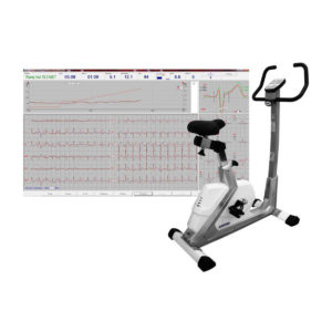 |  |  |  |
| SKU | SF1033560011-1 | SF1033560012-1 | SF1033560075-1 | SF1033560012-3 | SF1033560012-17 | SF1033560074-5 |
| Rating | ||||||
| Price |
| $6,380.00 | $4,202.00 | $9,350.00 | $5,500.00 |
|
| Stock | ||||||
| Availability | ||||||
| Add to cart | ||||||
| Description | Shipped from Abroad Our Neeo “C” arms are easy to place, use and are specifically designed to be used in orthopedics, traumatology, abdominal surgery, urology, cardiology and operating rooms. Delivery & Availability: Typically 21 working days – excluding furniture and heavy/bulky equipment. Please contact us for further information. | In Stock A Value Choice beyond Your Expectation. SonoScape’s trolley color Doppler system S11 redefines price and performance with practical design. The S11 will go beyond your expectations but not your budget. Delivery & Availability: Typically 2 working days – excluding furniture and heavy/bulky equipment. Please contact us for further information. | Shipped from Abroad Ergometer CRG 200 is dedicated for Exercise Stress Tests System CardioTEST. The Ergometer has been designed according to modern technologies. It is controlled from PC equipped in CardioTEST software. Load level is controlled by a microprocessor, therefore it does not depend on speed, which in turn can be adjusted according to patient’s individual needs. Ergometer is equipped with ECG mode recording 12 standard leads. Delivery & Availability: Typically 21 working days – excluding furniture and heavy/bulky equipment. Please contact us for further information. | Shipped from Abroad As SonoScape steps forward to add value and efficiency to ultrasound, the latest S22 was designed in a user-friendly platform to address current and future demanding needs. It represents an excellent mix in performance and price. Delivery & Availability: Typically 5-7 working days – excluding furniture and heavy/bulky equipment. Please contact us for further information. | Shipped from Abroad Sonoscape E2 portable ultrasound machine is a color Doppler ultrasound system that reaches beyond your expectations due to its compact and fashionable appearance. It fulfills GI, OB/GYN, Cardiac and POC applications to fit your routine scanning needs while its color mode will help you for more accurate and efficient diagnosis of lesions. E2 provides a wide range of applications to assist users with routine scanning. E2 provides automatic calculations to enhance your diagnostic confidence and save you time for patient communication. Delivery & Availability: Typically 14 working days – excluding furniture and heavy/bulky equipment. Please contact us for further information. | In Stock The GXR-SD Digital X-ray is a diagnostic digital radiography system that provides reliable high quality digital radiographic images with a reduced dose. The GXR-SD DR systems offer comprehensive digital solutions to all radiography needs, featuring ACQUIDR digital imaging system with stationary or portable digital flat-panel detectors as well as reliable high-frequency x-ray generators that are known worldwide for their excellent performance, lifetime and stability. Patient tables and wall stands are also offered. Delivery & Availability: Typically 21 working days – excluding furniture and heavy/bulky equipment. Please contact us for further information. |
| Content | Our Neeo “C” arms are easy to place, use and are specifically designed to be used in orthopedics, traumatology, abdominal surgery, urology, cardiology and operating rooms.
Using Neeo with the RTP (Real Time Processing) option it is possible to perform vascular, urological and cardiological diagnostics. One of the main functions, digital image subtraction, allows to see, as an example, the passage of contrast liquids in a tissue or in a venous or arterial duct; thanks to the possibility of looping, the acquired video can be reproduced several times to monitor more accurately the passage of the fluid within the area in question. Angiographic measurement is another useful function in the vascular field (QA Quantitative Angiography) that allows the measurement of stenoses. Finally, fluoroscopy allows the correct positioning of stents or expanders.
Neeo is used in various interventional and diagnostic procedures in traumatology and orthopedics wards and operating rooms as well. Thanks to low-dose fluoroscopy, it is possible to use the device for positioning bone or subcutaneous grafts, inserting K-wire (Kirschner wire) for stabilization of bone fragments or for the correct positioning of prostheses. The low dose emitted ensures safe use for both the patient and the surgeon or doctor on the operating field.
On the control panel there is a large touch screen display that allows to adjust the basic functions of the equipment. From this display it is possible to select and adjust the fluoroscopic data for the examination, activate or deactivate the laser pointer, select between pulsed, one shot or standard fluoroscopy, rotate the image and perform all operations on collimator. The four side buttons on the display offer the possibility to move the bow vertically thanks to an extremely silent motor.
Neeo has two 19 “medical grade monitors that can be positioned according to the needs of the medical practitioner. Work monitors and feedback monitors are separated to be managed independently. The possible movements are: rotation, revolution, tilting and possibility of height adjustment.
Features:
Click Here To Download Catalogue | DETAILS
SonoScape’s trolley colour Doppler system S11 redefines price and performance with practical design. The S11 will go beyond your expectations but not your budget. As an easy-to-use ultrasound system, the S11 is integrated with a new software platform, especially optimized for a smooth workflow and convenient operation. The system speeds up the exam process and makes file management easier.
SPECIFICATION
- 15-inch high definition LCD monitor with articulating arm
- Compact and agile trolley design
- 3 active transducer sockets available for a wide range of applications
- Duplex, Color Doppler, DPI, PW Doppler, tissue harmonic imaging, μ-scan speckle reduction imaging, compound imaging, trapezoidal imaging
- Customized settings based on your own working style
- Full patient database and image management solutions
Click Here To Download Catalogue | Ergometer CRG 200 is dedicated for Exercise Stress Tests System CardioTEST. The Ergometer has been designed according to modern technologies. It is controlled from PC equipped in CardioTEST software. Load level is controlled by a microprocessor, therefore it does not depend on speed, which in turn can be adjusted according to patient’s individual needs. Ergometer is equipped with ECG mode recording 12 standard leads.
Technical Specifications:
Click Here To Download Catalogue | DETAILS
As SonoScape steps forward to add value and efficiency to ultrasound, the latest S22 was designed in a user-friendly platform to address current and future demanding needs. It represents an excellent mix in performance and price.
S22, is a shared service ultrasound system with a slim and elegant package that has combined mobility with utility to fit in specific clinical situations including emergency department, ICU, operating room and so on. Furthermore, its ergonomic design, easy operating and flexible data management will give you a memorable experience.
SPECIFICATION
• Large high-resolution widescreen LED
• Sensitive touch screen
• Four transducer sockets plus one socket for pencil probe
• A comprehensive selection of probes: linear, Convex, Micro-convex, Volumetric, Endocavity, Bi-plane, Phased Array, TEE, Intraoperative, Pencil
• Premium application technology: 4D, μ-scan speckle reduction, compound imaging, Pulse Inversion Harmonic Imaging, Color M-Mode, Steer M-Mode, PDI, TDI, Real-time Panoramic Imaging, Trapezoid Imaging, Auto-IMT…
• Full patient database and image management solutions: DICOM 3.0, AVI/JPG, USB 2.0, HDD, DVD, PDF report
• Multi-Language Input Keyboard
• Built-in battery
Click Here To Download Catalogue | SONOSCAPE E2 DETAILS
Auto Image Optimization
A portable ultrasound machine with the press of a button, the image is automatically adjusted and optimized, saving you time with parameter adjustments. Additionally, with Auto Focus on, the focus area follows the depth of the ROI box as it is moved in the scanning field, providing users with excellent image quality in the desired area of interest.
Automated Calculation
Auto IMT is used when determining the level of vascular sclerosis present in the patient by automatically tracing the thickness of the carotid vessels.
Auto trace provides users sensitive and accurate wave tracing, avoiding the error of manual trace and giving out calculation result in no time
In-Build Battery pack
This portable ultrasound machine was equipped with an in-build battery pack which enable the user to perform image scanning when AC power is not available.
Click Here To Download Catalogue | DrGem GXR-SD 400mA Floor Mounted Digital X-ray system matches with a radiographic room which perfectly fits your workow and can be easily upgraded to DR system with the help of DR interface and PC interface in GXR generator as well as Bucky suitable to Flat Panel Detector. GXR X-ray system is equipped with a high frequency X-ray generator which consistently produces high quality radiograph in favor of high quality X-ray output with a very small kV ripple and accurate mA and mAs. GXR X-ray system is designed to provide convenience to operator and comfort to patient
Features of DrGem GXR-SD 400mA Floor Mounted Digital X-ray:
Click Here To Download Catalogue |
| Weight | N/A | N/A | N/A | N/A | N/A | N/A |
| Dimensions | N/A | N/A | N/A | N/A | N/A | N/A |
| Additional information |


Reviews
There are no reviews yet.