| Description | In Stock
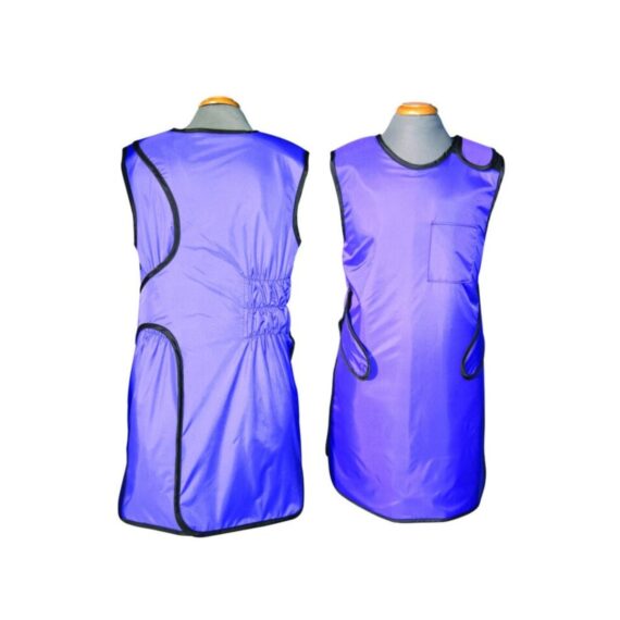
Overlap wrap around x-ray lead apron offers full body protection. Distributes weight between shoulders and hips. • 0.5mm Pb equivalency protection in the front and 0.25mm in the back. Comes with Hook & loop (Velcro) closure and built in 6" Elastic Back-Guard. Front belt assures snug fit and maximum support.
| In stock
- Length: 355 mm
- Height: 560 mm
- Diameter: 150 mm
- Power: 30 w
- Scattering diffusion: Greater than 0.9
Delivery & Availability:
Typically 5-7 working days – excluding furniture and heavy/bulky equipment. Please contact us for further information. | In Stock
A Value Choice beyond Your Expectation. SonoScape’s trolley color Doppler system S11 redefines price and performance with practical design. The S11 will go beyond your expectations but not your budget.
Delivery & Availability:
Typically 2 working days – excluding furniture and heavy/bulky equipment. Please contact us for further information. | In Stock
JADE is one of the lightest portable X-ray systems on the market, allowing it to be used in any imaginable way including bedside, operating rooms, intensive care units and in veterinary fields. With a simple, easy-to-use operator console, three-way control, two-step foldable stand and auto lock system, JADE is a user-friendly portable X-ray system.
Delivery & Availability:
Typically 21 working days – excluding furniture and heavy/bulky equipment. Please contact us for further information. | Shipped from Abroad
This Machine gives a possibility to perform computed tomography without any problems and on high quality level. This device is used to conduct exams of internal organs and their functioning. With its help, a physician has a possibility to assess the condition of the human body as a whole.
Delivery & Availability:
Typically 90 working days – excluding furniture and heavy/bulky equipment. Please contact us for further information. | Shipped from Abroad
OPENMARK 5000 is 0.51T MRI. It's approved by FDA and has CE mark. It adopts two-pillar magnet design with 280 degree openness and equipped with powerful
RF and gradient system, together with advanced imaging technology, making it as a high-end system which is comparable to high-field MRI.
Delivery & Availability:
Typically 90 working days – excluding furniture and heavy/bulky equipment. Please contact us for further information. |
| Content | Overlap wrap around x-ray lead apron offers full body protection. Distributes weight between shoulders and hips. • 0.5mm Pb equivalency protection in the front and 0.25mm in the back. Comes with Hook & loop (Velcro) closure and built in 6" Elastic Back-Guard. Front belt assures snug fit and maximum support. Our Protective Apparel unique 2-in-1 pocket design allows you to store like a cell phone or markers in the other.
|
- Length: 355 mm
- Height: 560 mm
- Diameter: 150 mm
- Power: 30 w
- Scattering diffusion: Greater than 0.9
| DETAILS
SonoScape’s trolley colour Doppler system S11 redefines price and performance with practical design. The S11 will go beyond your expectations but not your budget. As an easy-to-use ultrasound system, the S11 is integrated with a new software platform, especially optimized for a smooth workflow and convenient operation. The system speeds up the exam process and makes file management easier.
SPECIFICATION
- 15-inch high definition LCD monitor with articulating arm
- Compact and agile trolley design
- 3 active transducer sockets available for a wide range of applications
- Duplex, Color Doppler, DPI, PW Doppler, tissue harmonic imaging, μ-scan speckle reduction imaging, compound imaging, trapezoidal imaging
- Customized settings based on your own working style
- Full patient database and image management solutions
| JADE Mobile X-ray machine is one of the lightest portable X-ray systems on the market, allowing it to be used in any imaginable way including bedside, operating rooms, intensive care units and veterinary fields. With a simple, easy-to-use operator console, three-way control, two-step foldable stand and auto-lock system, the JADE Mobile X-ray machine is a user-friendly portable X-ray system.
Convenient & Intuitive Operation:
JADE is one of the lightest portable X-ray systems on the market, allowing it to be used in any imaginable way including bedside, operating rooms, intensive care units and in veterinary fields. With a simple, easy-to-use operator console, three-way control, two-step foldable stand and auto-lock system, JADE is a user-friendly portable X-ray system.
Compact & Powerful Design:
JADE Mobile X-ray machine is an innovative, highly versatile portable X-ray system suitable for a variety of clinical uses. Utilizing the unique technology used in DRGEM’s universally recognized X-ray generators, JADE is a compact but powerful unit with a 4kW output and thoughtfully designed components to increase efficiency and maximize workflow. The core part of X-ray source adopts high-quality tube assembly, X-ray collimator and high frequency X-ray generator with excellent performance, lifetime and stability.
Features:
- Vehicle loadable
- Wheel lock
- Automatic tube arm lock at any angle
- Storage space for cassettes or detectors
- User Programmable APR, save up to 9 APR settings
- Three way X-ray exposure
- USB interface & Bluetooth
- Remote control (Option)
- 5kg including X-ray unit, collimator and stand
- Maximum hight of 228.6cm
- Exposure Hand Switch
- Foldable, two-step stand
Technical Specification:
- Power Rating - 4kW,
- 100kHz, high frequency X-ray Generator
- kVp Range - Maximum 140kVp
- mAs Range - 0.1-250mAs
- mA Range – 10 to 100mA
- 330~2000mm FD
- Collimator with 30 seconds LED lamp timer
| This Machine gives a possibility to perform computed tomography without any problems and on high quality level. This device is used to conduct exams of internal organs and their functioning. With its help, a physician has a possibility to assess the condition of the human body as a whole.
Features:
- It is easy to use;
- Convenience;
- Multi functionality;
- Obtained images are of high definition;
- High-definition 3D images of the area under study;
- The procedure is pain-free;
- The data is processed fast;
- The image can be stored in the computer memory;
- The diagnostics does not take a lot of time;
- Acceptable radiation dose.
Technical Specifications:
| No. |
Technical Features |
Descriptions |
| 1 |
Gantry |
|
| 1.01 |
Gantry type |
Low voltage slip-ring |
| 1.02 |
Gantry driven type |
Strap-driven |
| 1.03 |
Patient opening |
70cm |
| 1.04 |
Gantry tilt mode |
Digital gantry tilt |
| 1.05 |
Digital tilt capability |
±50° |
| 1.06 |
Detector type |
OptiWave rare-earth ceramic detector |
| 1.07 |
Numbers of detector rows |
16 |
| 1.08 |
Width of Z-axle detector |
20mm |
| 1.09 |
Detector columns of channels per row |
848 |
| 1.10 |
Numbers of detector columns |
13568 |
| 1.11 |
Data-transfer type |
RF, optical fiber communication |
| 1.12 |
Distance of focus-ISO-center |
53cm |
| 1.13 |
Distance of focus-detector |
94cm |
| 1.14 |
3D laser orientation |
Provided |
| 1.15 |
13" integrated display panel |
Provided |
| 1.16 |
Adose automatic exposure control (mA
Modulation) |
Provided |
| 1.17 |
Auto-voice manager |
Breath Graphical Display
Hold Message (Record/Playback)
Breath Message (Record/Playback) |
| 1.18 |
AccuSaving energy conservation management |
Provided |
| 2 |
HVPS and X-ray tube |
|
| 2.01 |
Maximum continuous output of HVgenerator |
42kW |
| 2.02 |
Tube kV selections |
70kV, 80kV, 100 kV, 120 kV, 140 kV |
| 2.03 |
Tube mA range |
10~350mA |
| 2.04 |
Tube anode heat capacity |
3.5MHU |
| 2.05 |
Max. anode cooling rate |
735kHU/min |
| 2.06 |
Type of cooling |
Oil cooling + Air cooling |
| 2.07 |
Tube focus |
Large: 1.2mm×1.4mm
Small: 0.7mm×0.8mm |
| 2.08 |
Collimator width selection |
4-level election |
| 2.09 |
Focus spot tracking technology |
Provided |
| 3 |
Patient table |
|
| 3.01 |
Maximum horizontal-movable range |
1850mm |
| 3.02 |
Table horizontal-scannablerange |
1800mm |
| 3.03 |
Table horizontal-position repeatability |
±0.25mm |
| 3.04 |
Minimum height above floor |
430mm |
| 3.05 |
Maximum vertical-movable range |
500mm |
| 3.06 |
Maximum speed of vertical movement |
35mm |
| 3.07 |
Maximum speed of horizontal movement |
150mm/s |
| 3.08 |
Maximum patient weight |
205kg |
| 3.09 |
Foot pedal of patient table control |
Provided |
| 4 |
Computer |
|
| 4.01 |
CPU |
3.5GHz |
| 4.02 |
Memory |
32GB |
| 4.03 |
Storage of hard-disk |
1TB×2 |
| 4.04 |
Monitor |
24’’ LCD Monitor |
| 4.05 |
Resolution of monitor |
1920×1200 |
| 4.06 |
Image-data external storage type |
CD/DVD/USB |
| 4.07 |
Time of image reconstruction (512×512) |
33.3ms/image |
| 4.08 |
Speed of image reconstruction (512×12) |
30fps |
| 4.09 |
DICOM 3.0 interface |
Provided |
| 4.10 |
Printer DICOM 3.0 interface |
Provided |
| 4.11 |
Auto filming |
Provided |
| 4.12 |
Worklist function |
Provided |
| 5 |
Scan parameters |
|
| 5.01 |
Shortest 360 degree rotation time |
0.75s |
| 5.02 |
Allowed rotation times |
0.75s, 1.0s, 1.5s, 2.0s, 3.0s, 4.0s |
| 5.03 |
Maximum slice numbers per rotation |
32 |
| 5.04 |
Minimum slice thickness of scan |
1.25mm |
| 5.05 |
Minimum slice thickness of reconstruction |
0.625mm |
| 5.06 |
Maximum slice thickness of scan |
20mm |
| 5.07 |
Nominal reconstruction slice thickness |
0.625mm, 1.25mm, 2.5mm, 5.0mm, 7.5mm,
10mm, 20mm |
| 5.08 |
Speed of image reconstruction (512×512) |
30 frames/s |
| 5.09 |
Scan FOV |
50cm |
| 5.10 |
Image reconstruction matrix |
512×512, 1024×1024 (Optional) |
| 5.11 |
Image reconstruction matrix |
512×512, 1024×1024 (Optional) |
| 5.12 |
Image display matrix |
512×512, 1024×1024 (Optional) |
| 5.13 |
Maximum continuous scan duration |
120s |
| 5.14 |
Maximum continuous scan length |
180cm |
| 5.15 |
Direction of TOPO |
Front-back, Left-right |
| 5.16 |
Max. length of TOPO |
180cm |
| 5.17 |
Range of pitch |
0.5~1.5 |
| 5.18 |
Scan mode |
Scout scan
Axial scan
Helical scan
Cine scan |
| 6 |
Image Quality |
|
| 6.01 |
High contrast resolution |
21lp/cm@0%MTF |
| 6.02 |
Low contrast resolution |
2.0mm@0.30% |
| 6.03 |
Isotropic imaging resolution |
0.24mm |
| 6.04 |
Range of CT numbers |
-32767~32768 |
| 6.05 |
Image noise |
≤0.29@28mGy |
| 7 |
Advanced application |
|
| 7.01 |
Multi-Planar Reconstruction (MPR) |
Provided |
| 7.02 |
Curve Multi-Planar Reconstruction (CPR) |
Provided |
| 7.03 |
Surface Shaded Display (SSD) |
Provided |
| 7.04 |
Volume Rendering (VR) |
Provided |
| 7.05 |
Maximum Intensity Projection (MIP) |
Provided |
| 7.06 |
Minimum Intensity Projection (MinIP) |
Provided |
| 7.07 |
Virtual Endoscopy (VE) |
Provided |
| 7.08 |
CT angiography (CTA) |
Provided |
| 7.09 |
Tissue segmentation |
Provided |
| 7.10 |
One click bone remove |
Provided |
| 7.11 |
One click patient table remove |
Provided |
| 7.12 |
Bolus-tracking Technology |
Provided |
| 7.13 |
Spiral auto start |
Provided |
| 7.14 |
Cine display |
Provided |
| 7.15 |
AbastTM bone artifact suppression technology |
Provided |
| 7.16 |
AmastTM metal artifact suppression technology |
Provided |
| 7.17 |
Admir3D all-domain iterative reconstruction |
Provided |
| 7.18 |
Low-dose pediatric scan technology |
Provided |
| 7.19 |
Low-dose lung scan technology |
Provided |
| 7.20 |
AccuHead grey-white matter enhanced
technology |
Provided |
| 7.21 |
AccuOrgan lung high resolution scan technology |
Provided |
| 7.22 |
AccuOrgan inner-ear high resolution scan
technology |
Provided |
| 7.23 |
AccuOrgan body high resolution scan technology |
Provided |
| 7.24 |
AccuOrgan bone high resolution scan technology |
Provided |
| 7.25 |
AccuMatter dual-energy with Admir3D for new
application |
Provided |
| OPENMARK 5000 is 0.51T MRI. It's approved by FDA and has CE mark. It adopts two-pillar magnet design with 280 degree openness and equipped with powerful
RF and gradient system, together with advanced imaging technology, making it as a high-end system which is comparable to high-field MRI.
Features:
- With the highest system stability and the highest homogeneity of the
magnet field in permanent MRI
- Screens on both sides facilitate positioning; 280 degree two-pillar magnet
design ensures stable magnet structure and facilitates interventional
treatment.
- Active and passive shimming calibrate technology ensures the magnetic
field uniformity
- Motor-driven patient couch makes it easier for patients to access and for
positioning
- Powerful hardware and software platforms ensure the scan speed, image
quality and make it possible for advanced imaging functions
- Fast scan speed eliminates motion artifact
- Rich scan sequences, advanced imaging technology and powerful postprocessing
technology ensure image quality, extend more applications,
which can fully satisfy the clinical needs
- Intelligent user-friendly operating system ensures you easy operation
Technical Specifications:
| No. |
Technique Description |
Parameter |
| 1 |
Magnet System |
|
| 1.1 |
Magnet Type |
Permanent Magnet
Automatic constant temperature
system |
| 1.2 |
Field Strength |
0.51T |
| 1.3 |
Magnet Shape |
Dual-pillar shape |
| 1.4 |
Homogeneity(40cm,DSV,VRMS) |
≤1.6ppm |
| 1.5 |
Shim Method |
Active/Passive |
| 1.6 |
Magnet Vertical Gap (Cover) |
40cm |
| 1.7 |
Magnetic Pole Dia. (Exclude Cover) |
145cm |
| 1.8 |
Accessibility(Horizontal Opening Angle, |
280° |
| 1.9 |
5 Gauss fringe field |
X-axis:horizontal ≤2.5m
Y-axis:Vertical ≤2.5m
Z-axis:horizontal ≤2.5m |
| 2 |
Patient Couch and Communication |
|
| 2.1 |
Patient Couch Driven mode |
Motor-driven |
| 2.2 |
Max. Patient Weight |
≥200kg(440lbs) |
| 2.3 |
Patient Positioning Tools |
Laser Light Localizer for positioning of center slice Motor-driven transfer to center of imaging volume |
| 2.4 |
Position accuracy |
±1mm |
| 2.5 |
Emergency Call Key |
Yes |
| 2.6 |
Intercom System |
Yes |
| 3 |
Gradient System |
|
| 3.1 |
Gradient Field Strength(Single Axis) |
≥30mT/m |
| 3.2 |
Gradient Slew Rate (Single Axis) |
≥100mT/m/ms |
| 3.3 |
Rise Time |
≤0.3ms |
| 3.4 |
Gradient Cooling System ( Gradient coils
and Power electronics) |
Air Cooling |
| 4 |
RF System |
|
| 4.1 |
RF System Type |
Digital Transmit and
Receive signal |
| 4.2 |
Number of RF Channels |
4 |
| 4.3 |
Transmitter Amplifier Peak Power |
6kW |
| 4.4 |
RF Bandwidth of Receiver |
≥1.25MHz |
| 4.5 |
Head Coil |
Yes |
| 4.6 |
Neck Coil |
Yes |
| 4.7 |
Body/Spine Coil (17 inch) |
Yes |
| 4.8 |
Body/Spine Coil (21 inch) |
Yes |
| 4.9 |
Knee Coil |
Yes |
| 4.10 |
Shoulder Coil |
Yes |
| 4.11 |
Flexible Coil |
Optional |
| 4.12 |
Breast Coil |
Optional |
| 5 |
Computer System |
|
| 5.1 |
Host Computer |
DELL Computer (for MR) |
| 5.2 |
System Software |
Windows XP |
| 5.3 |
Operation Software |
APEX |
| 5.4 |
CPU Clock rate |
3.0GHz |
| 5.5 |
Main Memory |
4GB |
| 5.6 |
Color LCD Monitor |
19” |
| 5.7 |
Keyboard and Mouse |
Standard |
| 5.8 |
Image Reconstruction Speed(256 x 256
Matrix) |
200 frame/Sec. |
| 5.9 |
Hard Disk |
500GB |
| 5.10 |
Image Storage Capacity(256 x 256
Matrix) |
500,000 |
| 5.11 |
Media Driver |
DVD RW |
| 5.12 |
DICOM 3.0 |
Yes |
| 5.13 |
Ethernet |
Yes |
| 5.14 |
Operation Console |
Yes |
| 5.15 |
Operation Chair |
Yes |
| 6 |
Scanning Parameter |
|
| 6.1 |
Max. FOV |
410mm |
| 6.2 |
Min. FOV |
5mm |
| 6.3 |
Min. TE(SE) |
5ms |
| 6.4 |
Min. TR(SE) |
11ms |
| 6.5 |
Min. TE(GR) |
1ms |
| 6.6 |
Min. TR(GR) |
3ms |
| 6.7 |
Min. 2D Thickness |
1.0mm |
| 6.8 |
Min. 3D Thickness |
0.1mm |
| 6.9 |
Max. Image Matrix |
512x512 |
| 7 |
Scanning Sequence & Imaging Technique |
|
| 7.1 |
Spin Echo 2D/3D (SE 2D/3D) |
Yes |
| 7.2 |
DE/QE |
Yes |
| 7.3 |
Fast Spin Echo 2D/3D(FSE 2D/3D) |
Yes |
| 7.4 |
Single Shot FSE 2D/3D |
Yes |
| 7.5 |
Inversion Recovery(IR) |
Yes |
| 7.6 |
Fast Inversion Recovery(FIR) |
Yes |
| 7.7 |
Gradient Echo 2D/3D(GR 2D/3D) |
Yes |
| 7.8 |
Fast GR 2D/3D |
Yes |
| 7.9 |
SPGR |
Yes |
| 7.10 |
FLAIR |
Yes |
| 7.11 |
Fat Imaging |
Yes |
| 7.12 |
Fat Suppression imaging |
Yes |
| 7.13 |
Water-Fat Separation imaging |
Yes |
| 7.14 |
TOF MRA(2D/3D) |
Yes |
| 7.15 |
MRCP(2D/3D) |
Yes |
| 7.16 |
MRU (2D/3D) |
Yes |
| 7.17 |
MRM |
Yes |
| 7.18 |
Fast Hydrograph Imaging |
Yes |
| 7.19 |
Diffusion Weighted Imaging(DWI) |
Yes |
| 7.20 |
Max. b Value |
1000s/mm2 |
| 7.21 |
Breath Hold Technology |
Yes |
| 7.22 |
Magnetization Transfer Contrast(MTC) |
Yes |
| 7.23 |
Multi-slice and Angle-free Presaturation |
Yes |
| 7.24 |
Saturation Tracking |
Yes |
| 7.25 |
Maximum Intensity Projection(MIP) |
Yes |
| 7.26 |
Multi-Angle Projection(MAP) |
Yes |
| 7.27 |
3D Reconstruction |
Yes |
| 7.28 |
Multi-planar Reconstruction(MPR) |
Yes |
| 7.29 |
Multi-Artifacts Eliminating technology |
Yes |
| 7.30 |
Checking with Part Metal Implant |
Yes |
| 7.31 |
Online Image Filtration |
Yes |
| 7.32 |
Online Post Procession |
Yes |
| 7.33 |
3D Scout |
Yes |
| 7.34 |
Scanning Protocol Preset |
Yes |
| 7.35 |
Scanning Protocol Queue Waiting |
Yes |
| 7.36 |
Advanced Image Post Processing |
Yes |
| 7.37 |
Image Fusion Technology of Vascular |
Yes |
| 7.38 |
Image Fusion Technology of Spine |
Yes |
|


Reviews
There are no reviews yet.