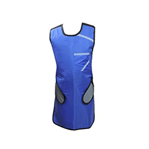
Lead Apron Single
$142.00
In Stock
Our x-ray lead aprons provide complete protection to people who are exposed to radiations for prolonged hours. X-ray lead aprons are a must since they reduce the chances of developing cancer which is a common result of radiation exposure. Lead aprons reduce the exposure to these harmful rays and thus the chances of getting cancer.
Description
Our x-ray lead aprons provide complete protection to people who are exposed to radiations for prolonged hours. X-ray lead aprons are a must since they reduce the chances of developing cancer which is a common result of radiation exposure. Lead aprons reduce the exposure to these harmful rays and thus the chances of getting cancer.
Features:
- 0.5mm Lead (pb) Equivalency Protection at 100 kVp of X-ray beam energy
- With ROBUST HANGER, Protection Material-Lead
- Front Velcro Closure, Includes Single Front pocket
- Padded Shoulders to Prevent Strain
- Stain Resistant and Washable Material for Minimum Maintenance
Quick Comparison
| Lead Apron Single remove | Single X-Ray Viewing Box remove | DrGem Diamond All-In-One Digital X-ray Machine remove | SIGNERS SUPiA X-ray Digitizer ( CR Scanner) remove | Sonoscape E1 Ultrasound Machine With Two Probes remove | Sonoscape S22 Ultrasound Machine remove | |||||||||||||||||||||||||
|---|---|---|---|---|---|---|---|---|---|---|---|---|---|---|---|---|---|---|---|---|---|---|---|---|---|---|---|---|---|---|
| Name | Lead Apron Single remove | Single X-Ray Viewing Box remove | DrGem Diamond All-In-One Digital X-ray Machine remove | SIGNERS SUPiA X-ray Digitizer ( CR Scanner) remove | Sonoscape E1 Ultrasound Machine With Two Probes remove | Sonoscape S22 Ultrasound Machine remove | ||||||||||||||||||||||||
| Image |  |  |  | 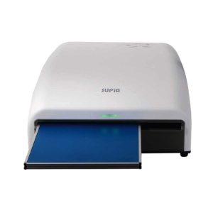 |  |  | ||||||||||||||||||||||||
| SKU | SF1033560071 | SF1033560084-203 | SF1033560074-3 | SF1033560050-01 | SF1033560012-20 | SF1033560012-3 | ||||||||||||||||||||||||
| Rating | ||||||||||||||||||||||||||||||
| Price | $142.00 | $95.20 |
| $6,930.00 | $4,620.00 | $9,350.00 | ||||||||||||||||||||||||
| Stock | ||||||||||||||||||||||||||||||
| Availability | ||||||||||||||||||||||||||||||
| Add to cart | ||||||||||||||||||||||||||||||
| Description | In Stock
Our x-ray lead aprons provide complete protection to people who are exposed to radiations for prolonged hours. X-ray lead aprons are a must since they reduce the chances of developing cancer which is a common result of radiation exposure. Lead aprons reduce the exposure to these harmful rays and thus the chances of getting cancer.
| In stock
| Shipped from Abroad DrGem Diamond All-In-One Digital X-ray Machine is a fully automatic digital radiography system providing state-of-the-art image quality, image processing and user interface. With a wide selection of anatomical studies on the imaging software, DIAMOND automatically sets up the x-ray generator’s preprogrammed exposure technique settings, motorized radiographic stand positioning, x-ray collimation and post-image processing for the selected study. Specifically designed to increase workflow, this fully digital system offers convenient auto-positioning and advanced image processing to achieve big performance with little effort. Delivery & Availability: Typically 21 working days – excluding furniture and heavy/bulky equipment. Please contact us for further information. | Shipped from Abroad SUPiA made by Signers offers such a better clinic environment with no chemicals, ideal space, high-resolution image quality, and affordability. Delivery & Availability: Typically 14 working days – excluding furniture and heavy/bulky equipment. Please contact us for further information. | Shipped from Abroad SonoScape has developed a new probe and function for the E1 Exp. With these additions the E1 Exp will bring users a more efficient examination experience with satisfying image quality and a smooth workflow. Delivery & Availability: Typically 5-7 working days – excluding furniture and heavy/bulky equipment. Please contact us for further information. | Shipped from Abroad As SonoScape steps forward to add value and efficiency to ultrasound, the latest S22 was designed in a user-friendly platform to address current and future demanding needs. It represents an excellent mix in performance and price. Delivery & Availability: Typically 5-7 working days – excluding furniture and heavy/bulky equipment. Please contact us for further information. | ||||||||||||||||||||||||
| Content | Our x-ray lead aprons provide complete protection to people who are exposed to radiations for prolonged hours. X-ray lead aprons are a must since they reduce the chances of developing cancer which is a common result of radiation exposure. Lead aprons reduce the exposure to these harmful rays and thus the chances of getting cancer.
Features:
|
| DrGem Diamond All-In-One Digital X-ray Machine is a fully automatic digital radiography system providing state-of-the-art image quality, image processing and user interface. With a wide selection of anatomical studies on the imaging software, DIAMOND automatically sets up the x-ray generator’s pre-programmed exposure technique settings, motorized radiographic stand positioning, x-ray collimation and post-image processing for the selected study. Specifically designed to increase workflow, this fully digital system offers convenient auto-positioning and advanced image processing to achieve big performance with little effort.
Features of DrGem Diamond All-In-One Digital X-ray Machine:
Outstanding Image Quality -
Digital radiography via at panel detector improves your workflow, exam speed and comfort with efficiency. Digital at panel detector with Csl screen provides excellent spatial resolution, MTF, DQE and stability based on ne pixel pitch. A 3-field ion-chamber is provided for AEC function.
Automatic Collimation –
Automatic x-ray eld size control of the motorized collimator corresponds to dierent SIDs. Includes user adjustable lamp timer with on/oswitch.
Automatic Positioning –
Click Here To Download Catalogue | SUPiA X-ray Digitizer made by Signers offers such a better clinic environment with no chemicals, ideal space, high-resolution image quality, and affordability
FEATURE
Rigid Type
• No damage or scratch on image plates during scanning & erasing
• Scanning & Erasing without a roller
• No cut-off image during winter and cold period
Durability
• Extremely simple structure design
• Strong aluminum base plate
• Flip covers preventing dust from inside scanner
Barcode System
• Automatically recognising cassette sizes(14x17", 10x12", 18x24cm) by barcode reader
Compact & lightweight design
Click Here To Download Catalogue | DETAILS
Efficient Diagnosis
μ-Scan, Speckle Reduction & Edge Enhancement
Spatial Compound Imaging
PIH - Pure Inversion Harmonic
Wide Scan - Enlarged Image Area
Tissue-Specific Imaging
SR Flow
Ergonomic Designs
Up to 2 Transducer Ports
Light Weight and Compact
15.6 inch Anti-flickering HD LED Screen
Tilting Monitor Angle Adjustment
Backlit Keyboard and Intelligent Panel
Long-lasting Battery for 90 mins
Ease of Use
Quick Boot Up
Auto-Brightness Adjustment
Auto Image Optimization
Auto IMT
Auto Trace
Equipped Accessories
Wi-Fi and Bluetooth Available
DICOM
500GB Hard Disk
Height Adjustable Trolley
Durable, Carry-on Site Suitcase
Click Here To Download Catalogue | DETAILS
As SonoScape steps forward to add value and efficiency to ultrasound, the latest S22 was designed in a user-friendly platform to address current and future demanding needs. It represents an excellent mix in performance and price.
S22, is a shared service ultrasound system with a slim and elegant package that has combined mobility with utility to fit in specific clinical situations including emergency department, ICU, operating room and so on. Furthermore, its ergonomic design, easy operating and flexible data management will give you a memorable experience.
SPECIFICATION
• Large high-resolution widescreen LED
• Sensitive touch screen
• Four transducer sockets plus one socket for pencil probe
• A comprehensive selection of probes: linear, Convex, Micro-convex, Volumetric, Endocavity, Bi-plane, Phased Array, TEE, Intraoperative, Pencil
• Premium application technology: 4D, μ-scan speckle reduction, compound imaging, Pulse Inversion Harmonic Imaging, Color M-Mode, Steer M-Mode, PDI, TDI, Real-time Panoramic Imaging, Trapezoid Imaging, Auto-IMT…
• Full patient database and image management solutions: DICOM 3.0, AVI/JPG, USB 2.0, HDD, DVD, PDF report
• Multi-Language Input Keyboard
• Built-in battery
Click Here To Download Catalogue | ||||||||||||||||||||||||
| Weight | N/A | N/A | N/A | N/A | N/A | N/A | ||||||||||||||||||||||||
| Dimensions | N/A | N/A | N/A | N/A | N/A | N/A | ||||||||||||||||||||||||
| Additional information |


Reviews
There are no reviews yet.