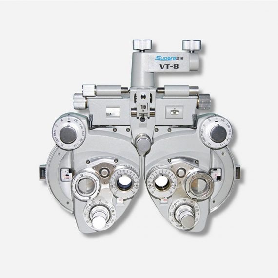
View Tester (Manual Phoropter)
Ask for Price$0.00
Ship from abroad
- Equipped with comprehensive measuring functions, it provides SPH, CYL, AXIS and pupil distance optometry
- Durable and easy to operate
- Easily and intuitively read the sphere focal scale value
- High eco-friendly materials
- Design fitting the face curve and no stimulation
Delivery & Availability:
Typically 14 working days – excluding furniture and heavy/bulky equipment. Please contact us for further information.
Description
Features:
- Equipped with comprehensive measuring functions, it provides SPH, CYL, AXIS and pupil distance optometry
- Durable and easy to operate
- Easily and intuitively read the sphere focal scale value
- High eco-friendly materials
- Design fitting the face curve and no stimulation
- Easy to take and clean
- Free switch between the cross-cylindrical lens and the rotary prism
- When the rotating risk is turning by the sphere, it can make sphere power adjust 3.00D for big scope.
- It is designed expediently and smartly for a particular cross cylinder. Supporting supplementary lens could increase scope of measurement.
Technical Specifications:
|
Sphere
|
Range:-19.00~+16.75m-1 Step: 0.25m-1, 3.00m-1
|
||
|
Cylinder
|
Range: 0.00~-6.00m-1(Measuring Range With Accessories0.00~-8.00m-1) Step: 0.25m-1
|
||
|
Cylinder Axis
|
Range: 0~180°, Step:5°
|
||
|
Distance of Optical center (also known as Pupil)
|
Range: 50~75mmStep: 1mm
|
||
|
Sight Switch
|
Range:∞~380mm (distance of Optical center is64mm)
|
||
|
Front Chin Test
|
Range: 0~16mm
|
||
|
Distance (from cornea vertex to the lens surface)
|
16mm
|
||
|
Standard Accessories Lens
|
two pieces of Auxiliary Cylinder -2.00m-1 and -0.12m-1 respectively
|
||
|
Standard Accessories
|
one piece of M2 Hexagon wrench , one piece of a Myopia Standard Card, two piece of Myopia Standard Card , one piece of standard card holder , a dust cover
|
||
|
Auxiliary Lens
|
“O”:Open aperture “R”:Retinoscope lens “R”:Retinoscope lens “R”:Retinoscope lens *Lens of +1.50m-1 ,It is suit for the distance of 67 centimeters “P”:Polaroid * it is used for examining the dioptric balance of eyes , Implicit strabismus and stereo vision “RMV”:Red Vertical maddox *Be used to examine Implicit strabismus “RMH”:Red horizontal maddox *Be used to examine Implicit strabismus “WMV”:Plane Vertical Maddox *Be used to examine Implicit strabismus “WMH”:Plane horizontal maddox *Be used to examine Implicit strabismus “RL”:Red lens *Be used to examine eye function, Blending function and Implicit strabismus “GL”:Green lens *Be used to examine eye function, Blending function and Implicit strabismus “+”:Test mark of optical center adjustment “+.12”:Dioptric of the Spherical Lens is +0.12m-1 *Be used for the semi-adjustment of sphere lens, 0.25m-1 “PH”:1mmPinhole lens *Be used to exclude visual non-refractive errors of the tested eye “6ΔU”:6ΔBottom-up prism *Be used to examine the rotating prism with the detection of nearly horizontal squint “10ΔI”:10ΔBottom-up prism *Be used to examine the rotating prism with the detection of nearly horizontal squint “±0.50”:Cross-cylindrical lens *Be used to examine the corrected dioptric of the Presbyopia and spherical lens “OC”:Black lens |
||
|
size
|
338(L)×99(W)×292(H)mm
|
||
|
NW
|
about 5kg
|
||
Quick Comparison
| Settings | View Tester (Manual Phoropter) remove | Sonoscape P20 Ultrasound Machine remove | ASPEL AsCARD Grey ECG Machine remove | DrGem Floor Mounted Analogue X-ray remove | DrGem Ceiling Mounted Digital X-ray remove | ASPEL Stress ECG with Treadmill and Software remove | ||||||||||||||||||||||||||||||||||||||||||||||||
|---|---|---|---|---|---|---|---|---|---|---|---|---|---|---|---|---|---|---|---|---|---|---|---|---|---|---|---|---|---|---|---|---|---|---|---|---|---|---|---|---|---|---|---|---|---|---|---|---|---|---|---|---|---|---|
| Name | View Tester (Manual Phoropter) remove | Sonoscape P20 Ultrasound Machine remove | ASPEL AsCARD Grey ECG Machine remove | DrGem Floor Mounted Analogue X-ray remove | DrGem Ceiling Mounted Digital X-ray remove | ASPEL Stress ECG with Treadmill and Software remove | ||||||||||||||||||||||||||||||||||||||||||||||||
| Image | 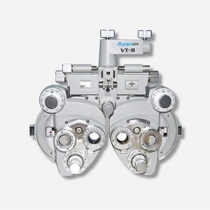 |  | 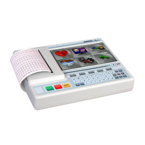 |  |  |  | ||||||||||||||||||||||||||||||||||||||||||||||||
| SKU | SF1033560107-26 | SF1033560012-9 | SF1033560075-5 | SF1033560074-6 | SF1033560074-4 | SF1033560075-2 | ||||||||||||||||||||||||||||||||||||||||||||||||
| Rating | ||||||||||||||||||||||||||||||||||||||||||||||||||||||
| Price | Ask for Price | Ask for Price | Ask for Price | $26,924.00 | $68,468.00 | Ask for Price | ||||||||||||||||||||||||||||||||||||||||||||||||
| Stock | ||||||||||||||||||||||||||||||||||||||||||||||||||||||
| Availability | ||||||||||||||||||||||||||||||||||||||||||||||||||||||
| Add to cart | ||||||||||||||||||||||||||||||||||||||||||||||||||||||
| Description | Ship from abroad
| Shipped from Abroad Incorporating innovative technologies, P20’s user-friendly design with a simple operation panel, intuitive user interface and a variety of intelligent auxiliary scanning tools, will significantly improve your daily examination experience. Besides general imaging applications, P20 has entitled with diagnostic 4D technology which has an extraordinary performance in obstetrics and gynecology applications. Delivery & Availability: Typically 5-7 working days – excluding furniture and heavy/bulky equipment. Please contact us for further information. | Shipped from Abroad Electrocardiograph AsCARD Grey v.07.225 - is a 1, 3, 6, 12 channel ECG unit which enables to make electrocardiogram in full 12 leads. It is intended to conduct ECG examinations of adults and paediatric patients in all types of health care centres. ECG examination may be recorded in manual or automatic mode, with the possibility of analysis and interpretation. The device can be powered from 100 V ÷ 240 V mains supply or by an internal battery. Delivery & Availability: Typically 10 working days – excluding furniture and heavy/bulky equipment. Please contact us for further information. | In Stock GXR Analogue X-ray system matches with a radiographic room which perfectly fits your workow and can be easily upgraded to DR system with the help of DR interface and PC interface in GXR generator as well as Bucky suitable to Flat Panel Detector. GXR X-ray system is equipped with a high frequency X-ray generator which consistently produces high quality radiograph in favor of high quality X-ray output with a very small kV ripple and accurate mA and mAs. GXR X-ray system is designed to provide convenience to operator and comfort to patient. Delivery & Availability: Typically 21 working days – excluding furniture and heavy/bulky equipment. Please contact us for further information. | In Stock The GXR-SD is a diagnostic digital radiography system that provides reliable high quality digital radiographic images with a reduced dose. The GXR-SD DR systems offer comprehensive digital solutions to all radiography needs, featuring ACQUIDR digital imaging system with stationary or portable digital flat-panel detectors as well as reliable high-frequency x-ray generators that are known worldwide for their excellent performance, lifetime and stability. Patient tables and wall stands are also offered. Delivery & Availability: Typically 21 working days – excluding furniture and heavy/bulky equipment. Please contact us for further information. | Shipped from Abroad It is a system with professional tool dedicated to exercise and resting ECG examination. Treadmill has 12 lead ECG modules. With ECG Analyzing Software. Delivery & Availability: Typically 21 working days – excluding furniture and heavy/bulky equipment. Please contact us for further information. | ||||||||||||||||||||||||||||||||||||||||||||||||
| Content | Features:
| DETAILS
Upgraded Images with More Clarity
SonoScape never stops making progress in improving the image quality of its ultrasound products to enhance the confidence of diagnosis for doctors. With extraordinary images provided by P20, the anatomy structures are clearer than ever.
C-Xlasto Imaging
With C-xlasto Imaging, P20 enables comprehensive quantitative elastic analysis. Meanwhile, C-xlasto on P20 is supported by linear, convex and transvaginal probes, to ensure good reproducibility and highly consistent quantitative elastic results.
S-Live
S-Live allows for detailed visualization of subtle anatomical features, thereby enabling intuitive diagnosis with real-time 3D images and enriching patient communication.
Pelvic Floor 4D
Transperineal 4D pelvic floor ultrasound can provide useful clinical values in assessing the vaginal delivery impact on the female anterior compartment, judging whether the pelvic organs are prolapsed or not and the extent, determining if the pelvic muscles were torn accurately.
Anatomic M Mode
Anatomic M Mode helps you observe the myocardial motion at different phases by freely placing sample lines. It accurately measures the myocardial thickness and the heart size of even difficult patients and supports the myocardial function and LV wall-motion assessment.
Tissue Doppler Imaging
P20 is endowed with Tissue Doppler Imaging which provides velocities and other clinical information on myocardial functions, facilitating clinical doctors with the ability to analyze and compare the motions of different parts of the patient's heart.
Click Here To Download Catalogue |
Electrocardiograph AsCARD Grey v.07.225 - is a 1, 3, 6, 12 channel ECG unit which enables to make electrocardiogram in full 12 leads. It is intended to conduct ECG examinations of adults and paediatric patients in all types of health care centres. ECG examination may be recorded in manual or automatic mode, with the possibility of analysis and interpretation. The device can be powered from 100 V ÷ 240 V mains supply or by an internal battery.
Technical Specification:1. Visualisation of 1, 3, 6 or 12 ECG waveforms, analysis results and interpretations, examinations stored in memory.
2. Recording of 12 standard leads.
3. Print out in 1, 3, 6 or 12 ECG waveforms mode. Printing of a selected group:
Click Here To Download Catalogue | DrGem GXR Floor Mounted Analogue X-ray system matches with a radiographic room which perfectly fits your workflow and can be easily upgraded to DR system with the help of DR interface and PC interface in GXR generator as well as Bucky suitable to Flat Panel Detector. GXR (Analogue X-ray)system is equipped with a high frequency X-ray generator which consistently produces high quality radiograph in favor of high quality X-ray output with a very small kV ripple and accurate mA and mAs. GXR (Analogue X-ray) system is designed to provide convenience to operator and comfort to patient.
Features of DrGem GXR Floor Mounted Analogue X-ray:
Click Here To Download Catalogue | DrGem Ceiling Mounted Digital X-ray is a diagnostic digital radiography system that provides reliable high quality digital radiographic images with a reduced dose. The GXR-SD DR systems offer comprehensive digital solutions to all radiography needs, featuring ACQUIDR digital imaging system with stationary or portable digital flat-panel detectors as well as reliable high-frequency x-ray generators that are known worldwide for their excellent performance, lifetime and stability. Patient tables and wall stands are also offered.
Features:
Click Here To Download Catalogue | It is a system with professional tool dedicated to exercise and resting ECG examination. Treadmill has 12 lead ECG modules. With ECG Analyzing Software.
Technical Specification:
Click Here To Download Catalogue | ||||||||||||||||||||||||||||||||||||||||||||||||
| Weight | N/A | N/A | N/A | N/A | N/A | N/A | ||||||||||||||||||||||||||||||||||||||||||||||||
| Dimensions | N/A | N/A | N/A | N/A | N/A | N/A | ||||||||||||||||||||||||||||||||||||||||||||||||
| Additional information | ||||||||||||||||||||||||||||||||||||||||||||||||||||||


Reviews
There are no reviews yet.