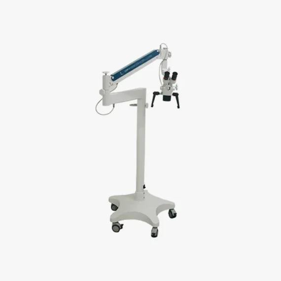
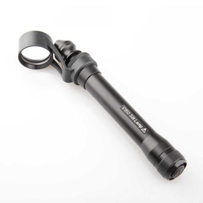
MICROSCOPE LINE 12
$0.00
Shipped From Abroad
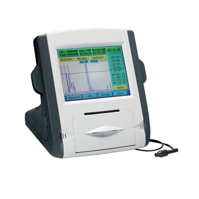
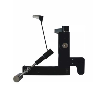
The Line 12 Microscopes have characteristics that meet different medical/dental specialties.
With excellence in optics and lighting, it provides a wide range of the observed field.
It allows the inclusion of various accessories that complement user experiences such as secondary assistants, video system, objective lenses and much more.It has the best value for money on the market!
With excellence in optics and lighting, it provides a wide range of the observed field.
It allows the inclusion of various accessories that complement user experiences such as secondary assistants, video system, objective lenses and much more.It has the best value for money on the market!
Delivery & Availability:
Typically 10-21 working days – excluding furniture and heavy/bulky equipment. Please contact us for further information.
Typically 10-21 working days – excluding furniture and heavy/bulky equipment. Please contact us for further information.
Description
Description
The Line 12 Microscopes have characteristics that meet different medical/dental specialties.
With excellence in optics and lighting, it provides a wide range of the observed field.
It allows the inclusion of various accessories that complement user experiences such as secondary assistants, video system, objective lenses and much more.
It has the best value for money on the market!
Quick Comparison
| MICROSCOPE LINE 12 remove | Handheld Digital Auto-refractometer remove | Slit Lamp with Workstation remove | View Tester (Manual Phoropter) remove | Manual lensmeter remove | Ear Irrigation and acumen removal remove | |||||||||||||||||||||||||||||||||||||||||||||||||||||||||||||||||||||||||||||||||||||||||||||||||||
|---|---|---|---|---|---|---|---|---|---|---|---|---|---|---|---|---|---|---|---|---|---|---|---|---|---|---|---|---|---|---|---|---|---|---|---|---|---|---|---|---|---|---|---|---|---|---|---|---|---|---|---|---|---|---|---|---|---|---|---|---|---|---|---|---|---|---|---|---|---|---|---|---|---|---|---|---|---|---|---|---|---|---|---|---|---|---|---|---|---|---|---|---|---|---|---|---|---|---|---|---|---|---|---|---|
| Name | MICROSCOPE LINE 12 remove | Handheld Digital Auto-refractometer remove | Slit Lamp with Workstation remove | View Tester (Manual Phoropter) remove | Manual lensmeter remove | Ear Irrigation and acumen removal remove | ||||||||||||||||||||||||||||||||||||||||||||||||||||||||||||||||||||||||||||||||||||||||||||||||||
| Image | 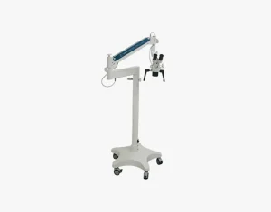 | 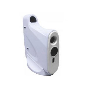 | 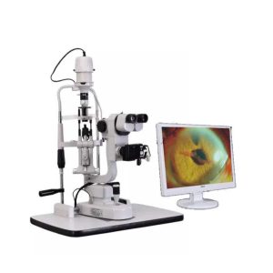 | 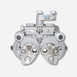 | 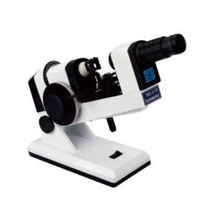 | 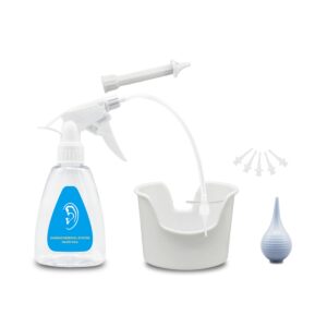 | ||||||||||||||||||||||||||||||||||||||||||||||||||||||||||||||||||||||||||||||||||||||||||||||||||
| SKU | SF103356013091-10 | SF1033560107-2 | SF1033560107-7 | SF1033560107-26 | SF1033560107-22 | SF103356013012 | ||||||||||||||||||||||||||||||||||||||||||||||||||||||||||||||||||||||||||||||||||||||||||||||||||
| Rating | ||||||||||||||||||||||||||||||||||||||||||||||||||||||||||||||||||||||||||||||||||||||||||||||||||||||||
| Price |
|
| $3,740.00 | $858.00 |
|
| ||||||||||||||||||||||||||||||||||||||||||||||||||||||||||||||||||||||||||||||||||||||||||||||||||
| Stock | ||||||||||||||||||||||||||||||||||||||||||||||||||||||||||||||||||||||||||||||||||||||||||||||||||||||||
| Availability | ||||||||||||||||||||||||||||||||||||||||||||||||||||||||||||||||||||||||||||||||||||||||||||||||||||||||
| Add to cart | ||||||||||||||||||||||||||||||||||||||||||||||||||||||||||||||||||||||||||||||||||||||||||||||||||||||||
| Description | Shipped From Abroad
The Line 12 Microscopes have characteristics that meet different medical/dental specialties.
With excellence in optics and lighting, it provides a wide range of the observed field.
It allows the inclusion of various accessories that complement user experiences such as secondary assistants, video system, objective lenses and much more.It has the best value for money on the market!
Delivery & Availability:
Typically 10-21 working days – excluding furniture and heavy/bulky equipment. Please contact us for further information.
| Shipped from abroad
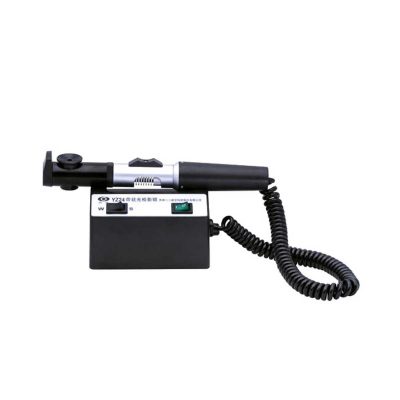
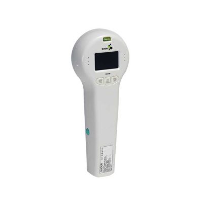
AutoSight 900 is a portable vision screener for patients at any age. Its working principle is the refraction of light.
| Shipped from abroad
| Ship from abroad
| Shipped from abroad
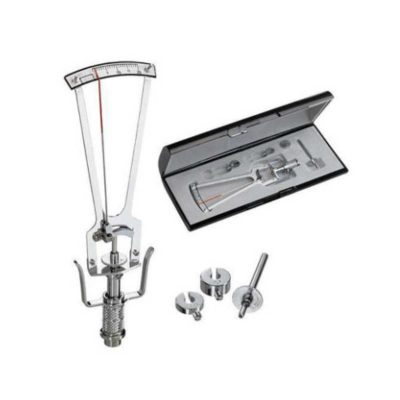
The NJC-4 Lensmeter is used to measure the diopters of spherical lens and cylinder lens, the axis of cylinder lens, the strength and the baseline direction of shuttle lens, it can also stamp the optical center of lens, the axis of cylinder len and the base direction of shuttle lens.
| In Stock
Features:
●Professional
Same ear wax removal tool as those used by doctors, you can easily eliminate ear wax buildup at home, really save your money and time on medical visiting. Safe and Environmentally Friendly.
●Quick & Easy
This ear wax removal kit is a quick, effective treatment for excess ear wax buildup. Fill the bottle with solution, Twist on the disposable tip, Use the trigger handle to spray solution into the ear canal. So Easy.
Delivery & Availability:
Typically 7-14 working days – excluding furniture and heavy/bulky equipment. Please contact us for further information.
| ||||||||||||||||||||||||||||||||||||||||||||||||||||||||||||||||||||||||||||||||||||||||||||||||||
| Content | Description
The Line 12 Microscopes have characteristics that meet different medical/dental specialties.
With excellence in optics and lighting, it provides a wide range of the observed field.
It allows the inclusion of various accessories that complement user experiences such as secondary assistants, video system, objective lenses and much more.
It has the best value for money on the market!
| Handheld Digital Auto-refractometer(AutoSight 900) is a portable vision screener for patients at any age. Its working principle is the refraction of light. Optical rays are focused on a sensor after passing through the eye's refractive system. The spherical power, cylindrical power, and axis of both eyes can be obtained by digital signal processing.
Features of Handheld Digital Auto-refractometer:
| Slit Lamp with Workstation Features:
| Features:
| The NJC-4 Lensmeter is used to measure the diopters of the spherical lens and cylinder lens, the axis of cylinder lens, the strength and the baseline direction of shuttle lens, it can also stamp the optical center of the lens, the axis of cylinder lens, and the base direction of shuttle lens. This instrument (Both Ac and Dc are permitted Two cells When Dc) has clear readings and graduations as well as high objective precision and reliabilities except that it can be operated easily and conveniently, AU the lenses and the made glasses can be measured by it, therefore, it is a required and ideal measuring instrument for glasses manufactures, glasses stores and ophthalmological hospitals.
Technical Specifications:
| Features: ●Professional Same ear wax removal tool as those used by doctors, you can easily eliminate ear wax buildup at home, really save your money and time on medical visiting. Safe and Environmentally Friendly. ●Quick & Easy This ear wax removal kit is a quick, effective treatment for excess ear wax buildup. Fill the bottle with solution, Twist on the disposable tip, Use the trigger handle to spray solution into the ear canal. So Easy. ●Standard Capacity of the ear cleaner solution bottle is 10.6Oz, it has the most suitable size to hold in hand. Working at condition 32-122℉(0-50℃). Recommend to fill 1/5 of the bottle with OTC hydrogen peroxide, and 4/5 with very warm water. ●Complete Ear Washer System Our earwax removal kit comes with 1× Ear Washer Bottle, 1× Wash Basin, 1× Rubber Bulb, 1× Short Injection Head, 1× Long Hose Injection Head, 5× Disposable Tip, 1× User Manual. | ||||||||||||||||||||||||||||||||||||||||||||||||||||||||||||||||||||||||||||||||||||||||||||||||||
| Weight | N/A | N/A | N/A | N/A | N/A | N/A | ||||||||||||||||||||||||||||||||||||||||||||||||||||||||||||||||||||||||||||||||||||||||||||||||||
| Dimensions | N/A | N/A | N/A | N/A | N/A | N/A | ||||||||||||||||||||||||||||||||||||||||||||||||||||||||||||||||||||||||||||||||||||||||||||||||||
| Additional information | ||||||||||||||||||||||||||||||||||||||||||||||||||||||||||||||||||||||||||||||||||||||||||||||||||||||||


Reviews
There are no reviews yet.