
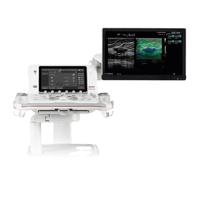
MyLab™9 Platform
$0.00
Shipped From Abroad
Shared service ultrasound system
MyLab™9 Platform ultrasound system is designed to support a full range of shared service diagnostic imaging environments. Take ultra-control of your images with unique visualization tools, and view results with clarity and sensitivity to help make more informed clinical decisions. Experience the ultra-comfort of Italian-designed ergonomics and an ultra-easy user interface that increases productivity.
Typically 10-21 working days – excluding furniture and heavy/bulky equipment. Please contact us for further information.
Description
The Esaote MyLab™9 Platform is a high-performance console system designed to bring “ultra” quality, comfort, and ease to shared-service diagnostic environments. The system provides superior image clarity, color, and contrast via its powerful Ultra-Engine architecture, Single Crystal probe technology, and an exceptional 24-inch Full HD monitor.
Ergonomics are maximized through Italian design, featuring a floating keyboard and the innovative APPLEPROBE design to reduce operator strain by up to 70%. Workflow is drastically accelerated by unique features like easyMODE, which intelligently optimizes over 40 imaging parameters in just three quick swipes, ensuring precision and speed in every examination.
Features
-
Ultra-Engine Platform: Powerful imaging backbone delivering precision, contrast, and depth.
-
Superior Visualization: Large 24-inch Full HD Medical-Grade Monitor (optional BARCO™) and a full HD tablet-style touchscreen for control.
-
Advanced Ergonomics: Italian-designed console with a floating keyboard, Opti-light integrated illumination, and APPLEPROBE technology for up to 70% reduction in musculoskeletal strain.
-
Workflow Automation: easyMODE unique touch-tool for intelligent real-time image optimization, and i-motion technology for sustained high frame rates.
-
Comprehensive Clinical Tools: Includes CnTI™ (Contrast Enhanced Ultrasound), QElaXto (Point and 2D Shearwave Elastography), microV (micro-vascular flow), XStrain™ 2D/4D (advanced cardiac analysis), and Virtual Navigator (fusion imaging).
-
High-Frequency Imaging: Supports imaging up to 24 MHz for high-resolution superficial and musculoskeletal applications.
-
Connectivity: Full DICOM support, PACS integration, and remote technical support.
-
Probes: Wide range of high-quality iQProbes, including Single Crystal technology.
Imaging & Visualization Technologies
The MyLab™X90 utilizes exclusive technologies to deliver outstanding image clarity and detail, even in challenging cases.
-
XCrystal Transducer Technology: This is Esaote’s latest generation of probes that dramatically increases sensitivity and penetration, resulting in sharper images and homogeneity from the near-field (superficial) to deeper areas. The technology supports high-frequency imaging up to 25 MHz.
-
ClearWave Architecture: This advanced signal processing framework combines XPower Beamforming and XSmart Postprocessing to set a new standard for image quality. It ensures superior detail enhancement and uniform image rendering.
-
eLED High Dynamic Range Monitor: The system features a large 23.8-inch Full HD monitor (developed with Barco) with Dual-Layer LCD technology. This offers:
-
Brilliant colors and infinite contrast.
-
A contrast resolution ratio up to 40 times higher than conventional LCDs.
-
Higher stability over time for reliable image viewing.
-
-
Opti-Light: An ambient light sensor and integrated backlit lighting feature that automatically optimizes display brightness to the scanning environment, improving comfort and visibility.
Clinical Applications & Advanced Tools
The MyLab™X90 is designed as a 360° shared-service solution, meaning it excels across numerous medical specialties and integrates AI tools—called Augmented Insight™—to automate complex tasks.
1. Cardiovascular (CV)
-
XStrain™: Provides zero-click detection of endocardial border profiles in the Left Ventricle (LV) and detailed exploration of strain in the LV, Right Ventricle (RV), and Left Atrium (LA).
-
AutoEF: Automatic AI-driven contouring of the left ventricle in 4 and 2 chambers to accurately assess the Ejection Fraction (EF).
-
HyperDoppler: Advanced tool for visualizing intracardiac flows and vortices, offering deeper insights for cardiac dynamics research.
2. General Imaging & Interventional (GI)
-
CnTI™ Clear: The latest evolution of Contrast Enhanced Ultrasound (CEUS) technology, which increases the persistence of contrast media and improves coverage of the deepest areas for microperfusion information (e.g., in liver and prostate).
-
LiverFusion & UroFusion: Fusion Imaging technologies that enable the real-time combination of ultrasound data with pre-acquired images (like MRI/CT) for targeted biopsies and navigation procedures, especially in the prostate and liver.
-
QAI (Quantification Attenuation Imaging): Provides mapping and quantification of sound attenuation along the liver depth to evaluate hepatic steatosis (fatty liver).
-
QElaXto 2D: Sophisticated Shear-Wave Elastography software for non-invasive assessment of tissue stiffness, widely used in liver and breast disease management.
3. Women’s Health (OB/GYN)
-
AutoOB: Integrates AI for primary fetal biometric measurements (like BPD, HC, AC, FL) with automatic plane recognition, speeding up the standard examination workflow.
-
3D/4D Imaging: Offers complete volumetric representation solutions for comprehensive fetal and gynecological examinations.
4. Breast & Small Parts
-
Breast Mass Analyser (BMA): Provides automatic AI-driven proposals for breast lesion classification in regions of interest.
-
Automatic Contouring: AI-based automatic contouring of thyroid and breast lesions in suspicious areas.
Probe Types & Compatibility
The MyLab™X90 platform supports a very wide variety of XCrystal and high-frequency probes for a multidisciplinary approach, with five transducer connectors for immediate switching.
The system is compatible with several probe types, including:
| Probe Type | Key Application Areas | Example Probes (General Type) |
| Linear Array | Vascular, Musculoskeletal (MSK), Small Parts (Thyroid, Breast), high-frequency superficial imaging (up to 25 MHz with the LMX 4-20 HD Single Crystal probe) | LMX, LX, L, IH |
| Convex & Microconvex | Abdominal, General Imaging, Obstetrics/Gynecology, Deep Vascular, Pediatrics | CX, C, mC |
| Phased Array | Cardiac (Adult & Pediatric), Transcranial Doppler (TCD) | PX, P, P2, TE |
| Volumetric | 3D/4D imaging for Obstetrics/Gynecology and volume acquisitions | SB |
| Endocavitary | Transvaginal (Gynecology/Obstetrics), Transrectal (Urology) | E, TLC |
Quick Comparison
| MyLab™9 Platform remove | Sonoscape E2 Ultrasound Machine remove | Sonoscape S11 Ultrasound Machine remove | FlowMir Disposable Turbine with Cardboard Mouthpiece remove | Sonoscape P20 Ultrasound Machine remove | Sonoscape P15 Ultrasound Machine With Four Probes remove | |||||||||||||||||||
|---|---|---|---|---|---|---|---|---|---|---|---|---|---|---|---|---|---|---|---|---|---|---|---|---|
| Name | MyLab™9 Platform remove | Sonoscape E2 Ultrasound Machine remove | Sonoscape S11 Ultrasound Machine remove | FlowMir Disposable Turbine with Cardboard Mouthpiece remove | Sonoscape P20 Ultrasound Machine remove | Sonoscape P15 Ultrasound Machine With Four Probes remove | ||||||||||||||||||
| Image |  |  |  | 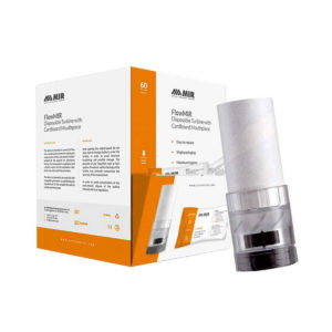 |  |  | ||||||||||||||||||
| SKU | SF1033560012-17 | SF1033560012-1 | SF1033560084-22 | SF1033560012-9 | SF1033560012-8 | |||||||||||||||||||
| Rating | ||||||||||||||||||||||||
| Price |
| $5,500.00 | $6,380.00 | $145.00 |
| $13,900.00 | ||||||||||||||||||
| Stock | ||||||||||||||||||||||||
| Availability | ||||||||||||||||||||||||
| Add to cart | ||||||||||||||||||||||||
| Description | Shipped From Abroad
Shared service ultrasound system
MyLab™9 Platform ultrasound system is designed to support a full range of shared service diagnostic imaging environments. Take ultra-control of your images with unique visualization tools, and view results with clarity and sensitivity to help make more informed clinical decisions. Experience the ultra-comfort of Italian-designed ergonomics and an ultra-easy user interface that increases productivity. Delivery & Availability:
Typically 10-21 working days – excluding furniture and heavy/bulky equipment. Please contact us for further information.
| Shipped from Abroad Sonoscape E2 portable ultrasound machine is a color Doppler ultrasound system that reaches beyond your expectations due to its compact and fashionable appearance. It fulfills GI, OB/GYN, Cardiac and POC applications to fit your routine scanning needs while its color mode will help you for more accurate and efficient diagnosis of lesions. E2 provides a wide range of applications to assist users with routine scanning. E2 provides automatic calculations to enhance your diagnostic confidence and save you time for patient communication. Delivery & Availability: Typically 14 working days – excluding furniture and heavy/bulky equipment. Please contact us for further information. | In Stock A Value Choice beyond Your Expectation. SonoScape’s trolley color Doppler system S11 redefines price and performance with practical design. The S11 will go beyond your expectations but not your budget. Delivery & Availability: Typically 2 working days – excluding furniture and heavy/bulky equipment. Please contact us for further information. | In stock Spirometry testing requires maximum accuracy and hygiene. Each Flowmir disposable turbine, which includes a cardboard mouthpiece, has been individually factory calibrated with a computerized system and it is packaged individually. FlowMIR® is an inexpensive alternative to a costly reusable flowmeter and replaces the need for an antibacterial filter Typically 2 working days – excluding furniture and heavy/bulky equipment. Please contact us for further information. | Shipped from Abroad Incorporating innovative technologies, P20’s user-friendly design with a simple operation panel, intuitive user interface and a variety of intelligent auxiliary scanning tools, will significantly improve your daily examination experience. Besides general imaging applications, P20 has entitled with diagnostic 4D technology which has an extraordinary performance in obstetrics and gynecology applications. Delivery & Availability: Typically 5-7 working days – excluding furniture and heavy/bulky equipment. Please contact us for further information. | In Stock A feature-rich system inheriting the Wi-Sono high-end platform, the P15 uses an array of advanced tools to help enhance the image quality. It's a cost-effective, simplified console with an intuitive user interface and multiple intelligent functions. Delivery & Availability: Typically 2 working days – excluding furniture and heavy/bulky equipment. Please contact us for further information. | ||||||||||||||||||
| Content | https://youtu.be/V0VhAaINKoM
The Esaote MyLab™9 Platform is a high-performance console system designed to bring "ultra" quality, comfort, and ease to shared-service diagnostic environments. The system provides superior image clarity, color, and contrast via its powerful Ultra-Engine architecture, Single Crystal probe technology, and an exceptional 24-inch Full HD monitor. Ergonomics are maximized through Italian design, featuring a floating keyboard and the innovative APPLEPROBE design to reduce operator strain by up to 70%. Workflow is drastically accelerated by unique features like easyMODE, which intelligently optimizes over 40 imaging parameters in just three quick swipes, ensuring precision and speed in every examination. Features
Imaging & Visualization Technologies The MyLab™X90 utilizes exclusive technologies to deliver outstanding image clarity and detail, even in challenging cases.
Clinical Applications & Advanced ToolsThe MyLab™X90 is designed as a 360° shared-service solution, meaning it excels across numerous medical specialties and integrates AI tools—called Augmented Insight™—to automate complex tasks. 1. Cardiovascular (CV)
Probe Types & Compatibility The MyLab™X90 platform supports a very wide variety of XCrystal and high-frequency probes for a multidisciplinary approach, with five transducer connectors for immediate switching. The system is compatible with several probe types, including:
| SONOSCAPE E2 DETAILS
Auto Image Optimization
A portable ultrasound machine with the press of a button, the image is automatically adjusted and optimized, saving you time with parameter adjustments. Additionally, with Auto Focus on, the focus area follows the depth of the ROI box as it is moved in the scanning field, providing users with excellent image quality in the desired area of interest.
Automated Calculation
Auto IMT is used when determining the level of vascular sclerosis present in the patient by automatically tracing the thickness of the carotid vessels.
Auto trace provides users sensitive and accurate wave tracing, avoiding the error of manual trace and giving out calculation result in no time
In-Build Battery pack
This portable ultrasound machine was equipped with an in-build battery pack which enable the user to perform image scanning when AC power is not available.
Click Here To Download Catalogue | DETAILS
SonoScape’s trolley colour Doppler system S11 redefines price and performance with practical design. The S11 will go beyond your expectations but not your budget. As an easy-to-use ultrasound system, the S11 is integrated with a new software platform, especially optimized for a smooth workflow and convenient operation. The system speeds up the exam process and makes file management easier.
SPECIFICATION
- 15-inch high definition LCD monitor with articulating arm
- Compact and agile trolley design
- 3 active transducer sockets available for a wide range of applications
- Duplex, Color Doppler, DPI, PW Doppler, tissue harmonic imaging, μ-scan speckle reduction imaging, compound imaging, trapezoidal imaging
- Customized settings based on your own working style
- Full patient database and image management solutions
Click Here To Download Catalogue |
| DETAILS
Upgraded Images with More Clarity
SonoScape never stops making progress in improving the image quality of its ultrasound products to enhance the confidence of diagnosis for doctors. With extraordinary images provided by P20, the anatomy structures are clearer than ever.
C-Xlasto Imaging
With C-xlasto Imaging, P20 enables comprehensive quantitative elastic analysis. Meanwhile, C-xlasto on P20 is supported by linear, convex and transvaginal probes, to ensure good reproducibility and highly consistent quantitative elastic results.
S-Live
S-Live allows for detailed visualization of subtle anatomical features, thereby enabling intuitive diagnosis with real-time 3D images and enriching patient communication.
Pelvic Floor 4D
Transperineal 4D pelvic floor ultrasound can provide useful clinical values in assessing the vaginal delivery impact on the female anterior compartment, judging whether the pelvic organs are prolapsed or not and the extent, determining if the pelvic muscles were torn accurately.
Anatomic M Mode
Anatomic M Mode helps you observe the myocardial motion at different phases by freely placing sample lines. It accurately measures the myocardial thickness and the heart size of even difficult patients and supports the myocardial function and LV wall-motion assessment.
Tissue Doppler Imaging
P20 is endowed with Tissue Doppler Imaging which provides velocities and other clinical information on myocardial functions, facilitating clinical doctors with the ability to analyze and compare the motions of different parts of the patient's heart.
Click Here To Download Catalogue | DETAILS
Super Wide-bandwidth Platform
Inheriting Wi-sono's ultra-wide system platform and with the advanced probe technology, high-resolution and deep penetration images are provided for precision medicine.
Spatial Compound Imaging
Spatial Compound Imaging utilizes several lines of sight for optimal contrast resolution, speckle reduction and border detection, with which P15 is ideal for superficial and abdominal imaging with better clarity and improved continuity of structures.
μ-Scan+
The new generation μ-Scan imaging technology gives you better image quality by reducing noise, improving signal strength and improving visualization.
Dynamic Color
Dynamic color improves upon already existing color Doppler technologies for a clearer capture of color flow and detailed visualization of even tiny veins with lower velocities.
Real-time Panoramic
With real-time panoramic, you can acquire an extended field of view for large organs or long vessels for easy measurement and diagnostic efficiency. Accomplished in real-time for the convenience of the sonographers, any mistake can also be easily back tracked and corrected without interrupting the scan.
3D/4D
Outstanding volume performance with speed and convenience makes P15 outshine others on volume imaging.
Tissue Doppler Imaging
Tissue Doppler Imaging allows clinical doctors to quantitatively evaluate local myocardial movements and functions, facilitating them with the ability to analyze and compare the motions of the different parts of the patient's heart.
Auto IMT
Quick measurement of intra-media vessel thickness ensures good reproducibility and high diagnostic efficiency.
Click Here To Download Catalogue | ||||||||||||||||||
| Weight | N/A | N/A | N/A | N/A | N/A | N/A | ||||||||||||||||||
| Dimensions | N/A | N/A | N/A | N/A | N/A | N/A | ||||||||||||||||||
| Additional information |


Reviews
There are no reviews yet.