
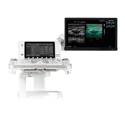
MyLab™9 Platform
$0.00
Shipped From Abroad
Shared service ultrasound system
MyLab™9 Platform ultrasound system is designed to support a full range of shared service diagnostic imaging environments. Take ultra-control of your images with unique visualization tools, and view results with clarity and sensitivity to help make more informed clinical decisions. Experience the ultra-comfort of Italian-designed ergonomics and an ultra-easy user interface that increases productivity.
Typically 10-21 working days – excluding furniture and heavy/bulky equipment. Please contact us for further information.
Description
The Esaote MyLab™9 Platform is a high-performance console system designed to bring “ultra” quality, comfort, and ease to shared-service diagnostic environments. The system provides superior image clarity, color, and contrast via its powerful Ultra-Engine architecture, Single Crystal probe technology, and an exceptional 24-inch Full HD monitor.
Ergonomics are maximized through Italian design, featuring a floating keyboard and the innovative APPLEPROBE design to reduce operator strain by up to 70%. Workflow is drastically accelerated by unique features like easyMODE, which intelligently optimizes over 40 imaging parameters in just three quick swipes, ensuring precision and speed in every examination.
Features
-
Ultra-Engine Platform: Powerful imaging backbone delivering precision, contrast, and depth.
-
Superior Visualization: Large 24-inch Full HD Medical-Grade Monitor (optional BARCO™) and a full HD tablet-style touchscreen for control.
-
Advanced Ergonomics: Italian-designed console with a floating keyboard, Opti-light integrated illumination, and APPLEPROBE technology for up to 70% reduction in musculoskeletal strain.
-
Workflow Automation: easyMODE unique touch-tool for intelligent real-time image optimization, and i-motion technology for sustained high frame rates.
-
Comprehensive Clinical Tools: Includes CnTI™ (Contrast Enhanced Ultrasound), QElaXto (Point and 2D Shearwave Elastography), microV (micro-vascular flow), XStrain™ 2D/4D (advanced cardiac analysis), and Virtual Navigator (fusion imaging).
-
High-Frequency Imaging: Supports imaging up to 24 MHz for high-resolution superficial and musculoskeletal applications.
-
Connectivity: Full DICOM support, PACS integration, and remote technical support.
-
Probes: Wide range of high-quality iQProbes, including Single Crystal technology.
Imaging & Visualization Technologies
The MyLab™X90 utilizes exclusive technologies to deliver outstanding image clarity and detail, even in challenging cases.
-
XCrystal Transducer Technology: This is Esaote’s latest generation of probes that dramatically increases sensitivity and penetration, resulting in sharper images and homogeneity from the near-field (superficial) to deeper areas. The technology supports high-frequency imaging up to 25 MHz.
-
ClearWave Architecture: This advanced signal processing framework combines XPower Beamforming and XSmart Postprocessing to set a new standard for image quality. It ensures superior detail enhancement and uniform image rendering.
-
eLED High Dynamic Range Monitor: The system features a large 23.8-inch Full HD monitor (developed with Barco) with Dual-Layer LCD technology. This offers:
-
Brilliant colors and infinite contrast.
-
A contrast resolution ratio up to 40 times higher than conventional LCDs.
-
Higher stability over time for reliable image viewing.
-
-
Opti-Light: An ambient light sensor and integrated backlit lighting feature that automatically optimizes display brightness to the scanning environment, improving comfort and visibility.
Clinical Applications & Advanced Tools
The MyLab™X90 is designed as a 360° shared-service solution, meaning it excels across numerous medical specialties and integrates AI tools—called Augmented Insight™—to automate complex tasks.
1. Cardiovascular (CV)
-
XStrain™: Provides zero-click detection of endocardial border profiles in the Left Ventricle (LV) and detailed exploration of strain in the LV, Right Ventricle (RV), and Left Atrium (LA).
-
AutoEF: Automatic AI-driven contouring of the left ventricle in 4 and 2 chambers to accurately assess the Ejection Fraction (EF).
-
HyperDoppler: Advanced tool for visualizing intracardiac flows and vortices, offering deeper insights for cardiac dynamics research.
2. General Imaging & Interventional (GI)
-
CnTI™ Clear: The latest evolution of Contrast Enhanced Ultrasound (CEUS) technology, which increases the persistence of contrast media and improves coverage of the deepest areas for microperfusion information (e.g., in liver and prostate).
-
LiverFusion & UroFusion: Fusion Imaging technologies that enable the real-time combination of ultrasound data with pre-acquired images (like MRI/CT) for targeted biopsies and navigation procedures, especially in the prostate and liver.
-
QAI (Quantification Attenuation Imaging): Provides mapping and quantification of sound attenuation along the liver depth to evaluate hepatic steatosis (fatty liver).
-
QElaXto 2D: Sophisticated Shear-Wave Elastography software for non-invasive assessment of tissue stiffness, widely used in liver and breast disease management.
3. Women’s Health (OB/GYN)
-
AutoOB: Integrates AI for primary fetal biometric measurements (like BPD, HC, AC, FL) with automatic plane recognition, speeding up the standard examination workflow.
-
3D/4D Imaging: Offers complete volumetric representation solutions for comprehensive fetal and gynecological examinations.
4. Breast & Small Parts
-
Breast Mass Analyser (BMA): Provides automatic AI-driven proposals for breast lesion classification in regions of interest.
-
Automatic Contouring: AI-based automatic contouring of thyroid and breast lesions in suspicious areas.
Probe Types & Compatibility
The MyLab™X90 platform supports a very wide variety of XCrystal and high-frequency probes for a multidisciplinary approach, with five transducer connectors for immediate switching.
The system is compatible with several probe types, including:
| Probe Type | Key Application Areas | Example Probes (General Type) |
| Linear Array | Vascular, Musculoskeletal (MSK), Small Parts (Thyroid, Breast), high-frequency superficial imaging (up to 25 MHz with the LMX 4-20 HD Single Crystal probe) | LMX, LX, L, IH |
| Convex & Microconvex | Abdominal, General Imaging, Obstetrics/Gynecology, Deep Vascular, Pediatrics | CX, C, mC |
| Phased Array | Cardiac (Adult & Pediatric), Transcranial Doppler (TCD) | PX, P, P2, TE |
| Volumetric | 3D/4D imaging for Obstetrics/Gynecology and volume acquisitions | SB |
| Endocavitary | Transvaginal (Gynecology/Obstetrics), Transrectal (Urology) | E, TLC |
Quick Comparison
| MyLab™9 Platform remove | Sonoscape P20 Ultrasound Machine remove | Sonoscape P50 Ultrasound Machine remove | ASPEL Stress ECG with Treadmill and Software remove | Sonoscape P10 Ultrasound Machine remove | Sonoscape S22 Ultrasound Machine remove | |||||||||||||||||||
|---|---|---|---|---|---|---|---|---|---|---|---|---|---|---|---|---|---|---|---|---|---|---|---|---|
| Name | MyLab™9 Platform remove | Sonoscape P20 Ultrasound Machine remove | Sonoscape P50 Ultrasound Machine remove | ASPEL Stress ECG with Treadmill and Software remove | Sonoscape P10 Ultrasound Machine remove | Sonoscape S22 Ultrasound Machine remove | ||||||||||||||||||
| Image |  |  |  |  |  |  | ||||||||||||||||||
| SKU | SF1033560012-9 | SF1033560012-11 | SF1033560075-2 | SF1033560012-7 | SF1033560012-3 | |||||||||||||||||||
| Rating | ||||||||||||||||||||||||
| Price |
|
|
| $6,542.00 | $9,350.00 | $9,350.00 | ||||||||||||||||||
| Stock | ||||||||||||||||||||||||
| Availability | ||||||||||||||||||||||||
| Add to cart | ||||||||||||||||||||||||
| Description | Shipped From Abroad
Shared service ultrasound system
MyLab™9 Platform ultrasound system is designed to support a full range of shared service diagnostic imaging environments. Take ultra-control of your images with unique visualization tools, and view results with clarity and sensitivity to help make more informed clinical decisions. Experience the ultra-comfort of Italian-designed ergonomics and an ultra-easy user interface that increases productivity. Delivery & Availability:
Typically 10-21 working days – excluding furniture and heavy/bulky equipment. Please contact us for further information.
| Shipped from Abroad Incorporating innovative technologies, P20’s user-friendly design with a simple operation panel, intuitive user interface and a variety of intelligent auxiliary scanning tools, will significantly improve your daily examination experience. Besides general imaging applications, P20 has entitled with diagnostic 4D technology which has an extraordinary performance in obstetrics and gynecology applications. Delivery & Availability: Typically 5-7 working days – excluding furniture and heavy/bulky equipment. Please contact us for further information. | Shipped from Abroad Easily accomplish more with SonoScape’s new P50 ultrasound system. Incorporating single crystal clarity, automatic corrections and calculation, and user defined flexibility promises a confident diagnostic experience as well as opening new doors of opportunity for ultrasound use. Delivery & Availability: Typically 7-14 working days – excluding furniture and heavy/bulky equipment. Please contact us for further information. | Shipped from Abroad It is a system with professional tool dedicated to exercise and resting ECG examination. Treadmill has 12 lead ECG modules. With ECG Analyzing Software. Delivery & Availability: Typically 21 working days – excluding furniture and heavy/bulky equipment. Please contact us for further information. | Shipped from Abroad The P10 color Doppler ultrasound system is a new generation product from SonoScape. It is designed to give high quality images, rich probe configurations, various clinical tools and automatic analysis software to provide you with comprehensive solutions for your growing demand for clinical applications. Delivery & Availability: Typically 5-7 working days – excluding furniture and heavy/bulky equipment. Please contact us for further information. | Shipped from Abroad As SonoScape steps forward to add value and efficiency to ultrasound, the latest S22 was designed in a user-friendly platform to address current and future demanding needs. It represents an excellent mix in performance and price. Delivery & Availability: Typically 5-7 working days – excluding furniture and heavy/bulky equipment. Please contact us for further information. | ||||||||||||||||||
| Content | https://youtu.be/V0VhAaINKoM
The Esaote MyLab™9 Platform is a high-performance console system designed to bring "ultra" quality, comfort, and ease to shared-service diagnostic environments. The system provides superior image clarity, color, and contrast via its powerful Ultra-Engine architecture, Single Crystal probe technology, and an exceptional 24-inch Full HD monitor. Ergonomics are maximized through Italian design, featuring a floating keyboard and the innovative APPLEPROBE design to reduce operator strain by up to 70%. Workflow is drastically accelerated by unique features like easyMODE, which intelligently optimizes over 40 imaging parameters in just three quick swipes, ensuring precision and speed in every examination. Features
Imaging & Visualization Technologies The MyLab™X90 utilizes exclusive technologies to deliver outstanding image clarity and detail, even in challenging cases.
Clinical Applications & Advanced ToolsThe MyLab™X90 is designed as a 360° shared-service solution, meaning it excels across numerous medical specialties and integrates AI tools—called Augmented Insight™—to automate complex tasks. 1. Cardiovascular (CV)
Probe Types & Compatibility The MyLab™X90 platform supports a very wide variety of XCrystal and high-frequency probes for a multidisciplinary approach, with five transducer connectors for immediate switching. The system is compatible with several probe types, including:
| DETAILS
Upgraded Images with More Clarity
SonoScape never stops making progress in improving the image quality of its ultrasound products to enhance the confidence of diagnosis for doctors. With extraordinary images provided by P20, the anatomy structures are clearer than ever.
C-Xlasto Imaging
With C-xlasto Imaging, P20 enables comprehensive quantitative elastic analysis. Meanwhile, C-xlasto on P20 is supported by linear, convex and transvaginal probes, to ensure good reproducibility and highly consistent quantitative elastic results.
S-Live
S-Live allows for detailed visualization of subtle anatomical features, thereby enabling intuitive diagnosis with real-time 3D images and enriching patient communication.
Pelvic Floor 4D
Transperineal 4D pelvic floor ultrasound can provide useful clinical values in assessing the vaginal delivery impact on the female anterior compartment, judging whether the pelvic organs are prolapsed or not and the extent, determining if the pelvic muscles were torn accurately.
Anatomic M Mode
Anatomic M Mode helps you observe the myocardial motion at different phases by freely placing sample lines. It accurately measures the myocardial thickness and the heart size of even difficult patients and supports the myocardial function and LV wall-motion assessment.
Tissue Doppler Imaging
P20 is endowed with Tissue Doppler Imaging which provides velocities and other clinical information on myocardial functions, facilitating clinical doctors with the ability to analyze and compare the motions of different parts of the patient's heart.
Click Here To Download Catalogue | DETAILS
Powerful Compact Precision
Taking into consideration the evolving expectations and needs for ultrasound, the P50 is a slim and unobtrusive trolley system that is comfortable in tight, congested spaces with little room to work in. Providing everything you need for a comfortable examination in a small space for both you and your patient.
Single Crystal Transducer
Wideband single crystal probes greatly improve the signal ratio, acquire stunning images and provide superior sensitivity and resolution for both the near and far-fields.
μ-Scan+
The new generation μ-Scan imaging technologies give you better image quality by reducing noise, improving signal strength and improving visualization.
Dynamic Color
Dynamic colour improves upon already existing colour Doppler technologies for clear capture of colour flow and detail visualization of even tiny veins with lower velocities.
Solution for Radiology
P50, is a leading-edge ultrasound system that can meet the demands of any clinical setting. You can experience a superior performance in multi-dimensional imaging for a full range of clinical applications – abdominal, breast and cardiovascular.
C-xlasto Imaging
By understanding that tissue stiffness varies depending on the type of tissue, we can use C-xlasto Imaging to easily find abnormalities and tumours within soft tissue. The differences in tissue responses are detected and visualized in real-time by the elastography algorithms through different representations, which can be particularly helpful in analyzing breast, thyroid and musculoskeletal structures. Predominately used only in linear probes, SonoScape’s new transvaginal and bi-plane probe for gynaecology and urology are breaking the mould and expanding elastography applications.
Real-time Color Panoramic
With the combination of colour flow and real-time panoramic, visualizing the blood flow of an entire vein or artery is now an easy task. Accomplished in real-time for the convenience of the sonographers, any mistakes can also be easily backtracked and corrected without interrupting the scan.
Contrast Imaging
Contrast Imaging on P50 makes full use of the infra harmonic signal and second harmonic signal to improve the image resolution and deep penetration. What’s more, the Dynamic Acoustic Control technology effectively controls the acoustic pressure for the contrast agent, decreasing the required agent dose and assures uniform image quality, guaranteeing longer contrast agent duration and better lesion perfusion of delayed phase observation.
Solution for OB/GYN
P50 has superior image quality, automated measurement tools, and a variety of volume technologies to provide ideal solutions for clinical examinations such as pregnancy examinations, and gynecologic disease diagnosis. With a new 4D transvaginal probe, P50 helps you to see and detect fetal abnormalities and significantly improves your diagnostic confidence during your examinations.
S-Live Silhouette
A unique transparent 3D anatomical image of the fetus for improved initial anatomical review. By using this new application, the system can create completely different fetal images from conventional ultrasound images, which can depict the fetal's intracorporeal anatomical structure.
Pelvic Floor 4D
Working in conjunction with SonoScape’s latest transvaginal probes, trans-perineal 4D pelvic floor ultrasound provides a useful clinical assessment of the impact of vaginal delivery on the female anterior compartment. Allowing doctors to judge whether the pelvic organs prolapsed or not, the extent of prolapse, and determining whether the pelvic muscles tore correctly.
S-Guide
S-Guide gives the user an extensive list of example obstetric ultrasound images as reference guides and a convenient checklist system to keep track of their progress during their obstetrics examination.
Auto Face
Automatically removes masking layers in front of the fetus’s face for a clearer vision of the fetus’s face.
AVC Follicle
AVC Follicle automatically identifies how many follicles are present and calculates their individual volumes.
Solution for Cardiology
P50 provides clear 2D clinical images and Doppler sensitivity to assess critical cardiac performance. Compatible with SonoScape’s single crystal probes, the P50 can provide images with better resolution and penetration in Cardiac diagnosis.
Tissue Doppler Imaging
Tissue Doppler Imaging allows clinical doctors to quantitatively evaluate local myocardial movements and functions, facilitating them with the ability to analyze and compare the motions of the different parts of the patient’s heart.
Stress Echo
Stress echocardiography is the combination of 2D echocardiography with physical, pharmacological or electrical stress of the patient. It also then provides users with report management tools such as configurable template editor, multiple loops to select one for storage, wall motion scoring, stress echo report, etc
Auto IMT
Auto IMT is used when determining the level of vascular sclerosis present in the patient by automatically tracing and calculating the thickness of the carotid vessels. What distinguishes the P50 is that it provides an instant and accurate Mean and Max index at the touch of a single button.
Auto EF
Automated 2D Cardiac Quantification is a fully intelligent trace function for endocardium with 19 easily-adjustable points providing rapid access to proven 2D EF and volumes.
Click Here To Download Catalogue | It is a system with professional tool dedicated to exercise and resting ECG examination. Treadmill has 12 lead ECG modules. With ECG Analyzing Software.
Technical Specification:
Click Here To Download Catalogue | DETAILS
B + Compound
B + Compound utilizes several lines of sight for optimal contrast resolution, speckle reduction and border detection, with which P10 is ideal for superficial and abdominal imaging with better clarity and improved continuity of structures.
μ-Scan
The new generation μ-Scan imaging technology gives you better image quality by reducing noise, improving signal strength and improving visualization.
P10 offers a comprehensive selection of electronic probes to maximize its capabilities to meet a wide range of applications including abdomen, pediatric, OB/GYN, cardiovascular, musculoskeletal, etc. The advanced probe technologies also effectively enhance the image quality and confidence in reaching clinical diagnoses, even in difficult patients.
Convex Probe 3C-A
Ideal for an abundant of application such as abdomen, gynecology, obstetrics, urology and even abdomen biopsy.
Linear Probe L741
This linear probe is designed to satisfy vascular, breast, thyroid, and other small parts diagnosis, and its adjustable parameters could also present users a clear view of MSK and deep vessels.
Phase Array Probe 3P-A
For the purpose of adult and pediatric cardiology and emergency, the phase array probe provides elaborate presets for different exam modes, even for difficult patients.
Intracavitary Probe 6V1
Intracavitary probe could face application of gynecology, urology, prostate, and its temperature detection technology not only protects the patient but also extends the service life.
Click Here To Download Catalogue | DETAILS
As SonoScape steps forward to add value and efficiency to ultrasound, the latest S22 was designed in a user-friendly platform to address current and future demanding needs. It represents an excellent mix in performance and price.
S22, is a shared service ultrasound system with a slim and elegant package that has combined mobility with utility to fit in specific clinical situations including emergency department, ICU, operating room and so on. Furthermore, its ergonomic design, easy operating and flexible data management will give you a memorable experience.
SPECIFICATION
• Large high-resolution widescreen LED
• Sensitive touch screen
• Four transducer sockets plus one socket for pencil probe
• A comprehensive selection of probes: linear, Convex, Micro-convex, Volumetric, Endocavity, Bi-plane, Phased Array, TEE, Intraoperative, Pencil
• Premium application technology: 4D, μ-scan speckle reduction, compound imaging, Pulse Inversion Harmonic Imaging, Color M-Mode, Steer M-Mode, PDI, TDI, Real-time Panoramic Imaging, Trapezoid Imaging, Auto-IMT…
• Full patient database and image management solutions: DICOM 3.0, AVI/JPG, USB 2.0, HDD, DVD, PDF report
• Multi-Language Input Keyboard
• Built-in battery
Click Here To Download Catalogue | ||||||||||||||||||
| Weight | N/A | N/A | N/A | N/A | N/A | N/A | ||||||||||||||||||
| Dimensions | N/A | N/A | N/A | N/A | N/A | N/A | ||||||||||||||||||
| Additional information |


Reviews
There are no reviews yet.