
ND-8S EEG Monitor
$0.00
Shipped From Abroad
Typically 10-21 working days – excluding furniture and heavy/bulky equipment. Please contact us for further information.
Description
Features
HIE multi-dimensional identification and application
1) According to the amplitude and continuity of the upper/lower boundary of aEEG, the neonatal EEG activity will be classified, and the Hellstrom-Westas background five-classification method will be used to improve the ability of medical staff to identify the aEEG waveforms;
2) Masimo Rainbow SET pulse oximetry monitoring, monitor the SpO2/PI/PR change in a non-invasive and continuous manner, effectively eliminate the exercise interference, and improve the measurement performance under low perfusion and body motion.
-
Rechargeable battery
Equipped with a rechargeable lithium-ion battery to support 2h independent working time, which is convenient for transfer of patient.
-
Real-time detection of electrode impedance
Provide the function of real-time detection of electrode impedance to ensure the validity of the detection information.
-
Stable integrated medical trolley
Compliant with the allowable load in IEC60601-1, more stable than ordinary trolleys, easy to move, and special for bedside monitoring.
- Epilepsy rapid identification and application
1) Envelope trend chart (ENVELOPE) is used to represent the change in the EEG amplitude, which can better eliminate the artifacts caused by interference and identify epileptic seizure events more accurately;
2) TP (total power), also known as total energy, reflects the change in the EEG amplitude and has high sensitivity and low specificity for clinically detecting epileptic seizures.
- Medical all-in-one machine – “intuitive display in the multi-trend chart”
1) Complies with the EU CE-related: EN60601-1-2: 2015 and EN60601-1: 2006 standards, with better hardware security, stability, durability, and anti-interference;
2) Equipped with a 19-inch color touch medical-grade display screen, displays each trend chart and corresponding values clearly and intuitively;
3) Provides a variety of trend chart displays and real-time EEG displays to help clinicians interpret data in time and ensure the accuracy of clinical diagnosis.
- Signal amplifier – “multi-channel signal acquisition”
1) Standard 4 channels and optional 8/12 channels, with impedance selection button, built-in SpO2 input channel, less interference from the environment, and more stable signal acquisition;
2) With strong anti-interference performance, it can also record high-quality electrophysiological signals in the complex NICU and operating room environments and during the transfer process.
- High-definition night-vision video monitoring
1) The optional high-definition camera with night vision function supports horizontal and vertical rotation, and the illumination distance can be up to 20m;
2) The physical movement of patients can be monitored in a real-time manner through the LCD screen, eliminate artifacts caused by various interference, and detects EEG signals accurately.
Quick Comparison
| ND-8S EEG Monitor remove | ASPEL Stress ECG with Treadmill and Software remove | Sonoscape S8 Exp Portable Ultrasound remove | Sonoscape P50 Ultrasound Machine remove | DrGem Floor Mounted Analogue X-ray remove | Sonoscape S11 Ultrasound Machine remove | |
|---|---|---|---|---|---|---|
| Name | ND-8S EEG Monitor remove | ASPEL Stress ECG with Treadmill and Software remove | Sonoscape S8 Exp Portable Ultrasound remove | Sonoscape P50 Ultrasound Machine remove | DrGem Floor Mounted Analogue X-ray remove | Sonoscape S11 Ultrasound Machine remove |
| Image | 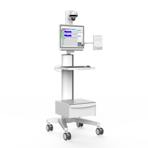 |  |  |  |  |  |
| SKU | SF1033560075-2 | SF1033560012-15 | SF1033560012-11 | SF1033560074-6 | SF1033560012-1 | |
| Rating | ||||||
| Price |
| $6,542.00 | $9,350.00 |
|
| $6,380.00 |
| Stock | ||||||
| Availability | ||||||
| Add to cart | ||||||
| Description | Shipped From Abroad
Delivery & Availability:
Typically 10-21 working days – excluding furniture and heavy/bulky equipment. Please contact us for further information.
| Shipped from Abroad It is a system with professional tool dedicated to exercise and resting ECG examination. Treadmill has 12 lead ECG modules. With ECG Analyzing Software. Delivery & Availability: Typically 21 working days – excluding furniture and heavy/bulky equipment. Please contact us for further information. | Shipped from Abroad With ultra-modern innovative design and the clinically-proven technologies, S8 Exp is portable ultrasound scanner well equipped as a low-physical-effort and enhanced-image-quality ultrasound scanner, which not only provides optimized solutions for versatile applications, but does help to improve the user-experience for both routine and non-traditional challenges. Delivery & Availability: Typically 5-7 working days – excluding furniture and heavy/bulky equipment. Please contact us for further information. | Shipped from Abroad Easily accomplish more with SonoScape’s new P50 ultrasound system. Incorporating single crystal clarity, automatic corrections and calculation, and user defined flexibility promises a confident diagnostic experience as well as opening new doors of opportunity for ultrasound use. Delivery & Availability: Typically 7-14 working days – excluding furniture and heavy/bulky equipment. Please contact us for further information. | In Stock GXR Analogue X-ray system matches with a radiographic room which perfectly fits your workow and can be easily upgraded to DR system with the help of DR interface and PC interface in GXR generator as well as Bucky suitable to Flat Panel Detector. GXR X-ray system is equipped with a high frequency X-ray generator which consistently produces high quality radiograph in favor of high quality X-ray output with a very small kV ripple and accurate mA and mAs. GXR X-ray system is designed to provide convenience to operator and comfort to patient. Delivery & Availability: Typically 21 working days – excluding furniture and heavy/bulky equipment. Please contact us for further information. | In Stock A Value Choice beyond Your Expectation. SonoScape’s trolley color Doppler system S11 redefines price and performance with practical design. The S11 will go beyond your expectations but not your budget. Delivery & Availability: Typically 2 working days – excluding furniture and heavy/bulky equipment. Please contact us for further information. |
| Content | FeaturesHIE multi-dimensional identification and application 1) According to the amplitude and continuity of the upper/lower boundary of aEEG, the neonatal EEG activity will be classified, and the Hellstrom-Westas background five-classification method will be used to improve the ability of medical staff to identify the aEEG waveforms; 2) Masimo Rainbow SET pulse oximetry monitoring, monitor the SpO2/PI/PR change in a non-invasive and continuous manner, effectively eliminate the exercise interference, and improve the measurement performance under low perfusion and body motion.
| It is a system with professional tool dedicated to exercise and resting ECG examination. Treadmill has 12 lead ECG modules. With ECG Analyzing Software.
Technical Specification:
Click Here To Download Catalogue | Sonoscape S8 Exp Portable Ultrasound scannerDETAILS Agile and Versatile With ultra-modern innovative design and the clinically-proven technologies, S8 Exp Portable Ultrasound scanner is well equipped as a low-physical-effort and enhanced-image-quality ultrasound scanner, which not only provides optimized solutions for versatile applications but does help to improve the user experience for both routine and non-traditional challenges. Working with S8 Exp, it will trigger your unlimited reverie and endow you with endless charm. Carrying forward the classical design of SonoScape's portable ultrasound products, S8 Exp successfully combines the best ergonomics, attractive design and ease of use. This charismatic identity is also enhanced by a sophisticated color palette—with sedate grey as its interior paint color and pearl white as exterior cover, S8 Exp reveals a style of aristocrat and strong character among SonoScape's ultrasound systems. Workflow The S8 Exp is a portable ultrasound scanner that adapts to your workflow, whether you are in the consulting room, at the bedside, or at a remote location. With easy-to-use new platform designed for sonographers' needs and full connection interfaces for easy connectivity and data sharing, S8 Exp leads to improved user comfort and clinical outcome as well as patient throughput and working efficiency. Powerful Platform Embedded with SonoScape's core imaging technologies such as μ-scan, PHI and Spatial Compound, S8 Exp boasts exceptional 2D image, sensitive spectral, Color and Power Doppler, displaying well-defined anatomy and pathology and facilitating a highly optimized diagnostic user environment for conclusive diagnoses. Besides, S8 Exp offers a comprehensive selection of electronic probes to maximally extend its capabilities to meet a wide range of applications including the abdomen, pediatric, OB/GYN, cardiovascular, musculoskeletal, etc. The advanced probe technologies also effectively enhance the image quality and confidence in reaching clinical diagnoses even in difficult patients.Click Here To Download Catalogue | DETAILS
Powerful Compact Precision
Taking into consideration the evolving expectations and needs for ultrasound, the P50 is a slim and unobtrusive trolley system that is comfortable in tight, congested spaces with little room to work in. Providing everything you need for a comfortable examination in a small space for both you and your patient.
Single Crystal Transducer
Wideband single crystal probes greatly improve the signal ratio, acquire stunning images and provide superior sensitivity and resolution for both the near and far-fields.
μ-Scan+
The new generation μ-Scan imaging technologies give you better image quality by reducing noise, improving signal strength and improving visualization.
Dynamic Color
Dynamic colour improves upon already existing colour Doppler technologies for clear capture of colour flow and detail visualization of even tiny veins with lower velocities.
Solution for Radiology
P50, is a leading-edge ultrasound system that can meet the demands of any clinical setting. You can experience a superior performance in multi-dimensional imaging for a full range of clinical applications – abdominal, breast and cardiovascular.
C-xlasto Imaging
By understanding that tissue stiffness varies depending on the type of tissue, we can use C-xlasto Imaging to easily find abnormalities and tumours within soft tissue. The differences in tissue responses are detected and visualized in real-time by the elastography algorithms through different representations, which can be particularly helpful in analyzing breast, thyroid and musculoskeletal structures. Predominately used only in linear probes, SonoScape’s new transvaginal and bi-plane probe for gynaecology and urology are breaking the mould and expanding elastography applications.
Real-time Color Panoramic
With the combination of colour flow and real-time panoramic, visualizing the blood flow of an entire vein or artery is now an easy task. Accomplished in real-time for the convenience of the sonographers, any mistakes can also be easily backtracked and corrected without interrupting the scan.
Contrast Imaging
Contrast Imaging on P50 makes full use of the infra harmonic signal and second harmonic signal to improve the image resolution and deep penetration. What’s more, the Dynamic Acoustic Control technology effectively controls the acoustic pressure for the contrast agent, decreasing the required agent dose and assures uniform image quality, guaranteeing longer contrast agent duration and better lesion perfusion of delayed phase observation.
Solution for OB/GYN
P50 has superior image quality, automated measurement tools, and a variety of volume technologies to provide ideal solutions for clinical examinations such as pregnancy examinations, and gynecologic disease diagnosis. With a new 4D transvaginal probe, P50 helps you to see and detect fetal abnormalities and significantly improves your diagnostic confidence during your examinations.
S-Live Silhouette
A unique transparent 3D anatomical image of the fetus for improved initial anatomical review. By using this new application, the system can create completely different fetal images from conventional ultrasound images, which can depict the fetal's intracorporeal anatomical structure.
Pelvic Floor 4D
Working in conjunction with SonoScape’s latest transvaginal probes, trans-perineal 4D pelvic floor ultrasound provides a useful clinical assessment of the impact of vaginal delivery on the female anterior compartment. Allowing doctors to judge whether the pelvic organs prolapsed or not, the extent of prolapse, and determining whether the pelvic muscles tore correctly.
S-Guide
S-Guide gives the user an extensive list of example obstetric ultrasound images as reference guides and a convenient checklist system to keep track of their progress during their obstetrics examination.
Auto Face
Automatically removes masking layers in front of the fetus’s face for a clearer vision of the fetus’s face.
AVC Follicle
AVC Follicle automatically identifies how many follicles are present and calculates their individual volumes.
Solution for Cardiology
P50 provides clear 2D clinical images and Doppler sensitivity to assess critical cardiac performance. Compatible with SonoScape’s single crystal probes, the P50 can provide images with better resolution and penetration in Cardiac diagnosis.
Tissue Doppler Imaging
Tissue Doppler Imaging allows clinical doctors to quantitatively evaluate local myocardial movements and functions, facilitating them with the ability to analyze and compare the motions of the different parts of the patient’s heart.
Stress Echo
Stress echocardiography is the combination of 2D echocardiography with physical, pharmacological or electrical stress of the patient. It also then provides users with report management tools such as configurable template editor, multiple loops to select one for storage, wall motion scoring, stress echo report, etc
Auto IMT
Auto IMT is used when determining the level of vascular sclerosis present in the patient by automatically tracing and calculating the thickness of the carotid vessels. What distinguishes the P50 is that it provides an instant and accurate Mean and Max index at the touch of a single button.
Auto EF
Automated 2D Cardiac Quantification is a fully intelligent trace function for endocardium with 19 easily-adjustable points providing rapid access to proven 2D EF and volumes.
Click Here To Download Catalogue | DrGem GXR Floor Mounted Analogue X-ray system matches with a radiographic room which perfectly fits your workflow and can be easily upgraded to DR system with the help of DR interface and PC interface in GXR generator as well as Bucky suitable to Flat Panel Detector. GXR (Analogue X-ray)system is equipped with a high frequency X-ray generator which consistently produces high quality radiograph in favor of high quality X-ray output with a very small kV ripple and accurate mA and mAs. GXR (Analogue X-ray) system is designed to provide convenience to operator and comfort to patient.
Features of DrGem GXR Floor Mounted Analogue X-ray:
Click Here To Download Catalogue | DETAILS
SonoScape’s trolley colour Doppler system S11 redefines price and performance with practical design. The S11 will go beyond your expectations but not your budget. As an easy-to-use ultrasound system, the S11 is integrated with a new software platform, especially optimized for a smooth workflow and convenient operation. The system speeds up the exam process and makes file management easier.
SPECIFICATION
- 15-inch high definition LCD monitor with articulating arm
- Compact and agile trolley design
- 3 active transducer sockets available for a wide range of applications
- Duplex, Color Doppler, DPI, PW Doppler, tissue harmonic imaging, μ-scan speckle reduction imaging, compound imaging, trapezoidal imaging
- Customized settings based on your own working style
- Full patient database and image management solutions
Click Here To Download Catalogue |
| Weight | N/A | N/A | N/A | N/A | N/A | N/A |
| Dimensions | N/A | N/A | N/A | N/A | N/A | N/A |
| Additional information |


Reviews
There are no reviews yet.