
Neuro MEP Micro-Portable EMG and NCS System
$0.00
Shipped From Abroad
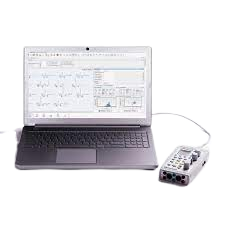
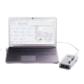
In coloproctology centers, EMG of external sphincter and pelvic floor muscles is often required. Portable EMG and NCS system Neuro-MEP-Micro packed with the necessary techniques is the right solution for these needs.
Typically 10-21 working days – excluding furniture and heavy/bulky equipment. Please contact us for further information.
Description
Features
Specific techniques for neurophysiological assessment of external sphincter and other pelvic floor neuromuscular structures
The system is used to perform various neurophysiological examinations of pelvic floor for diagnostic and scientific purposes, i.e.:
- bulbocavernosus and other sacral reflex tests with electrical stimulation of pudendal nerve to assess the conduction of reflex arcs (S2-S4);
- surface EMG of external sphincter and pelvic floor muscles to assess the level of tonic contraction;
- needle EMG with quantitative motor unit potential (MUP) analysis coupled with sacral reflex test to detect sacral denervation;
- stimulation EMG used with a disposable St. Mark’s electrode to stimulate the distal part of pudendal nerve;
- pudendal nerve somatosensory evoked potentials (pSEPs) especially in patients with preserved sacral reflexes and hypoesthesia of perineum;
- sympathetic skin responses (SSR) recorded from perineal area to assess the conduction velocity of sympathetic unmyelinated fibers and myelinated sensory nerve fibers;
- analysis of motor evoked potentials (MEP) in perineal muscles during the cortical and sacral magnetic stimulation (the assessment of corticospinal tract conduction with recording of pelvic floor motor evoked potentials (if the magnetic stimulator is available)).
Portable design
Neuro-MEP-Micro requires very little space and is connected to a computer via the USB cable which ensures data uploading and power supply of the device. If not powered from mains, the device can operate from the notebook battery.
Electrical stimulator with two outputs
Two software switchable outputs to plug in the electrical stimulator allow a specialist to place two pairs of stimulating electrodes on a patient and connect them to the device. Thus, there is no need to switch the electrodes as the stimulating electrode is software-defined.
Quick Comparison
| Settings | Neuro MEP Micro-Portable EMG and NCS System remove | Sonoscape P20 Ultrasound Machine remove | Ecleris C100-FID Binocular Colposcope remove | Bettermed BT666EPZ Multi-function Electric Delivery Bed remove | Sonoscape S22 Ultrasound Machine remove | Ecleris HD Digital Video Colposcope remove |
|---|---|---|---|---|---|---|
| Name | Neuro MEP Micro-Portable EMG and NCS System remove | Sonoscape P20 Ultrasound Machine remove | Ecleris C100-FID Binocular Colposcope remove | Bettermed BT666EPZ Multi-function Electric Delivery Bed remove | Sonoscape S22 Ultrasound Machine remove | Ecleris HD Digital Video Colposcope remove |
| Image |  |  | 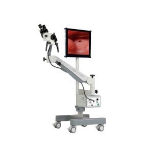 |  |  | 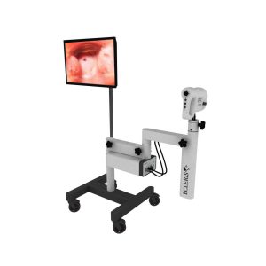 |
| SKU | SF1033560012-9 | SF1033560087-1 | SF1033560084-51 | SF1033560012-3 | SF1033560087-2 | |
| Rating | ||||||
| Price |
|
| $5,042.00 | $2,024.00 | $9,350.00 | $3,564.00 |
| Stock | ||||||
| Availability | ||||||
| Add to cart | ||||||
| Description | Shipped From Abroad
In coloproctology centers, EMG of external sphincter and pelvic floor muscles is often required. Portable EMG and NCS system Neuro-MEP-Micro packed with the necessary techniques is the right solution for these needs.
Delivery & Availability:
Typically 10-21 working days – excluding furniture and heavy/bulky equipment. Please contact us for further information.
| Shipped from Abroad Incorporating innovative technologies, P20’s user-friendly design with a simple operation panel, intuitive user interface and a variety of intelligent auxiliary scanning tools, will significantly improve your daily examination experience. Besides general imaging applications, P20 has entitled with diagnostic 4D technology which has an extraordinary performance in obstetrics and gynecology applications. Delivery & Availability: Typically 5-7 working days – excluding furniture and heavy/bulky equipment. Please contact us for further information. | Shipped from Abroad Content Includes: Forearm, Pantographic Arm, Floor Stand (H Shaped Base and Column), 5 Magnifications Head, Green Filter and Inclined Binocular, LED Light Source, Fiber Optic Cable, 110/220V Power Cable and User`s Guide, Stand for LCD Monitor and Printer, Digital Capturing System for Images, Videos and Sounds, USB 2.0. Includes Main Unit Processor with three Camera Inputs, USB Cable, Software, Hands Free Microphone and Footswitch. Delivery & Availability: Typically 10 working days – excluding furniture and heavy/bulky equipment. Please contact us for further information. | In Stock
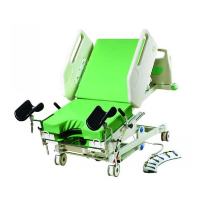
Function:Backrest tilting From 0°to 75° ±5°
Hi-lo function
Reverse:14°±1°
Trendelenburg:12°±1°
Delivery & Availability: Typically 2 working days – excluding furniture and heavy/bulky equipment. Please contact us for further information. | Shipped from Abroad As SonoScape steps forward to add value and efficiency to ultrasound, the latest S22 was designed in a user-friendly platform to address current and future demanding needs. It represents an excellent mix in performance and price. Delivery & Availability: Typically 5-7 working days – excluding furniture and heavy/bulky equipment. Please contact us for further information. | Shipped from Abroad Content Includes: Floor Mounted with Base with 4 Castors, Video Head, Positioning Arm, LED Light Included on Video Head, 110/220V Power Cable and User´s Guide, Stand For LCD Monitor and Printer, Digital Capturing System for Images, Videos and Sounds, USB 2.0. Includes Main Unit Processor with three Camera Inputs, USB Cable, Software, Hands Free Microphone and Footswitch. Delivery & Availability: Typically 10 working days – excluding furniture and heavy/bulky equipment. Please contact us for further information. |
| Content | FeaturesSpecific techniques for neurophysiological assessment of external sphincter and other pelvic floor neuromuscular structures The system is used to perform various neurophysiological examinations of pelvic floor for diagnostic and scientific purposes, i.e.:
| DETAILS
Upgraded Images with More Clarity
SonoScape never stops making progress in improving the image quality of its ultrasound products to enhance the confidence of diagnosis for doctors. With extraordinary images provided by P20, the anatomy structures are clearer than ever.
C-Xlasto Imaging
With C-xlasto Imaging, P20 enables comprehensive quantitative elastic analysis. Meanwhile, C-xlasto on P20 is supported by linear, convex and transvaginal probes, to ensure good reproducibility and highly consistent quantitative elastic results.
S-Live
S-Live allows for detailed visualization of subtle anatomical features, thereby enabling intuitive diagnosis with real-time 3D images and enriching patient communication.
Pelvic Floor 4D
Transperineal 4D pelvic floor ultrasound can provide useful clinical values in assessing the vaginal delivery impact on the female anterior compartment, judging whether the pelvic organs are prolapsed or not and the extent, determining if the pelvic muscles were torn accurately.
Anatomic M Mode
Anatomic M Mode helps you observe the myocardial motion at different phases by freely placing sample lines. It accurately measures the myocardial thickness and the heart size of even difficult patients and supports the myocardial function and LV wall-motion assessment.
Tissue Doppler Imaging
P20 is endowed with Tissue Doppler Imaging which provides velocities and other clinical information on myocardial functions, facilitating clinical doctors with the ability to analyze and compare the motions of different parts of the patient's heart.
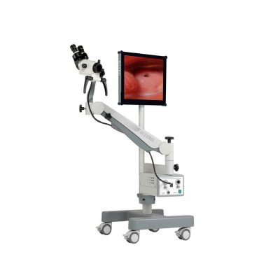
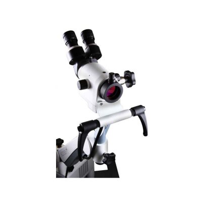
Click Here To Download Catalogue | Ecleris Colposcope Series C-100 was designed to cover all the diagnosis and therapeutic needs of modern gynecology. All models can be transformed into video colposcopes with our high resolution video camera. Digital handling of patients and image filing is achieved through the endoDIGI software which is easily adaptable to a portable PC or desktop computer. Great and accurate quality images, improved clearness, resolution and focal range can be obtained through our new C-100 Colposcope optic system. Floor and wall-mounted models include 5 magnifications (4, 6, 10, 16 and 25x).
The C-100 model, with its pantographic arm has enhanced maneuverability, as the arms are mounted on bearings and guarantee smooth movements and greater stability (WBS, weight balance system). Its state-of-the-art designed base allows for easy transport. We provide accessories that allow using the microscope light source and video camera also to carry out endoscopic studies, without need to have two light sources and two video cameras at the doctor’s office.
A new dimension in microsurgery
Ecleris 3D Splitter
Click Here To Download Catalogue |
| DETAILS
As SonoScape steps forward to add value and efficiency to ultrasound, the latest S22 was designed in a user-friendly platform to address current and future demanding needs. It represents an excellent mix in performance and price.
S22, is a shared service ultrasound system with a slim and elegant package that has combined mobility with utility to fit in specific clinical situations including emergency department, ICU, operating room and so on. Furthermore, its ergonomic design, easy operating and flexible data management will give you a memorable experience.
SPECIFICATION
• Large high-resolution widescreen LED
• Sensitive touch screen
• Four transducer sockets plus one socket for pencil probe
• A comprehensive selection of probes: linear, Convex, Micro-convex, Volumetric, Endocavity, Bi-plane, Phased Array, TEE, Intraoperative, Pencil
• Premium application technology: 4D, μ-scan speckle reduction, compound imaging, Pulse Inversion Harmonic Imaging, Color M-Mode, Steer M-Mode, PDI, TDI, Real-time Panoramic Imaging, Trapezoid Imaging, Auto-IMT…
• Full patient database and image management solutions: DICOM 3.0, AVI/JPG, USB 2.0, HDD, DVD, PDF report
• Multi-Language Input Keyboard
• Built-in battery
Click Here To Download Catalogue | The Ecleris ColpoHD Digital Video Colposcope offers clinicians an illuminated, high definition magnified view of the cervix and tissues of the vagina and vulva. As a screening tool for sexual assault victims or for the early detection of precancerous lesions, the ColpoHD provides insight into relevant pathology changes of tissue shape and color with magnification of up to 50x and a digital zoom up to 128x. The integrated electronic green filter delivers exceptional pathology visualization, with clear, true color imaging and superb depth of field. The swing arm mounted high-definition (HD) camera can be easily maneuvered into place. Optional integrated image capture is available for documentation along with archiving and printing of images.
A powerful tool for cervical cancer screening
The ColpoHD is precise and easy to handle in all examination situations. High quality optics along bright LED illumination and high-tech CMOS image sensors guarantee fantastic quality HD images are displayed for every patient.
The ColpoHD is also ideally suited for the examination of sexual assault victims who may be uncomfortable with a traditional Colposcope examination with the physician working through the standard microscope binoculars. The ColpoHD offers distance and a level of privacy for these patients who may be in a vulnerable frame of mind. The ColpoHD also offers optional image archiving and documentation of the examination in high definition image quality which is vital for sexual assault cases.
Features at a Glance
Click Here To Download Catalogue |
| Weight | N/A | N/A | N/A | N/A | N/A | N/A |
| Dimensions | N/A | N/A | N/A | N/A | N/A | N/A |
| Additional information |


Reviews
There are no reviews yet.