

Nio Color 5.8MP (MDNC‑6121)
$0.00
Shipped From Abroad
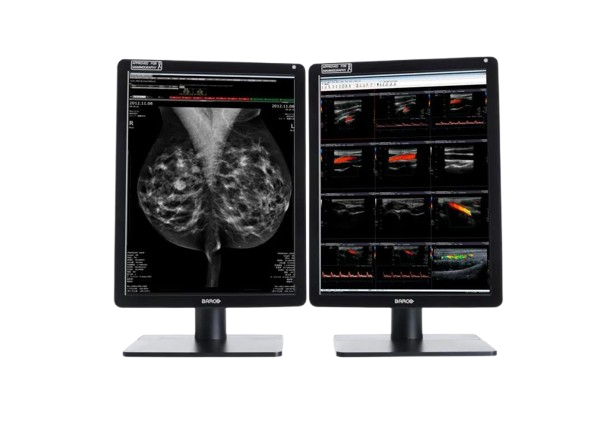
The Barco Nio Color 5MP (MDNC-6121) is a high-brightness, color medical display tailored for mammography and radiology. Offering 5.8 MP resolution, uniform color/gray, and advanced QA integration, it supports precise image review in breast, 3D mammography, and general diagnostic work.
Typically 10-21 working days – excluding furniture and heavy/bulky equipment. Please contact us for further information.
Description
The Barco Nio Color 5MP (MDNC-6121) display brings enhanced clarity and reliability to medical imaging workflows. With a resolution of 4200 × 2800 pixels (5.8 MP), it delivers high-fidelity color and grayscale rendering suited for mammography, tomosynthesis, and general radiology. The IPS-based LCD supports wide viewing angles and uniform luminance correction. Nominal power usage is approx. 60 W. Integrated performance tools, medical certifications, and display consistency make it ideal for diagnostic reading environments where both detail and color accuracy are critical.
Nio Color 5.8MP offers super bright, color-calibrated, and the most detailed medical images, including mammography and breast tomosynthesis. It’s how we help you improve your workflow and make more confident diagnoses.
No detail goes unnoticed
Barco’s Nio Color 5.8MP renders excellent color and grayscale images used in general radiology as well 2D and 3D mammography. Its high brightness and high contrast ratio help you discern the most subtle image details for an accurate diagnosis. And the additional resolution allows you to fit more of the image on the screen for reduced panning and zooming.
Using Barco’s integrated front sensor, the Nio Color 5.8MP works perfectly together with Barco’s QAWeb Enterprise solution for automated Quality Assurance and calibration. QAWeb Enterprise guarantees stable DICOM grayscale images and, with SteadyColor, consistent, calibrated color images throughout the display’s lifetime.
Work smarter
With the integrated smart features, you can easily take control and improve your productivity. SpotView™, for example, allows you to focus on an area of interest to unveil even more details. And with DimView™, auxiliary displays can be dimmed automatically so they don’t interfere with your reading experience.
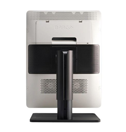
The Nio Color 5.8MP is an excellent solution for radiologists who want to angle their desktop: it lets you choose your preferred viewing angle and offers a highly ergonomic display configuration. It’s also possible to switch between Clearbase and Bluebase viewing modes on the fly. Whether to suit the image type or to change reading preferences, you decide which color you want, and when.
Ultimate peace of mind
Thanks to the high-performance LED backlights, the Nio Color 5.8MP has a positive impact on both maintenance and operational costs. The display is equipped with an integrated glass cover to safeguard your investment.
Barco is the only company that provides complete system solutions, from displays and controllers to workflow tools and calibration via QAWeb. All components are covered by our full 5-year warranty. At product release, we extensively test our displays’ compatibility with all major PACS applications.
Ensuring diagnostic confidence with MDR Class IIa
Our radiology displays are MDR-certified as Class IIa. Their product information has been reviewed and cleared by independent medical and technical experts, and is audited yearly. In other words, we ensure diagnostic confidence and peace of mind for our users.
An ecolabel for Nio Color 5.8MP
The Nio Color 5.8MP has been subjected to Barco’s ecoscoring protocol and has received an A rating. Some key factors that contributed to this rating are:
- Energy-efficient power supply, energy-efficient standby, and off modes
- Possibility to switch to standby mode when the device is not in use
- Halogen-free cables and plastics
- Use of recycled cardboard in packaging (>85% recycled content)
- Product design optimized for disassembly with common tools
Key Features
-
5.8 megapixel (4200 × 2800) resolution for high detail imaging
-
Bright, calibrated color and grayscale capability
-
Wide viewing angles (178° horizontal & vertical)
-
Uniform brightness correction for consistent image quality
-
Power-efficient design with nominal 60 W consumption
-
LCD technology optimized for medical imaging
-
Suitable for mammography, tomosynthesis, general radiology, and PACS integration
Specifications
| Category | Specification |
|---|---|
| Screen Technology | LCD |
| Active Screen Size | 21.3″ diagonal (541 mm); 324 × 433 mm (12.77″ × 17″) |
| Aspect Ratio | 3:4 per display (portrait), 3:2 overall |
| Resolution | 5.8 MP (2100 × 2800 pixels) |
| Pixel Pitch | 0.1545 mm |
| Color & Gray Imaging | Yes |
| Bit Depth | 30-bit |
| Viewing Angle | 178° (H/V) |
| Uniformity Correction | ULT |
| SteadyGray | Yes |
| SteadyColor | Yes (with Barco Display Controller, video driver, QAWeb Enterprise) |
| Color Gamut | NTSC: 72.2%, sRGB: 101.9%, DCI-P3: 75.5% |
| sRGB Delta E2000 | Avg < 3, Max < 5 |
| I-Luminate | Yes |
| Ambient Light Features | Presets and sensor; reading room selection |
| Front Sensor | Yes (I-Guard) |
| Max Luminance | 1560 cd/m² |
| DICOM Calibrated Luminance | 600 cd/m² |
| Contrast Ratio | 1400:1 |
| Response Time | 12.5 ms ((Tr + Tf)/2) |
| Housing Color | Black (RAL 9004) / White (RAL 9003) |
| Video Inputs | 2 × DisplayPort 1.4 |
| USB Ports | 2 × USB-B 2.0 upstream; 5 × USB-A 2.0 downstream (1 charging) |
| KVM Switch | Yes |
| Power Rating | 24 VDC, 5 A |
| Power Supply | AdapterTech ATM160T-P240 (100–240 Vac, 50–60 Hz, 1.8–0.9 A; Output: 24 Vdc, 6.6 A) |
| Power Consumption | 60 W (nominal); 0.4 W (hibernate/off) |
| Dimensions (with stand) | Portrait: 378 × 528~628 × 235 mm; Landscape: 491 × 472~572 × 235 mm |
| Dimensions (w/o stand) | Portrait: 378 × 491 × 84 mm; Landscape: 491 × 378 × 84 mm |
| Packaged Dimensions | 500 × 280 × 670 mm |
| Weight (with stand) | With cover: 11.9 kg; Without cover: 10.6 kg |
| Weight (w/o stand) | With cover: 6.9 kg; Without cover: 5.6 kg |
| Packaged Weight | With cover: 16.9 kg; Without cover: 15.6 kg |
| Tilt / Swivel / Pivot | Tilt: -10° to +30°; Swivel: ±45°; Pivot: 90° |
| Height Adjustment | 100 mm |
| Mounting Standard | VESA (100 mm) |
| Screen Protection | Optional anti-reflective glass |
| Recommended Modalities | All digital images, including digital mammography |
| Certifications | FDA 510(K), CE0123, CCC, KC, INMETRO, BIS, IEC/EN/AAMI/CSA standards |
| EMI Compliance | IEC/EN/FCC/ICES/VCCI standards |
| Environmental Compliance | EU RoHS, China RoHS, REACH, WEEE, Packaging Directive |
| Supplied Accessories | User guide, documentation disc, system sheet, DisplayPort cable, USB cable, PSU |
| Optional Accessories | Graphics board, QA software, QAWeb Enterprise |
| Warranty | 5 years (includes 40,000 hours backlight warranty) |
| Operating Conditions | Temp: 0–40 °C (specs: 15–30 °C); Humidity: 8–80%; Pressure: 70 kPa |
| Storage Conditions | Temp: -20–60 °C; Humidity: 5–85%; Pressure: 50–106 kPa |
Quick Comparison
| Nio Color 5.8MP (MDNC‑6121) remove | DrGem Ceiling Mounted Digital X-ray remove | Sonoscape P50 Ultrasound Machine remove | Sonoscape P10 Ultrasound Machine remove | DrGem Diamond All-In-One Digital X-ray Machine remove | Sonoscape S11 Ultrasound Machine remove | |||||||||||||||||||||||||||||||||||||||||||||||||||||||||||||||||||||||||||||||||||||||||||||||
|---|---|---|---|---|---|---|---|---|---|---|---|---|---|---|---|---|---|---|---|---|---|---|---|---|---|---|---|---|---|---|---|---|---|---|---|---|---|---|---|---|---|---|---|---|---|---|---|---|---|---|---|---|---|---|---|---|---|---|---|---|---|---|---|---|---|---|---|---|---|---|---|---|---|---|---|---|---|---|---|---|---|---|---|---|---|---|---|---|---|---|---|---|---|---|---|---|---|---|---|---|
| Name | Nio Color 5.8MP (MDNC‑6121) remove | DrGem Ceiling Mounted Digital X-ray remove | Sonoscape P50 Ultrasound Machine remove | Sonoscape P10 Ultrasound Machine remove | DrGem Diamond All-In-One Digital X-ray Machine remove | Sonoscape S11 Ultrasound Machine remove | ||||||||||||||||||||||||||||||||||||||||||||||||||||||||||||||||||||||||||||||||||||||||||||||
| Image | 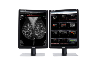 |  |  |  |  |  | ||||||||||||||||||||||||||||||||||||||||||||||||||||||||||||||||||||||||||||||||||||||||||||||
| SKU | SF1033560074-4 | SF1033560012-11 | SF1033560012-7 | SF1033560074-3 | SF1033560012-1 | |||||||||||||||||||||||||||||||||||||||||||||||||||||||||||||||||||||||||||||||||||||||||||||||
| Rating | ||||||||||||||||||||||||||||||||||||||||||||||||||||||||||||||||||||||||||||||||||||||||||||||||||||
| Price |
|
|
| $9,350.00 |
| $6,380.00 | ||||||||||||||||||||||||||||||||||||||||||||||||||||||||||||||||||||||||||||||||||||||||||||||
| Stock | ||||||||||||||||||||||||||||||||||||||||||||||||||||||||||||||||||||||||||||||||||||||||||||||||||||
| Availability | ||||||||||||||||||||||||||||||||||||||||||||||||||||||||||||||||||||||||||||||||||||||||||||||||||||
| Add to cart | ||||||||||||||||||||||||||||||||||||||||||||||||||||||||||||||||||||||||||||||||||||||||||||||||||||
| Description | Shipped From Abroad
The Barco Nio Color 5MP (MDNC-6121) is a high-brightness, color medical display tailored for mammography and radiology. Offering 5.8 MP resolution, uniform color/gray, and advanced QA integration, it supports precise image review in breast, 3D mammography, and general diagnostic work. Delivery & Availability:
Typically 10-21 working days – excluding furniture and heavy/bulky equipment. Please contact us for further information.
| In Stock The GXR-SD is a diagnostic digital radiography system that provides reliable high quality digital radiographic images with a reduced dose. The GXR-SD DR systems offer comprehensive digital solutions to all radiography needs, featuring ACQUIDR digital imaging system with stationary or portable digital flat-panel detectors as well as reliable high-frequency x-ray generators that are known worldwide for their excellent performance, lifetime and stability. Patient tables and wall stands are also offered. Delivery & Availability: Typically 21 working days – excluding furniture and heavy/bulky equipment. Please contact us for further information. | Shipped from Abroad Easily accomplish more with SonoScape’s new P50 ultrasound system. Incorporating single crystal clarity, automatic corrections and calculation, and user defined flexibility promises a confident diagnostic experience as well as opening new doors of opportunity for ultrasound use. Delivery & Availability: Typically 7-14 working days – excluding furniture and heavy/bulky equipment. Please contact us for further information. | Shipped from Abroad The P10 color Doppler ultrasound system is a new generation product from SonoScape. It is designed to give high quality images, rich probe configurations, various clinical tools and automatic analysis software to provide you with comprehensive solutions for your growing demand for clinical applications. Delivery & Availability: Typically 5-7 working days – excluding furniture and heavy/bulky equipment. Please contact us for further information. | Shipped from Abroad DrGem Diamond All-In-One Digital X-ray Machine is a fully automatic digital radiography system providing state-of-the-art image quality, image processing and user interface. With a wide selection of anatomical studies on the imaging software, DIAMOND automatically sets up the x-ray generator’s preprogrammed exposure technique settings, motorized radiographic stand positioning, x-ray collimation and post-image processing for the selected study. Specifically designed to increase workflow, this fully digital system offers convenient auto-positioning and advanced image processing to achieve big performance with little effort. Delivery & Availability: Typically 21 working days – excluding furniture and heavy/bulky equipment. Please contact us for further information. | In Stock A Value Choice beyond Your Expectation. SonoScape’s trolley color Doppler system S11 redefines price and performance with practical design. The S11 will go beyond your expectations but not your budget. Delivery & Availability: Typically 2 working days – excluding furniture and heavy/bulky equipment. Please contact us for further information. | ||||||||||||||||||||||||||||||||||||||||||||||||||||||||||||||||||||||||||||||||||||||||||||||
| Content | The Barco Nio Color 5MP (MDNC-6121) display brings enhanced clarity and reliability to medical imaging workflows. With a resolution of 4200 × 2800 pixels (5.8 MP), it delivers high-fidelity color and grayscale rendering suited for mammography, tomosynthesis, and general radiology. The IPS-based LCD supports wide viewing angles and uniform luminance correction. Nominal power usage is approx. 60 W. Integrated performance tools, medical certifications, and display consistency make it ideal for diagnostic reading environments where both detail and color accuracy are critical.
Nio Color 5.8MP offers super bright, color-calibrated, and the most detailed medical images, including mammography and breast tomosynthesis. It's how we help you improve your workflow and make more confident diagnoses. No detail goes unnoticed Barco's Nio Color 5.8MP renders excellent color and grayscale images used in general radiology as well 2D and 3D mammography. Its high brightness and high contrast ratio help you discern the most subtle image details for an accurate diagnosis. And the additional resolution allows you to fit more of the image on the screen for reduced panning and zooming. Using Barco’s integrated front sensor, the Nio Color 5.8MP works perfectly together with Barco’s QAWeb Enterprise solution for automated Quality Assurance and calibration. QAWeb Enterprise guarantees stable DICOM grayscale images and, with SteadyColor, consistent, calibrated color images throughout the display's lifetime. Work smarter With the integrated smart features, you can easily take control and improve your productivity. SpotView™, for example, allows you to focus on an area of interest to unveil even more details. And with DimView™, auxiliary displays can be dimmed automatically so they don't interfere with your reading experience. The Nio Color 5.8MP is an excellent solution for radiologists who want to angle their desktop: it lets you choose your preferred viewing angle and offers a highly ergonomic display configuration. It’s also possible to switch between Clearbase and Bluebase viewing modes on the fly. Whether to suit the image type or to change reading preferences, you decide which color you want, and when. Ultimate peace of mind Thanks to the high-performance LED backlights, the Nio Color 5.8MP has a positive impact on both maintenance and operational costs. The display is equipped with an integrated glass cover to safeguard your investment. Barco is the only company that provides complete system solutions, from displays and controllers to workflow tools and calibration via QAWeb. All components are covered by our full 5-year warranty. At product release, we extensively test our displays' compatibility with all major PACS applications. Ensuring diagnostic confidence with MDR Class IIa Our radiology displays are MDR-certified as Class IIa. Their product information has been reviewed and cleared by independent medical and technical experts, and is audited yearly. In other words, we ensure diagnostic confidence and peace of mind for our users. An ecolabel for Nio Color 5.8MP The Nio Color 5.8MP has been subjected to Barco’s ecoscoring protocol and has received an A rating. Some key factors that contributed to this rating are:
Key Features
Specifications
| DrGem Ceiling Mounted Digital X-ray is a diagnostic digital radiography system that provides reliable high quality digital radiographic images with a reduced dose. The GXR-SD DR systems offer comprehensive digital solutions to all radiography needs, featuring ACQUIDR digital imaging system with stationary or portable digital flat-panel detectors as well as reliable high-frequency x-ray generators that are known worldwide for their excellent performance, lifetime and stability. Patient tables and wall stands are also offered.
Features:
Click Here To Download Catalogue | DETAILS
Powerful Compact Precision
Taking into consideration the evolving expectations and needs for ultrasound, the P50 is a slim and unobtrusive trolley system that is comfortable in tight, congested spaces with little room to work in. Providing everything you need for a comfortable examination in a small space for both you and your patient.
Single Crystal Transducer
Wideband single crystal probes greatly improve the signal ratio, acquire stunning images and provide superior sensitivity and resolution for both the near and far-fields.
μ-Scan+
The new generation μ-Scan imaging technologies give you better image quality by reducing noise, improving signal strength and improving visualization.
Dynamic Color
Dynamic colour improves upon already existing colour Doppler technologies for clear capture of colour flow and detail visualization of even tiny veins with lower velocities.
Solution for Radiology
P50, is a leading-edge ultrasound system that can meet the demands of any clinical setting. You can experience a superior performance in multi-dimensional imaging for a full range of clinical applications – abdominal, breast and cardiovascular.
C-xlasto Imaging
By understanding that tissue stiffness varies depending on the type of tissue, we can use C-xlasto Imaging to easily find abnormalities and tumours within soft tissue. The differences in tissue responses are detected and visualized in real-time by the elastography algorithms through different representations, which can be particularly helpful in analyzing breast, thyroid and musculoskeletal structures. Predominately used only in linear probes, SonoScape’s new transvaginal and bi-plane probe for gynaecology and urology are breaking the mould and expanding elastography applications.
Real-time Color Panoramic
With the combination of colour flow and real-time panoramic, visualizing the blood flow of an entire vein or artery is now an easy task. Accomplished in real-time for the convenience of the sonographers, any mistakes can also be easily backtracked and corrected without interrupting the scan.
Contrast Imaging
Contrast Imaging on P50 makes full use of the infra harmonic signal and second harmonic signal to improve the image resolution and deep penetration. What’s more, the Dynamic Acoustic Control technology effectively controls the acoustic pressure for the contrast agent, decreasing the required agent dose and assures uniform image quality, guaranteeing longer contrast agent duration and better lesion perfusion of delayed phase observation.
Solution for OB/GYN
P50 has superior image quality, automated measurement tools, and a variety of volume technologies to provide ideal solutions for clinical examinations such as pregnancy examinations, and gynecologic disease diagnosis. With a new 4D transvaginal probe, P50 helps you to see and detect fetal abnormalities and significantly improves your diagnostic confidence during your examinations.
S-Live Silhouette
A unique transparent 3D anatomical image of the fetus for improved initial anatomical review. By using this new application, the system can create completely different fetal images from conventional ultrasound images, which can depict the fetal's intracorporeal anatomical structure.
Pelvic Floor 4D
Working in conjunction with SonoScape’s latest transvaginal probes, trans-perineal 4D pelvic floor ultrasound provides a useful clinical assessment of the impact of vaginal delivery on the female anterior compartment. Allowing doctors to judge whether the pelvic organs prolapsed or not, the extent of prolapse, and determining whether the pelvic muscles tore correctly.
S-Guide
S-Guide gives the user an extensive list of example obstetric ultrasound images as reference guides and a convenient checklist system to keep track of their progress during their obstetrics examination.
Auto Face
Automatically removes masking layers in front of the fetus’s face for a clearer vision of the fetus’s face.
AVC Follicle
AVC Follicle automatically identifies how many follicles are present and calculates their individual volumes.
Solution for Cardiology
P50 provides clear 2D clinical images and Doppler sensitivity to assess critical cardiac performance. Compatible with SonoScape’s single crystal probes, the P50 can provide images with better resolution and penetration in Cardiac diagnosis.
Tissue Doppler Imaging
Tissue Doppler Imaging allows clinical doctors to quantitatively evaluate local myocardial movements and functions, facilitating them with the ability to analyze and compare the motions of the different parts of the patient’s heart.
Stress Echo
Stress echocardiography is the combination of 2D echocardiography with physical, pharmacological or electrical stress of the patient. It also then provides users with report management tools such as configurable template editor, multiple loops to select one for storage, wall motion scoring, stress echo report, etc
Auto IMT
Auto IMT is used when determining the level of vascular sclerosis present in the patient by automatically tracing and calculating the thickness of the carotid vessels. What distinguishes the P50 is that it provides an instant and accurate Mean and Max index at the touch of a single button.
Auto EF
Automated 2D Cardiac Quantification is a fully intelligent trace function for endocardium with 19 easily-adjustable points providing rapid access to proven 2D EF and volumes.
Click Here To Download Catalogue | DETAILS
B + Compound
B + Compound utilizes several lines of sight for optimal contrast resolution, speckle reduction and border detection, with which P10 is ideal for superficial and abdominal imaging with better clarity and improved continuity of structures.
μ-Scan
The new generation μ-Scan imaging technology gives you better image quality by reducing noise, improving signal strength and improving visualization.
P10 offers a comprehensive selection of electronic probes to maximize its capabilities to meet a wide range of applications including abdomen, pediatric, OB/GYN, cardiovascular, musculoskeletal, etc. The advanced probe technologies also effectively enhance the image quality and confidence in reaching clinical diagnoses, even in difficult patients.
Convex Probe 3C-A
Ideal for an abundant of application such as abdomen, gynecology, obstetrics, urology and even abdomen biopsy.
Linear Probe L741
This linear probe is designed to satisfy vascular, breast, thyroid, and other small parts diagnosis, and its adjustable parameters could also present users a clear view of MSK and deep vessels.
Phase Array Probe 3P-A
For the purpose of adult and pediatric cardiology and emergency, the phase array probe provides elaborate presets for different exam modes, even for difficult patients.
Intracavitary Probe 6V1
Intracavitary probe could face application of gynecology, urology, prostate, and its temperature detection technology not only protects the patient but also extends the service life.
Click Here To Download Catalogue | DrGem Diamond All-In-One Digital X-ray Machine is a fully automatic digital radiography system providing state-of-the-art image quality, image processing and user interface. With a wide selection of anatomical studies on the imaging software, DIAMOND automatically sets up the x-ray generator’s pre-programmed exposure technique settings, motorized radiographic stand positioning, x-ray collimation and post-image processing for the selected study. Specifically designed to increase workflow, this fully digital system offers convenient auto-positioning and advanced image processing to achieve big performance with little effort.
Features of DrGem Diamond All-In-One Digital X-ray Machine:
Outstanding Image Quality -
Digital radiography via at panel detector improves your workflow, exam speed and comfort with efficiency. Digital at panel detector with Csl screen provides excellent spatial resolution, MTF, DQE and stability based on ne pixel pitch. A 3-field ion-chamber is provided for AEC function.
Automatic Collimation –
Automatic x-ray eld size control of the motorized collimator corresponds to dierent SIDs. Includes user adjustable lamp timer with on/oswitch.
Automatic Positioning –
Click Here To Download Catalogue | DETAILS
SonoScape’s trolley colour Doppler system S11 redefines price and performance with practical design. The S11 will go beyond your expectations but not your budget. As an easy-to-use ultrasound system, the S11 is integrated with a new software platform, especially optimized for a smooth workflow and convenient operation. The system speeds up the exam process and makes file management easier.
SPECIFICATION
- 15-inch high definition LCD monitor with articulating arm
- Compact and agile trolley design
- 3 active transducer sockets available for a wide range of applications
- Duplex, Color Doppler, DPI, PW Doppler, tissue harmonic imaging, μ-scan speckle reduction imaging, compound imaging, trapezoidal imaging
- Customized settings based on your own working style
- Full patient database and image management solutions
Click Here To Download Catalogue | ||||||||||||||||||||||||||||||||||||||||||||||||||||||||||||||||||||||||||||||||||||||||||||||
| Weight | N/A | N/A | N/A | N/A | N/A | N/A | ||||||||||||||||||||||||||||||||||||||||||||||||||||||||||||||||||||||||||||||||||||||||||||||
| Dimensions | N/A | N/A | N/A | N/A | N/A | N/A | ||||||||||||||||||||||||||||||||||||||||||||||||||||||||||||||||||||||||||||||||||||||||||||||
| Additional information |


Reviews
There are no reviews yet.