

Nio Color 5.8MP (MDNC‑6121)
$0.00
Shipped From Abroad
The Barco Nio Color 5MP (MDNC-6121) is a high-brightness, color medical display tailored for mammography and radiology. Offering 5.8 MP resolution, uniform color/gray, and advanced QA integration, it supports precise image review in breast, 3D mammography, and general diagnostic work.
Typically 10-21 working days – excluding furniture and heavy/bulky equipment. Please contact us for further information.
Description
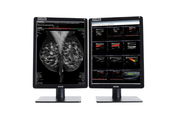
The Barco Nio Color 5MP (MDNC-6121) display brings enhanced clarity and reliability to medical imaging workflows. With a resolution of 4200 × 2800 pixels (5.8 MP), it delivers high-fidelity color and grayscale rendering suited for mammography, tomosynthesis, and general radiology. The IPS-based LCD supports wide viewing angles and uniform luminance correction. Nominal power usage is approx. 60 W. Integrated performance tools, medical certifications, and display consistency make it ideal for diagnostic reading environments where both detail and color accuracy are critical.
Nio Color 5.8MP offers super bright, color-calibrated, and the most detailed medical images, including mammography and breast tomosynthesis. It’s how we help you improve your workflow and make more confident diagnoses.
No detail goes unnoticed
Barco’s Nio Color 5.8MP renders excellent color and grayscale images used in general radiology as well 2D and 3D mammography. Its high brightness and high contrast ratio help you discern the most subtle image details for an accurate diagnosis. And the additional resolution allows you to fit more of the image on the screen for reduced panning and zooming.
Using Barco’s integrated front sensor, the Nio Color 5.8MP works perfectly together with Barco’s QAWeb Enterprise solution for automated Quality Assurance and calibration. QAWeb Enterprise guarantees stable DICOM grayscale images and, with SteadyColor, consistent, calibrated color images throughout the display’s lifetime.
Work smarter
With the integrated smart features, you can easily take control and improve your productivity. SpotView™, for example, allows you to focus on an area of interest to unveil even more details. And with DimView™, auxiliary displays can be dimmed automatically so they don’t interfere with your reading experience.
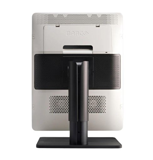
The Nio Color 5.8MP is an excellent solution for radiologists who want to angle their desktop: it lets you choose your preferred viewing angle and offers a highly ergonomic display configuration. It’s also possible to switch between Clearbase and Bluebase viewing modes on the fly. Whether to suit the image type or to change reading preferences, you decide which color you want, and when.
Ultimate peace of mind
Thanks to the high-performance LED backlights, the Nio Color 5.8MP has a positive impact on both maintenance and operational costs. The display is equipped with an integrated glass cover to safeguard your investment.
Barco is the only company that provides complete system solutions, from displays and controllers to workflow tools and calibration via QAWeb. All components are covered by our full 5-year warranty. At product release, we extensively test our displays’ compatibility with all major PACS applications.
Ensuring diagnostic confidence with MDR Class IIa
Our radiology displays are MDR-certified as Class IIa. Their product information has been reviewed and cleared by independent medical and technical experts, and is audited yearly. In other words, we ensure diagnostic confidence and peace of mind for our users.
An ecolabel for Nio Color 5.8MP
The Nio Color 5.8MP has been subjected to Barco’s ecoscoring protocol and has received an A rating. Some key factors that contributed to this rating are:
- Energy-efficient power supply, energy-efficient standby, and off modes
- Possibility to switch to standby mode when the device is not in use
- Halogen-free cables and plastics
- Use of recycled cardboard in packaging (>85% recycled content)
- Product design optimized for disassembly with common tools
Key Features
-
5.8 megapixel (4200 × 2800) resolution for high detail imaging
-
Bright, calibrated color and grayscale capability
-
Wide viewing angles (178° horizontal & vertical)
-
Uniform brightness correction for consistent image quality
-
Power-efficient design with nominal 60 W consumption
-
LCD technology optimized for medical imaging
-
Suitable for mammography, tomosynthesis, general radiology, and PACS integration
Specifications
| Category | Specification |
|---|---|
| Screen Technology | LCD |
| Active Screen Size | 21.3″ diagonal (541 mm); 324 × 433 mm (12.77″ × 17″) |
| Aspect Ratio | 3:4 per display (portrait), 3:2 overall |
| Resolution | 5.8 MP (2100 × 2800 pixels) |
| Pixel Pitch | 0.1545 mm |
| Color & Gray Imaging | Yes |
| Bit Depth | 30-bit |
| Viewing Angle | 178° (H/V) |
| Uniformity Correction | ULT |
| SteadyGray | Yes |
| SteadyColor | Yes (with Barco Display Controller, video driver, QAWeb Enterprise) |
| Color Gamut | NTSC: 72.2%, sRGB: 101.9%, DCI-P3: 75.5% |
| sRGB Delta E2000 | Avg < 3, Max < 5 |
| I-Luminate | Yes |
| Ambient Light Features | Presets and sensor; reading room selection |
| Front Sensor | Yes (I-Guard) |
| Max Luminance | 1560 cd/m² |
| DICOM Calibrated Luminance | 600 cd/m² |
| Contrast Ratio | 1400:1 |
| Response Time | 12.5 ms ((Tr + Tf)/2) |
| Housing Color | Black (RAL 9004) / White (RAL 9003) |
| Video Inputs | 2 × DisplayPort 1.4 |
| USB Ports | 2 × USB-B 2.0 upstream; 5 × USB-A 2.0 downstream (1 charging) |
| KVM Switch | Yes |
| Power Rating | 24 VDC, 5 A |
| Power Supply | AdapterTech ATM160T-P240 (100–240 Vac, 50–60 Hz, 1.8–0.9 A; Output: 24 Vdc, 6.6 A) |
| Power Consumption | 60 W (nominal); 0.4 W (hibernate/off) |
| Dimensions (with stand) | Portrait: 378 × 528~628 × 235 mm; Landscape: 491 × 472~572 × 235 mm |
| Dimensions (w/o stand) | Portrait: 378 × 491 × 84 mm; Landscape: 491 × 378 × 84 mm |
| Packaged Dimensions | 500 × 280 × 670 mm |
| Weight (with stand) | With cover: 11.9 kg; Without cover: 10.6 kg |
| Weight (w/o stand) | With cover: 6.9 kg; Without cover: 5.6 kg |
| Packaged Weight | With cover: 16.9 kg; Without cover: 15.6 kg |
| Tilt / Swivel / Pivot | Tilt: -10° to +30°; Swivel: ±45°; Pivot: 90° |
| Height Adjustment | 100 mm |
| Mounting Standard | VESA (100 mm) |
| Screen Protection | Optional anti-reflective glass |
| Recommended Modalities | All digital images, including digital mammography |
| Certifications | FDA 510(K), CE0123, CCC, KC, INMETRO, BIS, IEC/EN/AAMI/CSA standards |
| EMI Compliance | IEC/EN/FCC/ICES/VCCI standards |
| Environmental Compliance | EU RoHS, China RoHS, REACH, WEEE, Packaging Directive |
| Supplied Accessories | User guide, documentation disc, system sheet, DisplayPort cable, USB cable, PSU |
| Optional Accessories | Graphics board, QA software, QAWeb Enterprise |
| Warranty | 5 years (includes 40,000 hours backlight warranty) |
| Operating Conditions | Temp: 0–40 °C (specs: 15–30 °C); Humidity: 8–80%; Pressure: 70 kPa |
| Storage Conditions | Temp: -20–60 °C; Humidity: 5–85%; Pressure: 50–106 kPa |
Quick Comparison
| Nio Color 5.8MP (MDNC‑6121) remove | DrGem GXR-SD 400mA Floor Mounted Digital X-ray remove | Sonoscape P15 Ultrasound Machine With Four Probes remove | Sonoscape E1 Ultrasound Machine With Two Probes remove | Anke MRI Openmark 5000 Permanent System remove | Sonoscape S22 Ultrasound Machine remove | ||||||||||||||||||||||||||||||||||||||||||||||||||||||||||||||||||||||||||||||||||||||||||||||||||||||||||||||||||||||||||||||||||||||||||||||||||||||||||||||||||||||||||||||||||||||||||||||||||||||||||||||||||||||||||||||||||||||||||||||||||||||||||||||||||||||||||||||||||||||||||||||||||||||||||||||||||||||||||||||||||||||||||||||||||||||||||||||||||||||||||||||||||||||||||||||||||||||||||||||
|---|---|---|---|---|---|---|---|---|---|---|---|---|---|---|---|---|---|---|---|---|---|---|---|---|---|---|---|---|---|---|---|---|---|---|---|---|---|---|---|---|---|---|---|---|---|---|---|---|---|---|---|---|---|---|---|---|---|---|---|---|---|---|---|---|---|---|---|---|---|---|---|---|---|---|---|---|---|---|---|---|---|---|---|---|---|---|---|---|---|---|---|---|---|---|---|---|---|---|---|---|---|---|---|---|---|---|---|---|---|---|---|---|---|---|---|---|---|---|---|---|---|---|---|---|---|---|---|---|---|---|---|---|---|---|---|---|---|---|---|---|---|---|---|---|---|---|---|---|---|---|---|---|---|---|---|---|---|---|---|---|---|---|---|---|---|---|---|---|---|---|---|---|---|---|---|---|---|---|---|---|---|---|---|---|---|---|---|---|---|---|---|---|---|---|---|---|---|---|---|---|---|---|---|---|---|---|---|---|---|---|---|---|---|---|---|---|---|---|---|---|---|---|---|---|---|---|---|---|---|---|---|---|---|---|---|---|---|---|---|---|---|---|---|---|---|---|---|---|---|---|---|---|---|---|---|---|---|---|---|---|---|---|---|---|---|---|---|---|---|---|---|---|---|---|---|---|---|---|---|---|---|---|---|---|---|---|---|---|---|---|---|---|---|---|---|---|---|---|---|---|---|---|---|---|---|---|---|---|---|---|---|---|---|---|---|---|---|---|---|---|---|---|---|---|---|---|---|---|---|---|---|---|---|---|---|---|---|---|---|---|---|---|---|---|---|---|---|---|---|---|---|---|---|---|---|---|---|---|---|---|---|---|---|---|---|---|---|---|---|---|---|---|---|---|---|---|---|---|---|---|---|---|---|---|---|---|---|---|---|---|---|---|---|---|---|---|---|---|---|---|---|---|---|
| Name | Nio Color 5.8MP (MDNC‑6121) remove | DrGem GXR-SD 400mA Floor Mounted Digital X-ray remove | Sonoscape P15 Ultrasound Machine With Four Probes remove | Sonoscape E1 Ultrasound Machine With Two Probes remove | Anke MRI Openmark 5000 Permanent System remove | Sonoscape S22 Ultrasound Machine remove | |||||||||||||||||||||||||||||||||||||||||||||||||||||||||||||||||||||||||||||||||||||||||||||||||||||||||||||||||||||||||||||||||||||||||||||||||||||||||||||||||||||||||||||||||||||||||||||||||||||||||||||||||||||||||||||||||||||||||||||||||||||||||||||||||||||||||||||||||||||||||||||||||||||||||||||||||||||||||||||||||||||||||||||||||||||||||||||||||||||||||||||||||||||||||||||||||||||||||||||
| Image | 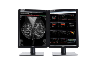 |  |  |  |  |  | |||||||||||||||||||||||||||||||||||||||||||||||||||||||||||||||||||||||||||||||||||||||||||||||||||||||||||||||||||||||||||||||||||||||||||||||||||||||||||||||||||||||||||||||||||||||||||||||||||||||||||||||||||||||||||||||||||||||||||||||||||||||||||||||||||||||||||||||||||||||||||||||||||||||||||||||||||||||||||||||||||||||||||||||||||||||||||||||||||||||||||||||||||||||||||||||||||||||||||||
| SKU | SF1033560074-5 | SF1033560012-8 | SF1033560012-20 | SF1033560092-3 | SF1033560012-3 | ||||||||||||||||||||||||||||||||||||||||||||||||||||||||||||||||||||||||||||||||||||||||||||||||||||||||||||||||||||||||||||||||||||||||||||||||||||||||||||||||||||||||||||||||||||||||||||||||||||||||||||||||||||||||||||||||||||||||||||||||||||||||||||||||||||||||||||||||||||||||||||||||||||||||||||||||||||||||||||||||||||||||||||||||||||||||||||||||||||||||||||||||||||||||||||||||||||||||||||||
| Rating | |||||||||||||||||||||||||||||||||||||||||||||||||||||||||||||||||||||||||||||||||||||||||||||||||||||||||||||||||||||||||||||||||||||||||||||||||||||||||||||||||||||||||||||||||||||||||||||||||||||||||||||||||||||||||||||||||||||||||||||||||||||||||||||||||||||||||||||||||||||||||||||||||||||||||||||||||||||||||||||||||||||||||||||||||||||||||||||||||||||||||||||||||||||||||||||||||||||||||||||||||||
| Price |
|
| $13,900.00 | $4,620.00 |
| $9,350.00 | |||||||||||||||||||||||||||||||||||||||||||||||||||||||||||||||||||||||||||||||||||||||||||||||||||||||||||||||||||||||||||||||||||||||||||||||||||||||||||||||||||||||||||||||||||||||||||||||||||||||||||||||||||||||||||||||||||||||||||||||||||||||||||||||||||||||||||||||||||||||||||||||||||||||||||||||||||||||||||||||||||||||||||||||||||||||||||||||||||||||||||||||||||||||||||||||||||||||||||||
| Stock | |||||||||||||||||||||||||||||||||||||||||||||||||||||||||||||||||||||||||||||||||||||||||||||||||||||||||||||||||||||||||||||||||||||||||||||||||||||||||||||||||||||||||||||||||||||||||||||||||||||||||||||||||||||||||||||||||||||||||||||||||||||||||||||||||||||||||||||||||||||||||||||||||||||||||||||||||||||||||||||||||||||||||||||||||||||||||||||||||||||||||||||||||||||||||||||||||||||||||||||||||||
| Availability | |||||||||||||||||||||||||||||||||||||||||||||||||||||||||||||||||||||||||||||||||||||||||||||||||||||||||||||||||||||||||||||||||||||||||||||||||||||||||||||||||||||||||||||||||||||||||||||||||||||||||||||||||||||||||||||||||||||||||||||||||||||||||||||||||||||||||||||||||||||||||||||||||||||||||||||||||||||||||||||||||||||||||||||||||||||||||||||||||||||||||||||||||||||||||||||||||||||||||||||||||||
| Add to cart | |||||||||||||||||||||||||||||||||||||||||||||||||||||||||||||||||||||||||||||||||||||||||||||||||||||||||||||||||||||||||||||||||||||||||||||||||||||||||||||||||||||||||||||||||||||||||||||||||||||||||||||||||||||||||||||||||||||||||||||||||||||||||||||||||||||||||||||||||||||||||||||||||||||||||||||||||||||||||||||||||||||||||||||||||||||||||||||||||||||||||||||||||||||||||||||||||||||||||||||||||||
| Description | Shipped From Abroad
The Barco Nio Color 5MP (MDNC-6121) is a high-brightness, color medical display tailored for mammography and radiology. Offering 5.8 MP resolution, uniform color/gray, and advanced QA integration, it supports precise image review in breast, 3D mammography, and general diagnostic work. Delivery & Availability:
Typically 10-21 working days – excluding furniture and heavy/bulky equipment. Please contact us for further information.
| In Stock The GXR-SD Digital X-ray is a diagnostic digital radiography system that provides reliable high quality digital radiographic images with a reduced dose. The GXR-SD DR systems offer comprehensive digital solutions to all radiography needs, featuring ACQUIDR digital imaging system with stationary or portable digital flat-panel detectors as well as reliable high-frequency x-ray generators that are known worldwide for their excellent performance, lifetime and stability. Patient tables and wall stands are also offered. Delivery & Availability: Typically 21 working days – excluding furniture and heavy/bulky equipment. Please contact us for further information. | In Stock A feature-rich system inheriting the Wi-Sono high-end platform, the P15 uses an array of advanced tools to help enhance the image quality. It's a cost-effective, simplified console with an intuitive user interface and multiple intelligent functions. Delivery & Availability: Typically 2 working days – excluding furniture and heavy/bulky equipment. Please contact us for further information. | Shipped from Abroad SonoScape has developed a new probe and function for the E1 Exp. With these additions the E1 Exp will bring users a more efficient examination experience with satisfying image quality and a smooth workflow. Delivery & Availability: Typically 5-7 working days – excluding furniture and heavy/bulky equipment. Please contact us for further information. | Shipped from Abroad
OPENMARK 5000 is 0.51T MRI. It's approved by FDA and has CE mark. It adopts two-pillar magnet design with 280 degree openness and equipped with powerful
RF and gradient system, together with advanced imaging technology, making it as a high-end system which is comparable to high-field MRI.
Delivery & Availability: Typically 90 working days – excluding furniture and heavy/bulky equipment. Please contact us for further information. | Shipped from Abroad As SonoScape steps forward to add value and efficiency to ultrasound, the latest S22 was designed in a user-friendly platform to address current and future demanding needs. It represents an excellent mix in performance and price. Delivery & Availability: Typically 5-7 working days – excluding furniture and heavy/bulky equipment. Please contact us for further information. | |||||||||||||||||||||||||||||||||||||||||||||||||||||||||||||||||||||||||||||||||||||||||||||||||||||||||||||||||||||||||||||||||||||||||||||||||||||||||||||||||||||||||||||||||||||||||||||||||||||||||||||||||||||||||||||||||||||||||||||||||||||||||||||||||||||||||||||||||||||||||||||||||||||||||||||||||||||||||||||||||||||||||||||||||||||||||||||||||||||||||||||||||||||||||||||||||||||||||||||
| Content | The Barco Nio Color 5MP (MDNC-6121) display brings enhanced clarity and reliability to medical imaging workflows. With a resolution of 4200 × 2800 pixels (5.8 MP), it delivers high-fidelity color and grayscale rendering suited for mammography, tomosynthesis, and general radiology. The IPS-based LCD supports wide viewing angles and uniform luminance correction. Nominal power usage is approx. 60 W. Integrated performance tools, medical certifications, and display consistency make it ideal for diagnostic reading environments where both detail and color accuracy are critical.
Nio Color 5.8MP offers super bright, color-calibrated, and the most detailed medical images, including mammography and breast tomosynthesis. It's how we help you improve your workflow and make more confident diagnoses. No detail goes unnoticed Barco's Nio Color 5.8MP renders excellent color and grayscale images used in general radiology as well 2D and 3D mammography. Its high brightness and high contrast ratio help you discern the most subtle image details for an accurate diagnosis. And the additional resolution allows you to fit more of the image on the screen for reduced panning and zooming. Using Barco’s integrated front sensor, the Nio Color 5.8MP works perfectly together with Barco’s QAWeb Enterprise solution for automated Quality Assurance and calibration. QAWeb Enterprise guarantees stable DICOM grayscale images and, with SteadyColor, consistent, calibrated color images throughout the display's lifetime. Work smarter With the integrated smart features, you can easily take control and improve your productivity. SpotView™, for example, allows you to focus on an area of interest to unveil even more details. And with DimView™, auxiliary displays can be dimmed automatically so they don't interfere with your reading experience. The Nio Color 5.8MP is an excellent solution for radiologists who want to angle their desktop: it lets you choose your preferred viewing angle and offers a highly ergonomic display configuration. It’s also possible to switch between Clearbase and Bluebase viewing modes on the fly. Whether to suit the image type or to change reading preferences, you decide which color you want, and when. Ultimate peace of mind Thanks to the high-performance LED backlights, the Nio Color 5.8MP has a positive impact on both maintenance and operational costs. The display is equipped with an integrated glass cover to safeguard your investment. Barco is the only company that provides complete system solutions, from displays and controllers to workflow tools and calibration via QAWeb. All components are covered by our full 5-year warranty. At product release, we extensively test our displays' compatibility with all major PACS applications. Ensuring diagnostic confidence with MDR Class IIa Our radiology displays are MDR-certified as Class IIa. Their product information has been reviewed and cleared by independent medical and technical experts, and is audited yearly. In other words, we ensure diagnostic confidence and peace of mind for our users. An ecolabel for Nio Color 5.8MP The Nio Color 5.8MP has been subjected to Barco’s ecoscoring protocol and has received an A rating. Some key factors that contributed to this rating are:
Key Features
Specifications
| DrGem GXR-SD 400mA Floor Mounted Digital X-ray system matches with a radiographic room which perfectly fits your workow and can be easily upgraded to DR system with the help of DR interface and PC interface in GXR generator as well as Bucky suitable to Flat Panel Detector. GXR X-ray system is equipped with a high frequency X-ray generator which consistently produces high quality radiograph in favor of high quality X-ray output with a very small kV ripple and accurate mA and mAs. GXR X-ray system is designed to provide convenience to operator and comfort to patient
Features of DrGem GXR-SD 400mA Floor Mounted Digital X-ray:
Click Here To Download Catalogue | DETAILS
Super Wide-bandwidth Platform
Inheriting Wi-sono's ultra-wide system platform and with the advanced probe technology, high-resolution and deep penetration images are provided for precision medicine.
Spatial Compound Imaging
Spatial Compound Imaging utilizes several lines of sight for optimal contrast resolution, speckle reduction and border detection, with which P15 is ideal for superficial and abdominal imaging with better clarity and improved continuity of structures.
μ-Scan+
The new generation μ-Scan imaging technology gives you better image quality by reducing noise, improving signal strength and improving visualization.
Dynamic Color
Dynamic color improves upon already existing color Doppler technologies for a clearer capture of color flow and detailed visualization of even tiny veins with lower velocities.
Real-time Panoramic
With real-time panoramic, you can acquire an extended field of view for large organs or long vessels for easy measurement and diagnostic efficiency. Accomplished in real-time for the convenience of the sonographers, any mistake can also be easily back tracked and corrected without interrupting the scan.
3D/4D
Outstanding volume performance with speed and convenience makes P15 outshine others on volume imaging.
Tissue Doppler Imaging
Tissue Doppler Imaging allows clinical doctors to quantitatively evaluate local myocardial movements and functions, facilitating them with the ability to analyze and compare the motions of the different parts of the patient's heart.
Auto IMT
Quick measurement of intra-media vessel thickness ensures good reproducibility and high diagnostic efficiency.
Click Here To Download Catalogue | DETAILS
Efficient Diagnosis
μ-Scan, Speckle Reduction & Edge Enhancement
Spatial Compound Imaging
PIH - Pure Inversion Harmonic
Wide Scan - Enlarged Image Area
Tissue-Specific Imaging
SR Flow
Ergonomic Designs
Up to 2 Transducer Ports
Light Weight and Compact
15.6 inch Anti-flickering HD LED Screen
Tilting Monitor Angle Adjustment
Backlit Keyboard and Intelligent Panel
Long-lasting Battery for 90 mins
Ease of Use
Quick Boot Up
Auto-Brightness Adjustment
Auto Image Optimization
Auto IMT
Auto Trace
Equipped Accessories
Wi-Fi and Bluetooth Available
DICOM
500GB Hard Disk
Height Adjustable Trolley
Durable, Carry-on Site Suitcase
Click Here To Download Catalogue | OPENMARK 5000 is 0.51T MRI. It's approved by FDA and has CE mark. It adopts two-pillar magnet design with 280 degree openness and equipped with powerful
RF and gradient system, together with advanced imaging technology, making it as a high-end system which is comparable to high-field MRI.
Features:
Click Here To Download Catalogue | DETAILS
As SonoScape steps forward to add value and efficiency to ultrasound, the latest S22 was designed in a user-friendly platform to address current and future demanding needs. It represents an excellent mix in performance and price.
S22, is a shared service ultrasound system with a slim and elegant package that has combined mobility with utility to fit in specific clinical situations including emergency department, ICU, operating room and so on. Furthermore, its ergonomic design, easy operating and flexible data management will give you a memorable experience.
SPECIFICATION
• Large high-resolution widescreen LED
• Sensitive touch screen
• Four transducer sockets plus one socket for pencil probe
• A comprehensive selection of probes: linear, Convex, Micro-convex, Volumetric, Endocavity, Bi-plane, Phased Array, TEE, Intraoperative, Pencil
• Premium application technology: 4D, μ-scan speckle reduction, compound imaging, Pulse Inversion Harmonic Imaging, Color M-Mode, Steer M-Mode, PDI, TDI, Real-time Panoramic Imaging, Trapezoid Imaging, Auto-IMT…
• Full patient database and image management solutions: DICOM 3.0, AVI/JPG, USB 2.0, HDD, DVD, PDF report
• Multi-Language Input Keyboard
• Built-in battery
Click Here To Download Catalogue | |||||||||||||||||||||||||||||||||||||||||||||||||||||||||||||||||||||||||||||||||||||||||||||||||||||||||||||||||||||||||||||||||||||||||||||||||||||||||||||||||||||||||||||||||||||||||||||||||||||||||||||||||||||||||||||||||||||||||||||||||||||||||||||||||||||||||||||||||||||||||||||||||||||||||||||||||||||||||||||||||||||||||||||||||||||||||||||||||||||||||||||||||||||||||||||||||||||||||||||
| Weight | N/A | N/A | N/A | N/A | N/A | N/A | |||||||||||||||||||||||||||||||||||||||||||||||||||||||||||||||||||||||||||||||||||||||||||||||||||||||||||||||||||||||||||||||||||||||||||||||||||||||||||||||||||||||||||||||||||||||||||||||||||||||||||||||||||||||||||||||||||||||||||||||||||||||||||||||||||||||||||||||||||||||||||||||||||||||||||||||||||||||||||||||||||||||||||||||||||||||||||||||||||||||||||||||||||||||||||||||||||||||||||||
| Dimensions | N/A | N/A | N/A | N/A | N/A | N/A | |||||||||||||||||||||||||||||||||||||||||||||||||||||||||||||||||||||||||||||||||||||||||||||||||||||||||||||||||||||||||||||||||||||||||||||||||||||||||||||||||||||||||||||||||||||||||||||||||||||||||||||||||||||||||||||||||||||||||||||||||||||||||||||||||||||||||||||||||||||||||||||||||||||||||||||||||||||||||||||||||||||||||||||||||||||||||||||||||||||||||||||||||||||||||||||||||||||||||||||
| Additional information |
|


Reviews
There are no reviews yet.