Nio Color 8MP (MDNC‑8132)
$0.00
Shipped From Abroad
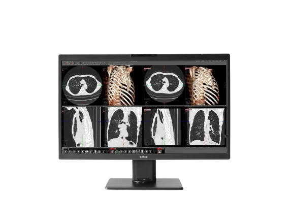
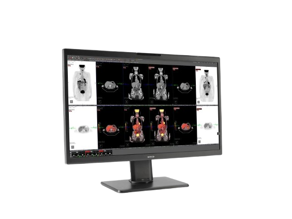
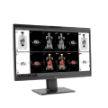
The Barco Nio Color 8MP (MDNC-8132) is a 32-inch IPS diagnostic display offering native 8-megapixel resolution, high luminance, wide color gamut, built-in QA functions, and image uniformity — ideal for radiology, mammography, and multi-modality imaging.
Typically 10-21 working days – excluding furniture and heavy/bulky equipment. Please contact us for further information.
Description
The Nio Color 8MP (MDNC-8132) by Barco is a premium diagnostic monitor designed to consolidate multiple images and clinical data on a single screen without losing detail. With a native resolution of 6848 × 4560 on a 32-inch IPS panel, it enables radiologists to view mammography, CT, MRI, and PACS images together. The display supports advanced calibration, uniformity correction (PPU), ambient light adaptation, and built-in QA via I-Guard and Barco’s QAWeb. High brightness, color/gray fidelity, and ergonomic design make this display suitable for critical diagnostic environments.
Barco’s Nio Color 8MP display is the ideal choice for radiology image interpretation in various settings. It seamlessly merges our acclaimed image quality and detail with built-in features that make collaboration with peers – from any location – easy and clutter-free.
Excellent 8MP image quality on a large 32” screen
Whether you’re working in an office, at home, or any other location, a solid setup is crucial. With its remarkable 8MP resolution and generous 32” screen, Nio Color 8MP empowers you to combine the information and images you need on a single screen.
The monitor includes our Intuitive Workflow Tools and cutting-edge technologies:
- SteadyGray for precise and consistent representation of grays
- SteadyColor for accurate and consistent representation of colors
- I-Guard for stable performance across the display’s full lifetime
- Uniform Luminance Technology for screen-wide uniformity correction
- Ambient Light Compensation for optimal viewing in various lighting conditions
- RapidFrame for sharp and fast image rendering when scrolling through cine images
Your perfect fit for reading radiology images, anywhere
In addition to the Nio Color 8MP’s exceptional core features, you also get:
- Integrated camera, speaker, and microphone
- Our advanced screensharing tool, Multi-Display Confer, for smooth virtual collaboration
- KVM functionality for effortless switching between multiple workstations
- Daisy chaining to keep your desk free of cable clutter
Stable performance, remotely managed
Enjoy automated QA and compliance for a stable performance anywhere, during the full lifetime of your monitor, thanks to QAWeb Enterprise. Finally, Barco’s all-inclusive warranty assures you a full return on investment over at least 5 to 7 years of your display.
A+ ecolabel for Nio Color 8MP
The Nio Color 8MP has been subjected to Barco’s ecoscoring protocol and has received an A+ rating. Some key factors that contributed to this rating are:
- Automatic standby mode when the device is not in use
- Use of plastics containing 35% post-consumer recycled content
- >50% halogen-free cables & >65% halogen-free PCBs
- >90% recyclable packaging
- 5 years all-inclusive warranty, extendable to 7 years
Key Features
-
8MP resolution (6848 × 4560) on a 32-inch IPS
-
Supports both color and grayscale imaging
-
Uniformity correction via PPU (Pixel Perception Uniformity)
-
SteadyGray™ and SteadyColor™ for stable luminance and color (with matching Barco controller/driver)
-
RapidFrame™ support for smooth display of motion/cine images
-
I-Guard front sensor for automatic QA / calibration
-
Ambient light presets and front light sensor
-
High color gamuts: 117% NTSC, 140% sRGB, 107% DCI-P3
-
Max Panel Brightness: ~2300 cd/m², calibrated 1200 cd/m² DICOM
-
Connectivity with 4× DisplayPort 1.4
-
Medical/electrical safety certifications
Specifications
| Category | Specification |
|---|---|
| Screen Technology | IPS |
| Active Screen Size | 32.0″ (812.80 mm); 708.48 × 398.52 mm (27.9 × 15.7″) |
| Aspect Ratio | 16:9 |
| Resolution | 8 MP (3840 × 2160 pixels @ 60 Hz) |
| Pixel Pitch | 0.1845 mm |
| Color Imaging | Yes |
| Gray Imaging | Yes |
| Bit Depth | 30-bit |
| Viewing Angle | 170° (H/V) |
| Uniformity Correction | ULT |
| SteadyGray | Yes |
| SteadyColor | Yes (with MXRT Display Controller & QAWeb Enterprise) |
| Pathology Setting | Yes |
| Color Gamut | NTSC: 94%, sRGB: 132% |
| sRGB Delta E2000 | Average < 3, Max < 5 |
| RapidFrame | Yes |
| Ambient Light Presets | Yes, reading room selection |
| Ambient Light Sensor | Yes |
| Front Sensor | Yes (I-Guard) |
| Presence Sensor | Yes |
| Maximum Luminance | 850 cd/m² |
| DICOM Calibrated Luminance | 420 cd/m² |
| Contrast Ratio | 1350:1 |
| Response Time | 12.7 ms ((Tr + Tf)/2) |
| Housing Color | Black (RAL 9004) / White (RAL 9003) |
| Video Input Signals | 2 × DisplayPort 1.4 |
| Video Output Signals | 1 × DisplayPort (MST) |
| USB Ports | 2 × USB-B 2.0 upstream; 5 × USB-A 2.0 downstream (1 charging) |
| KVM Switch | Yes |
| Power Rating | 24 VDC, 8.3 A |
| Power Requirements | Adapter Tech ATM200T-P240 (100–240 Vac, 50–60 Hz, 2.5–0.9 A; Output: 24 VDC, 8.3 A) |
| Power Consumption | 60 W (nominal); <0.35 W (hibernate/off) |
| Dimensions with Stand | 743 × 518~618 × 238 mm |
| Dimensions without Stand | 743 × 459 × 63 mm |
| Packaged Dimensions | 898 × 752 × 358 mm |
| Net Weight with Stand | 13 kg |
| Net Weight without Stand | 8.4 kg |
| Packaged Weight | 21 kg (without optional accessories) |
| Tilt / Swivel / Pivot | Tilt: -5° to +25°; Swivel: ±30°; Pivot: N/A |
| Height Adjustment Range | 100 mm |
| Mounting Standard | VESA (100 mm) |
| Screen Protection | N/A |
| Recommended Modalities | All digital images, except digital mammography |
| Certifications | CE0123, FDA 510(k) K233693, CCC, KC, INMETRO, BIS, EAC (Pending) |
| Safety Standards | IEC/EN/AAMI/CSA 60950-1, 62368-1, 60601-1 |
| EMI Compliance | IEC/EN 60601-1-2, FCC Part 15 Class B, ICES-001 Level B, VCCI (Pending) |
| Environmental Compliance | EU RoHS, China RoHS, REACH, Canada Health, WEEE, Packaging Directive |
| Supplied Accessories | User Guide, Quick Install Sheet, Documentation Disc, System Sheet, Video & USB cables, External Power Supply |
| Optional Accessories | Display Controller, Touch Pad, QA Software, QAWeb Enterprise |
| Warranty | 5 years, including 20,000 hours backlight warranty |
| Operating Temperature | 0–35 °C (specs: 20–30 °C) |
| Storage Temperature | -20–60 °C |
| Operating Humidity | 8–70% (non-condensing) |
| Storage Humidity | 5–70% (non-condensing) |
| Operating Pressure | 70 kPa |
| Storage Pressure | 50–106 kPa |
Quick Comparison
| Nio Color 8MP (MDNC‑8132) remove | Sonoscape E1 Ultrasound Machine With Two Probes remove | Sonoscape P20 Ultrasound Machine remove | Lab/Ward Coat remove | DrGem Ceiling Mounted Digital X-ray remove | SIGNERS SUPiA X-ray Digitizer ( CR Scanner) remove | |||||||||||||||||||||||||||||||||||||||||||||||||||||||||||||||||||||||||||||||||||||||||||||||||||||||||||||||||||||||||||||||||||||||||||
|---|---|---|---|---|---|---|---|---|---|---|---|---|---|---|---|---|---|---|---|---|---|---|---|---|---|---|---|---|---|---|---|---|---|---|---|---|---|---|---|---|---|---|---|---|---|---|---|---|---|---|---|---|---|---|---|---|---|---|---|---|---|---|---|---|---|---|---|---|---|---|---|---|---|---|---|---|---|---|---|---|---|---|---|---|---|---|---|---|---|---|---|---|---|---|---|---|---|---|---|---|---|---|---|---|---|---|---|---|---|---|---|---|---|---|---|---|---|---|---|---|---|---|---|---|---|---|---|---|---|---|---|---|---|---|---|---|---|---|---|---|---|---|---|---|
| Name | Nio Color 8MP (MDNC‑8132) remove | Sonoscape E1 Ultrasound Machine With Two Probes remove | Sonoscape P20 Ultrasound Machine remove | Lab/Ward Coat remove | DrGem Ceiling Mounted Digital X-ray remove | SIGNERS SUPiA X-ray Digitizer ( CR Scanner) remove | ||||||||||||||||||||||||||||||||||||||||||||||||||||||||||||||||||||||||||||||||||||||||||||||||||||||||||||||||||||||||||||||||||||||||||
| Image | 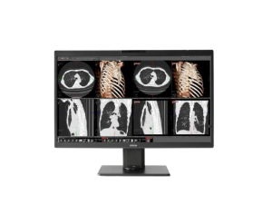 |  |  |  |  | 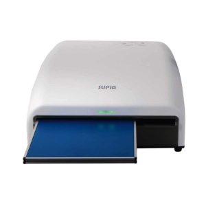 | ||||||||||||||||||||||||||||||||||||||||||||||||||||||||||||||||||||||||||||||||||||||||||||||||||||||||||||||||||||||||||||||||||||||||||
| SKU | SF1033560012-20 | SF1033560012-9 | SF1033560084-222 | SF1033560074-4 | SF1033560050-01 | |||||||||||||||||||||||||||||||||||||||||||||||||||||||||||||||||||||||||||||||||||||||||||||||||||||||||||||||||||||||||||||||||||||||||||
| Rating | ||||||||||||||||||||||||||||||||||||||||||||||||||||||||||||||||||||||||||||||||||||||||||||||||||||||||||||||||||||||||||||||||||||||||||||||||
| Price |
| $4,620.00 |
| $11.00 |
| $6,930.00 | ||||||||||||||||||||||||||||||||||||||||||||||||||||||||||||||||||||||||||||||||||||||||||||||||||||||||||||||||||||||||||||||||||||||||||
| Stock | ||||||||||||||||||||||||||||||||||||||||||||||||||||||||||||||||||||||||||||||||||||||||||||||||||||||||||||||||||||||||||||||||||||||||||||||||
| Availability | ||||||||||||||||||||||||||||||||||||||||||||||||||||||||||||||||||||||||||||||||||||||||||||||||||||||||||||||||||||||||||||||||||||||||||||||||
| Add to cart | ||||||||||||||||||||||||||||||||||||||||||||||||||||||||||||||||||||||||||||||||||||||||||||||||||||||||||||||||||||||||||||||||||||||||||||||||
| Description | Shipped From Abroad
The Barco Nio Color 8MP (MDNC-8132) is a 32-inch IPS diagnostic display offering native 8-megapixel resolution, high luminance, wide color gamut, built-in QA functions, and image uniformity — ideal for radiology, mammography, and multi-modality imaging.
Delivery & Availability:
Typically 10-21 working days – excluding furniture and heavy/bulky equipment. Please contact us for further information.
| Shipped from Abroad SonoScape has developed a new probe and function for the E1 Exp. With these additions the E1 Exp will bring users a more efficient examination experience with satisfying image quality and a smooth workflow. Delivery & Availability: Typically 5-7 working days – excluding furniture and heavy/bulky equipment. Please contact us for further information. | Shipped from Abroad Incorporating innovative technologies, P20’s user-friendly design with a simple operation panel, intuitive user interface and a variety of intelligent auxiliary scanning tools, will significantly improve your daily examination experience. Besides general imaging applications, P20 has entitled with diagnostic 4D technology which has an extraordinary performance in obstetrics and gynecology applications. Delivery & Availability: Typically 5-7 working days – excluding furniture and heavy/bulky equipment. Please contact us for further information. | In stock
| In Stock The GXR-SD is a diagnostic digital radiography system that provides reliable high quality digital radiographic images with a reduced dose. The GXR-SD DR systems offer comprehensive digital solutions to all radiography needs, featuring ACQUIDR digital imaging system with stationary or portable digital flat-panel detectors as well as reliable high-frequency x-ray generators that are known worldwide for their excellent performance, lifetime and stability. Patient tables and wall stands are also offered. Delivery & Availability: Typically 21 working days – excluding furniture and heavy/bulky equipment. Please contact us for further information. | Shipped from Abroad SUPiA made by Signers offers such a better clinic environment with no chemicals, ideal space, high-resolution image quality, and affordability. Delivery & Availability: Typically 14 working days – excluding furniture and heavy/bulky equipment. Please contact us for further information. | ||||||||||||||||||||||||||||||||||||||||||||||||||||||||||||||||||||||||||||||||||||||||||||||||||||||||||||||||||||||||||||||||||||||||||
| Content | https://youtu.be/TIO_tmvFinU?si=87M87Jzx_3sJzLEY
The Nio Color 8MP (MDNC-8132) by Barco is a premium diagnostic monitor designed to consolidate multiple images and clinical data on a single screen without losing detail. With a native resolution of 6848 × 4560 on a 32-inch IPS panel, it enables radiologists to view mammography, CT, MRI, and PACS images together. The display supports advanced calibration, uniformity correction (PPU), ambient light adaptation, and built-in QA via I-Guard and Barco’s QAWeb. High brightness, color/gray fidelity, and ergonomic design make this display suitable for critical diagnostic environments.
Barco's Nio Color 8MP display is the ideal choice for radiology image interpretation in various settings. It seamlessly merges our acclaimed image quality and detail with built-in features that make collaboration with peers – from any location – easy and clutter-free. Excellent 8MP image quality on a large 32” screen Whether you're working in an office, at home, or any other location, a solid setup is crucial. With its remarkable 8MP resolution and generous 32” screen, Nio Color 8MP empowers you to combine the information and images you need on a single screen. The monitor includes our Intuitive Workflow Tools and cutting-edge technologies:
Key Features
Specifications
| DETAILS
Efficient Diagnosis
μ-Scan, Speckle Reduction & Edge Enhancement
Spatial Compound Imaging
PIH - Pure Inversion Harmonic
Wide Scan - Enlarged Image Area
Tissue-Specific Imaging
SR Flow
Ergonomic Designs
Up to 2 Transducer Ports
Light Weight and Compact
15.6 inch Anti-flickering HD LED Screen
Tilting Monitor Angle Adjustment
Backlit Keyboard and Intelligent Panel
Long-lasting Battery for 90 mins
Ease of Use
Quick Boot Up
Auto-Brightness Adjustment
Auto Image Optimization
Auto IMT
Auto Trace
Equipped Accessories
Wi-Fi and Bluetooth Available
DICOM
500GB Hard Disk
Height Adjustable Trolley
Durable, Carry-on Site Suitcase
Click Here To Download Catalogue | DETAILS
Upgraded Images with More Clarity
SonoScape never stops making progress in improving the image quality of its ultrasound products to enhance the confidence of diagnosis for doctors. With extraordinary images provided by P20, the anatomy structures are clearer than ever.
C-Xlasto Imaging
With C-xlasto Imaging, P20 enables comprehensive quantitative elastic analysis. Meanwhile, C-xlasto on P20 is supported by linear, convex and transvaginal probes, to ensure good reproducibility and highly consistent quantitative elastic results.
S-Live
S-Live allows for detailed visualization of subtle anatomical features, thereby enabling intuitive diagnosis with real-time 3D images and enriching patient communication.
Pelvic Floor 4D
Transperineal 4D pelvic floor ultrasound can provide useful clinical values in assessing the vaginal delivery impact on the female anterior compartment, judging whether the pelvic organs are prolapsed or not and the extent, determining if the pelvic muscles were torn accurately.
Anatomic M Mode
Anatomic M Mode helps you observe the myocardial motion at different phases by freely placing sample lines. It accurately measures the myocardial thickness and the heart size of even difficult patients and supports the myocardial function and LV wall-motion assessment.
Tissue Doppler Imaging
P20 is endowed with Tissue Doppler Imaging which provides velocities and other clinical information on myocardial functions, facilitating clinical doctors with the ability to analyze and compare the motions of different parts of the patient's heart.
Click Here To Download Catalogue |
| DrGem Ceiling Mounted Digital X-ray is a diagnostic digital radiography system that provides reliable high quality digital radiographic images with a reduced dose. The GXR-SD DR systems offer comprehensive digital solutions to all radiography needs, featuring ACQUIDR digital imaging system with stationary or portable digital flat-panel detectors as well as reliable high-frequency x-ray generators that are known worldwide for their excellent performance, lifetime and stability. Patient tables and wall stands are also offered.
Features:
Click Here To Download Catalogue | SUPiA X-ray Digitizer made by Signers offers such a better clinic environment with no chemicals, ideal space, high-resolution image quality, and affordability
FEATURE
Rigid Type
• No damage or scratch on image plates during scanning & erasing
• Scanning & Erasing without a roller
• No cut-off image during winter and cold period
Durability
• Extremely simple structure design
• Strong aluminum base plate
• Flip covers preventing dust from inside scanner
Barcode System
• Automatically recognising cassette sizes(14x17", 10x12", 18x24cm) by barcode reader
Compact & lightweight design
Click Here To Download Catalogue | ||||||||||||||||||||||||||||||||||||||||||||||||||||||||||||||||||||||||||||||||||||||||||||||||||||||||||||||||||||||||||||||||||||||||||
| Weight | N/A | N/A | N/A | N/A | N/A | N/A | ||||||||||||||||||||||||||||||||||||||||||||||||||||||||||||||||||||||||||||||||||||||||||||||||||||||||||||||||||||||||||||||||||||||||||
| Dimensions | N/A | N/A | N/A | N/A | N/A | N/A | ||||||||||||||||||||||||||||||||||||||||||||||||||||||||||||||||||||||||||||||||||||||||||||||||||||||||||||||||||||||||||||||||||||||||||
| Additional information |


Reviews
There are no reviews yet.