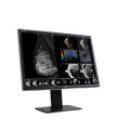
Nio Fusion 12MP (MDNC‑12130)
$0.00
Shipped From Abroad
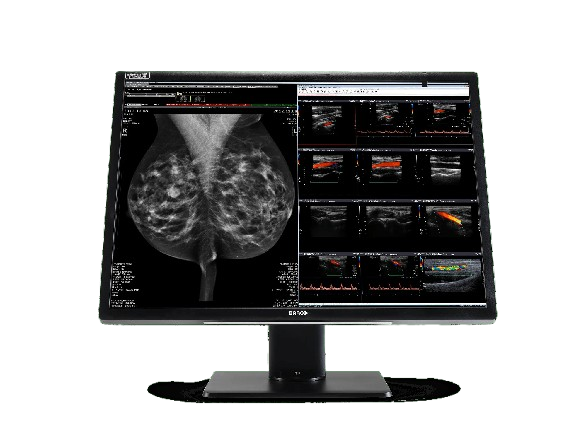
The Nio Fusion 12MP (MDNC-12130) display is designed to combine PACS and breast images on one workstation, so you don’t need to work on a cluttered desk with complex configurations and multiple portrait displays. A Nio Fusion 12MP will represent both 2D and 3D images fluidly, brightly and in detail, further helping you to speed up your reading sessions. A set of unique integrated tools improve reading ergonomics and support efficient workflow for static and dynamic imaging.
Typically 10-21 working days – excluding furniture and heavy/bulky equipment. Please contact us for further information.
Description
The Nio Fusion 12MP (MDNC-12130) display is designed to combine PACS and breast images on one workstation, so you don’t need to work on a cluttered desk with complex configurations and multiple portrait displays. A Nio Fusion 12MP will represent both 2D and 3D images fluidly, brightly, and in detail, further helping you to speed up your reading sessions. A set of unique integrated tools improves reading ergonomics and supports efficient workflow for static and dynamic imaging.
- Medical display
- Excellent uniformity correction
- Perfect representation of calibrated colors and greyscales
Enjoy consistent and compliant colors and grayscales
With a 12MP resolution, you’ll fit multiple images on one screen and enjoy every single one in extremely sharp and precise quality, with less panning and zooming. Nio Fusion 12MP displays are calibrated to meet the DICOM standard for grayscales. And thanks to the SteadyColor™ technology, you can also confidently rely on perceptually linear colors.
Barco’s QAWeb Enterprise software, included in the display, guarantees consistent image quality through automated calibration and QA, and also enables compliance with the latest regional and international regulations for image quality.
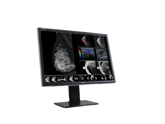
Read on a flexible display, with optimal comfort
The Nio Fusion 12MP is surprisingly thin and light. It mirrors most of a human’s natural field of vision and was designed to reduce head, hand, and eye movements to a minimum. You can even switch between two workstations in no time, at the touch of a button, with integrated KVM (Keyboard-Video-Mouse).
- A reflection-free surface enhances image sharpness
- SoftGlow ambient lighting reduces eye fatigue
- Uniform Luminance Technology ensures constant luminance in all regions of the screen
- Ambient Light Sensor and Compensation provide consistent images in any lighting conditions
A future-proof investment that lasts
The Nio Fusion 12MP is an all-in-one imaging solution for both PACS and breast imaging, which will make it possible for you to save operational costs. Its smooth, fast system was designed to support you in your workflow, enabling you to see more patients. And last but not least, thanks to its long lifetime, the display can be your companion for years to come. All its components are warranted for 5 years.
Ensuring diagnostic confidence with MDR Class IIa
Our radiology displays are MDR-certified as Class IIa. Their product information has been reviewed and cleared by independent medical and technical experts, and is audited yearly. In other words, we ensure diagnostic confidence and peace of mind for our users.
Technologies that enhance image quality:
- Uniform Luminance Technology to ensure that all regions of the screen have an even luminance
- SteadyColor™ calibration technology to meet the DICOM standard for grayscales and to guarantee consistent, perceptually linear color
- SteadyGray™ ensures that all gray values closely match the selected white tint. This can be a blue base, a clear base, or some other preferred white tint
- QAWeb Enterprise, a cloud-based technology for automated calibration and Quality Assurance
- I-Guard™ front sensor to ensure 24/7 compliance with image quality standards and guidelines
- Efficient DuraLight™ backlights for a long lifetime of brighter images
Technologies that enhance productivity:
- RapidFrame™ to ensure crisp and in-focus moving images, with up to 10% higher detection of small details in moving images*
- Conference CloneView™ software to project and control images on a large screen with ease
- SoftGlow™ task and wall lighting to improve reading room conditions
- SpotView™ to highlight subtle details in a region of interest
- KVM to switch effortlessly between two workstations
A+ ecolabel for Nio Fusion 12MP
The Nio Fusion 12MP has been subjected to Barco’s ecoscoring protocol and has received an A+ rating. Some key factors that contributed to this rating are:
- Automatic standby mode when the device is not in use
- 100% halogen-free PCBs, internal cables, and plastic parts >25g
- Packaging optimized for logistics
- Product design optimized for disassembly
- Large plastic parts unpainted
Specifications
| Category | Specification |
|---|---|
| Screen Technology | LCD |
| Active Screen Size (Diagonal) | 784 mm (30.9″) |
| Active Screen Size (H × V) | 653 × 435 mm (25.7″ × 17.1″) |
| Aspect Ratio | 3:2 |
| Resolution | Native 12MP (4200 × 2800 pixels); Configurable to 2 × 5.8MP (2100 × 2800 pixels) |
| Pixel Pitch | 0.1554 mm |
| Color Imaging / Gray Imaging | Yes / Yes |
| Bit Depth | 30-bit |
| Viewing Angle (H/V) | 178° |
| Uniformity Correction | ULT |
| SteadyGray / SteadyColor | Yes (in display, with system components as outlined in user guide) |
| Ambient Light Presets / Sensor | Yes / Yes |
| Front Sensor | Yes |
| Maximum Luminance (Typical) | 1200 cd/m² |
| DICOM Calibrated Luminance | MDNC-12130: 600 cd/m² |
| Contrast Ratio | 1500:1 |
| Response Time | 10 ms (average, all transitions within 1 frame period) |
| Housing Color | Black / White |
| Video Input Signals | 2 × DisplayPort 1.2 |
| Video Output Signals | N/A |
| USB Ports | 2 × USB-B 2.0 upstream (switchable endpoint); 2 × USB-A 2.0 downstream |
| KVM Switch | Yes |
| Power Rating | 100–240 Vac, 50/60 Hz, 3.6–1.6 A |
| Power Consumption | 105 W (nominal); <0.5 W (hibernate/standby) |
| Dimensions with Stand (W × H × D) | 695 × 528~628 × 239 mm |
| Dimensions without Stand (W × H × D) | 695 × 483 × 74 mm |
| Dimensions Packaged (W × H × D) | 800 × 650 × 295 mm |
| Net Weight with Stand | 16.6 kg |
| Net Weight without Stand | 12.0 kg |
| Net Weight Packaged | 21.3 kg (without optional accessories) |
| Tilt / Swivel / Pivot | -5° to +25° / ±30° / N/A |
| Height Adjustment Range | 100 mm |
| Mounting Standard | VESA (100 mm) |
| Screen Protection | N/A |
| Recommended Modalities | All digital images, including digital mammography and breast tomosynthesis |
| Certifications | CE0123, FDA 510(K) K203106, CCC, KC, BIS, EAC |
| Safety Standards | IEC/EN/UL/CSA 60950-1, 62368-1, 60601-1, AAMI ES 60601-1 |
| EMI Standards | IEC/EN 60601-1-2, FCC Part 15 Class B, ICES-001 Level B, VCCI |
| Environmental Compliance | EU RoHS, China RoHS, Korea e-Standby, REACH, WEEE, Packaging Directive |
| Supplied Accessories | User guide, Documentation disc, System sheet, Video cables, USB cables, Mains cables |
| Optional Accessories | Display controller, QA software (QAWeb) |
| Warranty | 5 years (includes 40,000 hrs backlight warranty) |
| Operating Temperature | 0–35°C (specs: 20–30°C) |
| Storage Temperature | -20–60°C |
| Operating Humidity | 10–70% RH (non-condensing) |
| Storage Humidity | 10–70% RH (max. 70% at 40°C) |
| Operating Pressure | ≥62 kPa |
| Storage Pressure | 50–106 kPa |
Let me know if you’d like this formatted for a datasheet, compared with another model, or exported into a document.
Quick Comparison
| Nio Fusion 12MP (MDNC‑12130) remove | DrGem Floor Mounted Analogue X-ray remove | Lab/Ward Coat remove | Sonoscape S8 Exp Portable Ultrasound remove | Anke Anatom 64 Clarity Multi-Slice Spiral CT Scan remove | DrGem GXR-SD 400mA Floor Mounted Digital X-ray remove | |||||||||||||||||||||||||||||||||||||||||||||||||||||||||||||||||||||||||||||||||||||||||||||||||||||||||||||||||||||||||||||||||||||||||||||||||||||||||||||||||||||||||||||||||||||||||||||||||||||||||||||||||||||||||||||||||||||||||||||||||||||||||||||||||||||||||||||||||||||||||||||||||||||||||||||||||||||||||||||||||||||||||||||||||||||||||||||||||||||||||||||||||||||||||||||
|---|---|---|---|---|---|---|---|---|---|---|---|---|---|---|---|---|---|---|---|---|---|---|---|---|---|---|---|---|---|---|---|---|---|---|---|---|---|---|---|---|---|---|---|---|---|---|---|---|---|---|---|---|---|---|---|---|---|---|---|---|---|---|---|---|---|---|---|---|---|---|---|---|---|---|---|---|---|---|---|---|---|---|---|---|---|---|---|---|---|---|---|---|---|---|---|---|---|---|---|---|---|---|---|---|---|---|---|---|---|---|---|---|---|---|---|---|---|---|---|---|---|---|---|---|---|---|---|---|---|---|---|---|---|---|---|---|---|---|---|---|---|---|---|---|---|---|---|---|---|---|---|---|---|---|---|---|---|---|---|---|---|---|---|---|---|---|---|---|---|---|---|---|---|---|---|---|---|---|---|---|---|---|---|---|---|---|---|---|---|---|---|---|---|---|---|---|---|---|---|---|---|---|---|---|---|---|---|---|---|---|---|---|---|---|---|---|---|---|---|---|---|---|---|---|---|---|---|---|---|---|---|---|---|---|---|---|---|---|---|---|---|---|---|---|---|---|---|---|---|---|---|---|---|---|---|---|---|---|---|---|---|---|---|---|---|---|---|---|---|---|---|---|---|---|---|---|---|---|---|---|---|---|---|---|---|---|---|---|---|---|---|---|---|---|---|---|---|---|---|---|---|---|---|---|---|---|---|---|---|---|---|---|---|---|---|---|---|---|---|---|---|---|---|---|---|---|---|---|---|---|---|---|---|---|---|---|---|---|---|---|---|---|---|---|---|---|---|---|---|---|---|---|---|---|---|---|---|---|---|---|---|---|---|---|---|---|---|---|---|---|---|---|---|---|---|---|---|---|---|---|---|---|---|---|---|---|
| Name | Nio Fusion 12MP (MDNC‑12130) remove | DrGem Floor Mounted Analogue X-ray remove | Lab/Ward Coat remove | Sonoscape S8 Exp Portable Ultrasound remove | Anke Anatom 64 Clarity Multi-Slice Spiral CT Scan remove | DrGem GXR-SD 400mA Floor Mounted Digital X-ray remove | ||||||||||||||||||||||||||||||||||||||||||||||||||||||||||||||||||||||||||||||||||||||||||||||||||||||||||||||||||||||||||||||||||||||||||||||||||||||||||||||||||||||||||||||||||||||||||||||||||||||||||||||||||||||||||||||||||||||||||||||||||||||||||||||||||||||||||||||||||||||||||||||||||||||||||||||||||||||||||||||||||||||||||||||||||||||||||||||||||||||||||||||||||||||||||||
| Image |  |  |  |  |  | |||||||||||||||||||||||||||||||||||||||||||||||||||||||||||||||||||||||||||||||||||||||||||||||||||||||||||||||||||||||||||||||||||||||||||||||||||||||||||||||||||||||||||||||||||||||||||||||||||||||||||||||||||||||||||||||||||||||||||||||||||||||||||||||||||||||||||||||||||||||||||||||||||||||||||||||||||||||||||||||||||||||||||||||||||||||||||||||||||||||||||||||||||||||||||||
| SKU | SF1033560074-6 | SF1033560084-222 | SF1033560012-15 | SF1033560092-2 | SF1033560074-5 | |||||||||||||||||||||||||||||||||||||||||||||||||||||||||||||||||||||||||||||||||||||||||||||||||||||||||||||||||||||||||||||||||||||||||||||||||||||||||||||||||||||||||||||||||||||||||||||||||||||||||||||||||||||||||||||||||||||||||||||||||||||||||||||||||||||||||||||||||||||||||||||||||||||||||||||||||||||||||||||||||||||||||||||||||||||||||||||||||||||||||||||||||||||||||||||
| Rating | ||||||||||||||||||||||||||||||||||||||||||||||||||||||||||||||||||||||||||||||||||||||||||||||||||||||||||||||||||||||||||||||||||||||||||||||||||||||||||||||||||||||||||||||||||||||||||||||||||||||||||||||||||||||||||||||||||||||||||||||||||||||||||||||||||||||||||||||||||||||||||||||||||||||||||||||||||||||||||||||||||||||||||||||||||||||||||||||||||||||||||||||||||||||||||||||||||
| Price |
|
| $11.00 | $9,350.00 |
|
| ||||||||||||||||||||||||||||||||||||||||||||||||||||||||||||||||||||||||||||||||||||||||||||||||||||||||||||||||||||||||||||||||||||||||||||||||||||||||||||||||||||||||||||||||||||||||||||||||||||||||||||||||||||||||||||||||||||||||||||||||||||||||||||||||||||||||||||||||||||||||||||||||||||||||||||||||||||||||||||||||||||||||||||||||||||||||||||||||||||||||||||||||||||||||||||
| Stock | ||||||||||||||||||||||||||||||||||||||||||||||||||||||||||||||||||||||||||||||||||||||||||||||||||||||||||||||||||||||||||||||||||||||||||||||||||||||||||||||||||||||||||||||||||||||||||||||||||||||||||||||||||||||||||||||||||||||||||||||||||||||||||||||||||||||||||||||||||||||||||||||||||||||||||||||||||||||||||||||||||||||||||||||||||||||||||||||||||||||||||||||||||||||||||||||||||
| Availability | ||||||||||||||||||||||||||||||||||||||||||||||||||||||||||||||||||||||||||||||||||||||||||||||||||||||||||||||||||||||||||||||||||||||||||||||||||||||||||||||||||||||||||||||||||||||||||||||||||||||||||||||||||||||||||||||||||||||||||||||||||||||||||||||||||||||||||||||||||||||||||||||||||||||||||||||||||||||||||||||||||||||||||||||||||||||||||||||||||||||||||||||||||||||||||||||||||
| Add to cart | ||||||||||||||||||||||||||||||||||||||||||||||||||||||||||||||||||||||||||||||||||||||||||||||||||||||||||||||||||||||||||||||||||||||||||||||||||||||||||||||||||||||||||||||||||||||||||||||||||||||||||||||||||||||||||||||||||||||||||||||||||||||||||||||||||||||||||||||||||||||||||||||||||||||||||||||||||||||||||||||||||||||||||||||||||||||||||||||||||||||||||||||||||||||||||||||||||
| Description | Shipped From Abroad
The Nio Fusion 12MP (MDNC-12130) display is designed to combine PACS and breast images on one workstation, so you don’t need to work on a cluttered desk with complex configurations and multiple portrait displays. A Nio Fusion 12MP will represent both 2D and 3D images fluidly, brightly and in detail, further helping you to speed up your reading sessions. A set of unique integrated tools improve reading ergonomics and support efficient workflow for static and dynamic imaging.
Delivery & Availability:
Typically 10-21 working days – excluding furniture and heavy/bulky equipment. Please contact us for further information.
| In Stock GXR Analogue X-ray system matches with a radiographic room which perfectly fits your workow and can be easily upgraded to DR system with the help of DR interface and PC interface in GXR generator as well as Bucky suitable to Flat Panel Detector. GXR X-ray system is equipped with a high frequency X-ray generator which consistently produces high quality radiograph in favor of high quality X-ray output with a very small kV ripple and accurate mA and mAs. GXR X-ray system is designed to provide convenience to operator and comfort to patient. Delivery & Availability: Typically 21 working days – excluding furniture and heavy/bulky equipment. Please contact us for further information. | In stock
| Shipped from Abroad With ultra-modern innovative design and the clinically-proven technologies, S8 Exp is portable ultrasound scanner well equipped as a low-physical-effort and enhanced-image-quality ultrasound scanner, which not only provides optimized solutions for versatile applications, but does help to improve the user-experience for both routine and non-traditional challenges. Delivery & Availability: Typically 5-7 working days – excluding furniture and heavy/bulky equipment. Please contact us for further information. | Shipped from Abroad
The ANATOM 64 CT scanner is the latest innovation for cardiac imaging based on Precision Platform system. The excellent design of Ahart technology which innovatively combined single spiral scan + gated imaging + mA modulation for easy heart imaging at extremely low radiation dose. We provide you ANATOM 64 Clarity/Precision of two models which are low/high configurations for preferences. It also offers you conventional clinical applications of low dose, better image quality and faster exams.
Delivery & Availability: Typically 90 working days – excluding furniture and heavy/bulky equipment. Please contact us for further information. | In Stock The GXR-SD Digital X-ray is a diagnostic digital radiography system that provides reliable high quality digital radiographic images with a reduced dose. The GXR-SD DR systems offer comprehensive digital solutions to all radiography needs, featuring ACQUIDR digital imaging system with stationary or portable digital flat-panel detectors as well as reliable high-frequency x-ray generators that are known worldwide for their excellent performance, lifetime and stability. Patient tables and wall stands are also offered. Delivery & Availability: Typically 21 working days – excluding furniture and heavy/bulky equipment. Please contact us for further information. | ||||||||||||||||||||||||||||||||||||||||||||||||||||||||||||||||||||||||||||||||||||||||||||||||||||||||||||||||||||||||||||||||||||||||||||||||||||||||||||||||||||||||||||||||||||||||||||||||||||||||||||||||||||||||||||||||||||||||||||||||||||||||||||||||||||||||||||||||||||||||||||||||||||||||||||||||||||||||||||||||||||||||||||||||||||||||||||||||||||||||||||||||||||||||||||
| Content | The Nio Fusion 12MP (MDNC-12130) display is designed to combine PACS and breast images on one workstation, so you don’t need to work on a cluttered desk with complex configurations and multiple portrait displays. A Nio Fusion 12MP will represent both 2D and 3D images fluidly, brightly, and in detail, further helping you to speed up your reading sessions. A set of unique integrated tools improves reading ergonomics and supports efficient workflow for static and dynamic imaging.
Enjoy consistent and compliant colors and grayscalesWith a 12MP resolution, you’ll fit multiple images on one screen and enjoy every single one in extremely sharp and precise quality, with less panning and zooming. Nio Fusion 12MP displays are calibrated to meet the DICOM standard for grayscales. And thanks to the SteadyColor™ technology, you can also confidently rely on perceptually linear colors. Barco's QAWeb Enterprise software, included in the display, guarantees consistent image quality through automated calibration and QA, and also enables compliance with the latest regional and international regulations for image quality.Read on a flexible display, with optimal comfortThe Nio Fusion 12MP is surprisingly thin and light. It mirrors most of a human's natural field of vision and was designed to reduce head, hand, and eye movements to a minimum. You can even switch between two workstations in no time, at the touch of a button, with integrated KVM (Keyboard-Video-Mouse).
A future-proof investment that lastsThe Nio Fusion 12MP is an all-in-one imaging solution for both PACS and breast imaging, which will make it possible for you to save operational costs. Its smooth, fast system was designed to support you in your workflow, enabling you to see more patients. And last but not least, thanks to its long lifetime, the display can be your companion for years to come. All its components are warranted for 5 years.Ensuring diagnostic confidence with MDR Class IIaOur radiology displays are MDR-certified as Class IIa. Their product information has been reviewed and cleared by independent medical and technical experts, and is audited yearly. In other words, we ensure diagnostic confidence and peace of mind for our users.Technologies that enhance image quality:
Technologies that enhance productivity:
A+ ecolabel for Nio Fusion 12MPThe Nio Fusion 12MP has been subjected to Barco’s ecoscoring protocol and has received an A+ rating. Some key factors that contributed to this rating are:
Specifications
Let me know if you'd like this formatted for a datasheet, compared with another model, or exported into a document. | DrGem GXR Floor Mounted Analogue X-ray system matches with a radiographic room which perfectly fits your workflow and can be easily upgraded to DR system with the help of DR interface and PC interface in GXR generator as well as Bucky suitable to Flat Panel Detector. GXR (Analogue X-ray)system is equipped with a high frequency X-ray generator which consistently produces high quality radiograph in favor of high quality X-ray output with a very small kV ripple and accurate mA and mAs. GXR (Analogue X-ray) system is designed to provide convenience to operator and comfort to patient.
Features of DrGem GXR Floor Mounted Analogue X-ray:
Click Here To Download Catalogue |
| Sonoscape S8 Exp Portable Ultrasound scannerDETAILS Agile and Versatile With ultra-modern innovative design and the clinically-proven technologies, S8 Exp Portable Ultrasound scanner is well equipped as a low-physical-effort and enhanced-image-quality ultrasound scanner, which not only provides optimized solutions for versatile applications but does help to improve the user experience for both routine and non-traditional challenges. Working with S8 Exp, it will trigger your unlimited reverie and endow you with endless charm. Carrying forward the classical design of SonoScape's portable ultrasound products, S8 Exp successfully combines the best ergonomics, attractive design and ease of use. This charismatic identity is also enhanced by a sophisticated color palette—with sedate grey as its interior paint color and pearl white as exterior cover, S8 Exp reveals a style of aristocrat and strong character among SonoScape's ultrasound systems. Workflow The S8 Exp is a portable ultrasound scanner that adapts to your workflow, whether you are in the consulting room, at the bedside, or at a remote location. With easy-to-use new platform designed for sonographers' needs and full connection interfaces for easy connectivity and data sharing, S8 Exp leads to improved user comfort and clinical outcome as well as patient throughput and working efficiency. Powerful Platform Embedded with SonoScape's core imaging technologies such as μ-scan, PHI and Spatial Compound, S8 Exp boasts exceptional 2D image, sensitive spectral, Color and Power Doppler, displaying well-defined anatomy and pathology and facilitating a highly optimized diagnostic user environment for conclusive diagnoses. Besides, S8 Exp offers a comprehensive selection of electronic probes to maximally extend its capabilities to meet a wide range of applications including the abdomen, pediatric, OB/GYN, cardiovascular, musculoskeletal, etc. The advanced probe technologies also effectively enhance the image quality and confidence in reaching clinical diagnoses even in difficult patients.Click Here To Download Catalogue | The ANATOM 64 CT scanner is the latest innovation for cardiac imaging based on Precision Platform system. The excellent design of Ahart technology which innovatively combined single spiral scan + gated imaging + mA modulation for easy heart imaging at extremely low radiation dose. We provide you ANATOM 64 Clarity/Precision of two models which are low/high configurations for preferences. It also offers you conventional clinical applications of low dose, better image quality and faster exams.
Features:
Click Here To Download Catalogue | DrGem GXR-SD 400mA Floor Mounted Digital X-ray system matches with a radiographic room which perfectly fits your workow and can be easily upgraded to DR system with the help of DR interface and PC interface in GXR generator as well as Bucky suitable to Flat Panel Detector. GXR X-ray system is equipped with a high frequency X-ray generator which consistently produces high quality radiograph in favor of high quality X-ray output with a very small kV ripple and accurate mA and mAs. GXR X-ray system is designed to provide convenience to operator and comfort to patient
Features of DrGem GXR-SD 400mA Floor Mounted Digital X-ray:
Click Here To Download Catalogue | ||||||||||||||||||||||||||||||||||||||||||||||||||||||||||||||||||||||||||||||||||||||||||||||||||||||||||||||||||||||||||||||||||||||||||||||||||||||||||||||||||||||||||||||||||||||||||||||||||||||||||||||||||||||||||||||||||||||||||||||||||||||||||||||||||||||||||||||||||||||||||||||||||||||||||||||||||||||||||||||||||||||||||||||||||||||||||||||||||||||||||||||||||||||||||||
| Weight | N/A | N/A | N/A | N/A | N/A | N/A | ||||||||||||||||||||||||||||||||||||||||||||||||||||||||||||||||||||||||||||||||||||||||||||||||||||||||||||||||||||||||||||||||||||||||||||||||||||||||||||||||||||||||||||||||||||||||||||||||||||||||||||||||||||||||||||||||||||||||||||||||||||||||||||||||||||||||||||||||||||||||||||||||||||||||||||||||||||||||||||||||||||||||||||||||||||||||||||||||||||||||||||||||||||||||||||
| Dimensions | N/A | N/A | N/A | N/A | N/A | N/A | ||||||||||||||||||||||||||||||||||||||||||||||||||||||||||||||||||||||||||||||||||||||||||||||||||||||||||||||||||||||||||||||||||||||||||||||||||||||||||||||||||||||||||||||||||||||||||||||||||||||||||||||||||||||||||||||||||||||||||||||||||||||||||||||||||||||||||||||||||||||||||||||||||||||||||||||||||||||||||||||||||||||||||||||||||||||||||||||||||||||||||||||||||||||||||||
| Additional information |
|


Reviews
There are no reviews yet.