| Description | | Shipped from Abroad
The ANATOM 64 CT scanner is the latest innovation for cardiac imaging based on Precision Platform system. The excellent design of Ahart technology which innovatively combined single spiral scan + gated imaging + mA modulation for easy heart imaging at extremely low radiation dose. We provide you ANATOM 64 Clarity/Precision of two models which are low/high configurations for preferences. It also offers you conventional clinical applications of low dose, better image quality and faster exams.
Delivery & Availability:
Typically 90 working days – excluding furniture and heavy/bulky equipment. Please contact us for further information. | In Stock
JADE is one of the lightest portable X-ray systems on the market, allowing it to be used in any imaginable way including bedside, operating rooms, intensive care units and in veterinary fields. With a simple, easy-to-use operator console, three-way control, two-step foldable stand and auto lock system, JADE is a user-friendly portable X-ray system.
Delivery & Availability:
Typically 21 working days – excluding furniture and heavy/bulky equipment. Please contact us for further information. | Shipped from Abroad
OPENMARK 5000 is 0.51T MRI. It's approved by FDA and has CE mark. It adopts two-pillar magnet design with 280 degree openness and equipped with powerful
RF and gradient system, together with advanced imaging technology, making it as a high-end system which is comparable to high-field MRI.
Delivery & Availability:
Typically 90 working days – excluding furniture and heavy/bulky equipment. Please contact us for further information. | In Stock
The GXR-SD Digital X-ray is a diagnostic digital radiography system that provides reliable high quality digital radiographic images with a reduced dose. The GXR-SD DR systems offer comprehensive digital solutions to all radiography needs, featuring ACQUIDR digital imaging system with stationary or portable digital flat-panel detectors as well as reliable high-frequency x-ray generators that are known worldwide for their excellent performance, lifetime and stability. Patient tables and wall stands are also offered.
Delivery & Availability:
Typically 21 working days – excluding furniture and heavy/bulky equipment. Please contact us for further information. | In stock
- Length: 355 mm
- Height: 560 mm
- Diameter: 150 mm
- Power: 30 w
- Scattering diffusion: Greater than 0.9
Delivery & Availability:
Typically 5-7 working days – excluding furniture and heavy/bulky equipment. Please contact us for further information. |
| Content | For gastroenterology
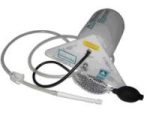
PNEUMOCOLON COMPLETE FOR EXAMINATION IN DOUBLE CONTRAST WALTER KREBS
Bock sealed composed of a tank for the contrast, pear Richardson, a hose and a field for carrying
Out the examinations of the colon in double contrast.
For routine barium irrigation or double contrast enema examination...
Pneumocolon is a lightweigh apparatus which is easy to handle....
Examinations with double contrasts are carried out simply by turning the apparatus....
Constant pressure gives a controlled flow of barium meal… | The ANATOM 64 CT scanner is the latest innovation for cardiac imaging based on Precision Platform system. The excellent design of Ahart technology which innovatively combined single spiral scan + gated imaging + mA modulation for easy heart imaging at extremely low radiation dose. We provide you ANATOM 64 Clarity/Precision of two models which are low/high configurations for preferences. It also offers you conventional clinical applications of low dose, better image quality and faster exams.
Features:
- Modularized OptiWave HD detector features low-cost & easy maintenance, high spatial resolution and long lifetime
- Admir3D iterative technology delivers optimal dose efficiency and noise reduction without compromising image quality
- High configurations of main components ensure the best results and maximum patient throughput
- Uniquely and creatively uses 140kV and 80kV dual energy scan mode for brain imaging on 16-slice CT to offers you extraordinary image quality both in low and high density resolutions
- AdoseTM mA modulation ensures you low dose imaging without compromising image quality particularly useful in pediatric applications
- Equipped with dedicated Abast and Amast for bone and metal artifacts
- The brilliant Ahart technology enables you to experience so easy and low-dose cardiac imaging applications
Technical Specifications:
| Model |
ANATOM 64 Precision |
ANATOM 64 Fit |
| Rack type |
Low pressure slip ring |
Low pressure slip ring |
| Scan aperture |
70cm |
70cm |
| Rack Physical inclination |
± 30 ° |
N.A |
| Rack digital inclination |
± 50 ° |
± 50 ° |
| cooling method |
Air-cooled |
Air-cooled |
| Focus to the center distance |
56 cm |
53 cm |
| |
|
|
| Maximum power (non-equivalent) |
80kW |
42kW |
| Votage (kV) |
80kV / 100kV / 120kV / 140kV |
70kV / 80kV / 100kV / 120kV /
140kV |
| |
|
|
| Heat capacity |
8MHU |
5.0MHU |
| Heat dissipation rate |
931 kHU / min |
748kHU / min |
| cooling method |
Oil cool |
Oil cool |
| Large focus size |
1.1mm × 1.2mm |
1.2mm × 1.4mm |
| Small focus size |
0.6mm × 1.2mm |
0.7mm × 0.8mm |
| Tube current range |
10-670mA |
10-350mA |
| |
|
|
| Detector type |
Optiwave detectors |
Optiwave detectors |
| Number of Z-axis |
32 |
32 |
| The width of the Z-axis |
20mm |
20mm |
| The number of elements per row |
912 |
848 |
| Total number of detectors |
29184 |
27136 |
| Acquisition mode |
64x0.625, 32x0.625,
16x0.625 |
64x0.625, 32x0.625,
16x0.625 |
| |
|
|
| Scanning range |
1800mm |
1800mm |
| Horizontal positioning accuracy |
± 0.25mm |
± 0.25mm |
| weight capacity |
205kg |
205kg |
| Minimum height |
43cm |
43cm |
| Anti - collision protection device |
Yes |
Yes |
| Foot control switch |
Yes |
Yes |
| IV rack |
Yes |
Yes |
| |
|
|
| CPU |
3.5GHz |
3.5GHz |
| RAM |
16 GB × 4 |
16 GB × 4 |
| Hard drive capacity |
1T × 2 |
1T × 2 |
| Display size |
24 inch LCD monitor |
24 inch LCD monitor |
| Display resolution |
1920 × 1200 |
1920 × 1200 |
| Computer operating system |
Windows 7 |
Windows 7 |
| Image reconstruction speed |
65 frames/ second |
65 frames/ second |
| Number of image store |
1000000 |
1000000 |
| Data external storage mode |
CD / DVD / USB |
CD / DVD / USB |
| |
|
|
| Minimum Scan Time of 360 degree |
0.39sec |
0.75sec |
| Sub-millimeter acquisition layers |
64 |
64 |
| Double sub-millimeter acquisition
layers |
64 |
64 |
| Thinnest acquisition thickness |
0.625mm |
0.625mm |
| The thinnest reconstruction
thickness |
0.3125mm |
0.625mm |
| Conventional reconstruction
thickness (mm) |
0.3125 mm, 0.625 mm, 1.25 mm, 2.5
mm, 5.0 mm, 7.5 mm, 10 mm |
0.625 mm, 1.25 mm, 2.5 mm, 5.0
mm, 7.5 mm, 10 mm |
| The reconstruction matrix |
512 x 512, 1024 x 1024 |
512 x 512, 1024 x 1024 |
| Display matrix |
1024 × 1024 |
1024 × 1024 |
| Max FOV |
52cm |
50cm |
| The maximum display field of view |
70cm |
50cm |
| Maximum scan length |
1800mm |
1800mm |
| Maximum continuous helix scan
time |
120s |
120s |
| Pitch range |
0.5-1.5 |
0.5, 1.0, 1.5 |
| |
|
|
| High contrast resolution |
21 Lp / cm @ 0% MTF |
21 Lp / cm @ 0% MTF |
| Low contrast resolution |
2mm @ 0.3% |
2mm @ 0.3% |
| Image noise |
≤ 0.25 |
≤ 0.29 |
| |
|
|
| MPR |
Yes |
Yes |
| CPR |
Yes |
Yes |
| SSD |
Yes |
Yes |
| VR |
Yes |
Yes |
| MIP |
Yes |
Yes |
| MinIP |
Yes |
Yes |
| Virtual endoscopy |
Yes |
Yes |
| CT angiography |
Yes |
Yes |
| Tissue segmentation |
Yes |
Yes |
| One-key bone removal |
Yes |
Yes |
| Automatically patient table removal |
Yes |
Yes |
| Contrast Agent Automatic Tracking
Technology- bolus tracking |
Yes |
Yes |
| Automatic linkage trigger
technology |
Yes |
Yes |
| Cine mode display |
Yes |
Yes |
| Bone artifact suppression technique |
AbastTM |
AbastTM |
| Metal artifact suppression technique |
AbastTM |
AbastTM |
| Iterative reconstruction technique |
Admir3D global iteration |
Admir3D full-domain iteration |
| Low - dose children 's scanning
technology |
Yes |
Yes |
| Low - dose lung scan |
Yes |
Yes |
| Gray matter enhancement
technology |
AccuHead |
AccuHead |
| High resolution imaging of the lung |
AccuLung |
AccuLung |
| Inner ear high resolution imaging |
AccuOtica |
AccuOtica |
| Body high resolution imaging |
AccuBody |
AccuBody |
| Bone high resolution imaging |
AccuBone |
AccuBone |
| Head dual-energy imaging |
Ahead |
Ahead |
| CT perfusion imaging |
Optional |
Optional |
| Quantitative analysis of blood
vessels |
Optional |
Optional |
| Heart coronary artery imaging |
Aheart |
N.A |
| ECG gated |
Yes |
N.A |
| Low dose cardiac scan |
Yes |
N.A |
| |
|
|
| Green energy saving technology |
AccuSaving |
AccuSaving |
| Dual-energy scan technology |
Optional |
Optional |
| JADE Mobile X-ray machine is one of the lightest portable X-ray systems on the market, allowing it to be used in any imaginable way including bedside, operating rooms, intensive care units and veterinary fields. With a simple, easy-to-use operator console, three-way control, two-step foldable stand and auto-lock system, the JADE Mobile X-ray machine is a user-friendly portable X-ray system.
Convenient & Intuitive Operation:
JADE is one of the lightest portable X-ray systems on the market, allowing it to be used in any imaginable way including bedside, operating rooms, intensive care units and in veterinary fields. With a simple, easy-to-use operator console, three-way control, two-step foldable stand and auto-lock system, JADE is a user-friendly portable X-ray system.
Compact & Powerful Design:
JADE Mobile X-ray machine is an innovative, highly versatile portable X-ray system suitable for a variety of clinical uses. Utilizing the unique technology used in DRGEM’s universally recognized X-ray generators, JADE is a compact but powerful unit with a 4kW output and thoughtfully designed components to increase efficiency and maximize workflow. The core part of X-ray source adopts high-quality tube assembly, X-ray collimator and high frequency X-ray generator with excellent performance, lifetime and stability.
Features:
- Vehicle loadable
- Wheel lock
- Automatic tube arm lock at any angle
- Storage space for cassettes or detectors
- User Programmable APR, save up to 9 APR settings
- Three way X-ray exposure
- USB interface & Bluetooth
- Remote control (Option)
- 5kg including X-ray unit, collimator and stand
- Maximum hight of 228.6cm
- Exposure Hand Switch
- Foldable, two-step stand
Technical Specification:
- Power Rating - 4kW,
- 100kHz, high frequency X-ray Generator
- kVp Range - Maximum 140kVp
- mAs Range - 0.1-250mAs
- mA Range – 10 to 100mA
- 330~2000mm FD
- Collimator with 30 seconds LED lamp timer
| OPENMARK 5000 is 0.51T MRI. It's approved by FDA and has CE mark. It adopts two-pillar magnet design with 280 degree openness and equipped with powerful
RF and gradient system, together with advanced imaging technology, making it as a high-end system which is comparable to high-field MRI.
Features:
- With the highest system stability and the highest homogeneity of the
magnet field in permanent MRI
- Screens on both sides facilitate positioning; 280 degree two-pillar magnet
design ensures stable magnet structure and facilitates interventional
treatment.
- Active and passive shimming calibrate technology ensures the magnetic
field uniformity
- Motor-driven patient couch makes it easier for patients to access and for
positioning
- Powerful hardware and software platforms ensure the scan speed, image
quality and make it possible for advanced imaging functions
- Fast scan speed eliminates motion artifact
- Rich scan sequences, advanced imaging technology and powerful postprocessing
technology ensure image quality, extend more applications,
which can fully satisfy the clinical needs
- Intelligent user-friendly operating system ensures you easy operation
Technical Specifications:
| No. |
Technique Description |
Parameter |
| 1 |
Magnet System |
|
| 1.1 |
Magnet Type |
Permanent Magnet
Automatic constant temperature
system |
| 1.2 |
Field Strength |
0.51T |
| 1.3 |
Magnet Shape |
Dual-pillar shape |
| 1.4 |
Homogeneity(40cm,DSV,VRMS) |
≤1.6ppm |
| 1.5 |
Shim Method |
Active/Passive |
| 1.6 |
Magnet Vertical Gap (Cover) |
40cm |
| 1.7 |
Magnetic Pole Dia. (Exclude Cover) |
145cm |
| 1.8 |
Accessibility(Horizontal Opening Angle, |
280° |
| 1.9 |
5 Gauss fringe field |
X-axis:horizontal ≤2.5m
Y-axis:Vertical ≤2.5m
Z-axis:horizontal ≤2.5m |
| 2 |
Patient Couch and Communication |
|
| 2.1 |
Patient Couch Driven mode |
Motor-driven |
| 2.2 |
Max. Patient Weight |
≥200kg(440lbs) |
| 2.3 |
Patient Positioning Tools |
Laser Light Localizer for positioning of center slice Motor-driven transfer to center of imaging volume |
| 2.4 |
Position accuracy |
±1mm |
| 2.5 |
Emergency Call Key |
Yes |
| 2.6 |
Intercom System |
Yes |
| 3 |
Gradient System |
|
| 3.1 |
Gradient Field Strength(Single Axis) |
≥30mT/m |
| 3.2 |
Gradient Slew Rate (Single Axis) |
≥100mT/m/ms |
| 3.3 |
Rise Time |
≤0.3ms |
| 3.4 |
Gradient Cooling System ( Gradient coils
and Power electronics) |
Air Cooling |
| 4 |
RF System |
|
| 4.1 |
RF System Type |
Digital Transmit and
Receive signal |
| 4.2 |
Number of RF Channels |
4 |
| 4.3 |
Transmitter Amplifier Peak Power |
6kW |
| 4.4 |
RF Bandwidth of Receiver |
≥1.25MHz |
| 4.5 |
Head Coil |
Yes |
| 4.6 |
Neck Coil |
Yes |
| 4.7 |
Body/Spine Coil (17 inch) |
Yes |
| 4.8 |
Body/Spine Coil (21 inch) |
Yes |
| 4.9 |
Knee Coil |
Yes |
| 4.10 |
Shoulder Coil |
Yes |
| 4.11 |
Flexible Coil |
Optional |
| 4.12 |
Breast Coil |
Optional |
| 5 |
Computer System |
|
| 5.1 |
Host Computer |
DELL Computer (for MR) |
| 5.2 |
System Software |
Windows XP |
| 5.3 |
Operation Software |
APEX |
| 5.4 |
CPU Clock rate |
3.0GHz |
| 5.5 |
Main Memory |
4GB |
| 5.6 |
Color LCD Monitor |
19” |
| 5.7 |
Keyboard and Mouse |
Standard |
| 5.8 |
Image Reconstruction Speed(256 x 256
Matrix) |
200 frame/Sec. |
| 5.9 |
Hard Disk |
500GB |
| 5.10 |
Image Storage Capacity(256 x 256
Matrix) |
500,000 |
| 5.11 |
Media Driver |
DVD RW |
| 5.12 |
DICOM 3.0 |
Yes |
| 5.13 |
Ethernet |
Yes |
| 5.14 |
Operation Console |
Yes |
| 5.15 |
Operation Chair |
Yes |
| 6 |
Scanning Parameter |
|
| 6.1 |
Max. FOV |
410mm |
| 6.2 |
Min. FOV |
5mm |
| 6.3 |
Min. TE(SE) |
5ms |
| 6.4 |
Min. TR(SE) |
11ms |
| 6.5 |
Min. TE(GR) |
1ms |
| 6.6 |
Min. TR(GR) |
3ms |
| 6.7 |
Min. 2D Thickness |
1.0mm |
| 6.8 |
Min. 3D Thickness |
0.1mm |
| 6.9 |
Max. Image Matrix |
512x512 |
| 7 |
Scanning Sequence & Imaging Technique |
|
| 7.1 |
Spin Echo 2D/3D (SE 2D/3D) |
Yes |
| 7.2 |
DE/QE |
Yes |
| 7.3 |
Fast Spin Echo 2D/3D(FSE 2D/3D) |
Yes |
| 7.4 |
Single Shot FSE 2D/3D |
Yes |
| 7.5 |
Inversion Recovery(IR) |
Yes |
| 7.6 |
Fast Inversion Recovery(FIR) |
Yes |
| 7.7 |
Gradient Echo 2D/3D(GR 2D/3D) |
Yes |
| 7.8 |
Fast GR 2D/3D |
Yes |
| 7.9 |
SPGR |
Yes |
| 7.10 |
FLAIR |
Yes |
| 7.11 |
Fat Imaging |
Yes |
| 7.12 |
Fat Suppression imaging |
Yes |
| 7.13 |
Water-Fat Separation imaging |
Yes |
| 7.14 |
TOF MRA(2D/3D) |
Yes |
| 7.15 |
MRCP(2D/3D) |
Yes |
| 7.16 |
MRU (2D/3D) |
Yes |
| 7.17 |
MRM |
Yes |
| 7.18 |
Fast Hydrograph Imaging |
Yes |
| 7.19 |
Diffusion Weighted Imaging(DWI) |
Yes |
| 7.20 |
Max. b Value |
1000s/mm2 |
| 7.21 |
Breath Hold Technology |
Yes |
| 7.22 |
Magnetization Transfer Contrast(MTC) |
Yes |
| 7.23 |
Multi-slice and Angle-free Presaturation |
Yes |
| 7.24 |
Saturation Tracking |
Yes |
| 7.25 |
Maximum Intensity Projection(MIP) |
Yes |
| 7.26 |
Multi-Angle Projection(MAP) |
Yes |
| 7.27 |
3D Reconstruction |
Yes |
| 7.28 |
Multi-planar Reconstruction(MPR) |
Yes |
| 7.29 |
Multi-Artifacts Eliminating technology |
Yes |
| 7.30 |
Checking with Part Metal Implant |
Yes |
| 7.31 |
Online Image Filtration |
Yes |
| 7.32 |
Online Post Procession |
Yes |
| 7.33 |
3D Scout |
Yes |
| 7.34 |
Scanning Protocol Preset |
Yes |
| 7.35 |
Scanning Protocol Queue Waiting |
Yes |
| 7.36 |
Advanced Image Post Processing |
Yes |
| 7.37 |
Image Fusion Technology of Vascular |
Yes |
| 7.38 |
Image Fusion Technology of Spine |
Yes |
| DrGem GXR-SD 400mA Floor Mounted Digital X-ray system matches with a radiographic room which perfectly fits your workow and can be easily upgraded to DR system with the help of DR interface and PC interface in GXR generator as well as Bucky suitable to Flat Panel Detector. GXR X-ray system is equipped with a high frequency X-ray generator which consistently produces high quality radiograph in favor of high quality X-ray output with a very small kV ripple and accurate mA and mAs. GXR X-ray system is designed to provide convenience to operator and comfort to patient
Features of DrGem GXR-SD 400mA Floor Mounted Digital X-ray:
- PBT-6 is a 4 way Motorized Tabletop with Elevating feature (66cm). A large tabletop with extended travel enables all radiography studies with minimal patient movement. Fully fat tabletop without a frame on the edge makes cleanliness and odors free
- Automatic Stitching - GXR-SD system provides outstanding automatic stitching function with Source tilting method
- Digital Flat Panel Detector (FPD) – Wireless 17X14 (Csl, 4336W) with Auto Exposure Detection (AED) function, there is no DR trigger cable between detector and generator.
- Full Featured Imaging Software & Excellent Digital Image Processing:
- Provides convenient user interface and easy operation
- Anatomical view-based digital image processing automatically optimizes and enhances the quality of the captured image for the pictured anatomy.
- Radiographic stand & automatic collimator control function
- DICOM 3.0 networking interface includes Worklist, Print, Store, Query for integration with any PACS or RIS
- Included – Software, HP Laptop Computer
- CPU≥3.2GHz
- Memory capacity:≥4GB
- Hard drive capacity :≥500 GB
- Resolution: 1280 x 1024
- Display size: 21 inch color LCD screen
- 64 bit Windows 10 operation system
- Core: i5
Technical Specifications of DrGem GXR-SD 400mA Floor Mounted Digital X-ray:
- Power Rating - 32KW
- Generator - GXR-32S
- Rotor - Dual Speed Starter(DSS)
- Input Power - 400/480VAC, Three phase
- Line Frequency - 50/60Hz
- X-ray tube - DXT-12M, (0.6/1.2mm, 300kHU)
- Tube Voltage - 40 to 150kV, 1kV Step
- Tube Current – 10 to 640mA
- Output - 640mA@81kV, 500mA@104kV, 400mA@130kV, 320mA@150kV
- Time Range - 1ms to 10s
- mAs Range - 0.1 to 800mAs
- Reproducibility - Coecient of Variation : kV < 0.005, Time < 0.005,mAs < 0.01
- Accuracy - kV < ±(1%+1kV), mA < ±(3%+1mA), Time <±(1%+0.5ms), mAs < ±(3%+0.1mAs)
- Linearity - Coecient of Linearity < 0.01 : CL = (X1-X2)/(X1+X2), where X is mR/mAs
|
- Length: 355 mm
- Height: 560 mm
- Diameter: 150 mm
- Power: 30 w
- Scattering diffusion: Greater than 0.9
|


Reviews
There are no reviews yet.