
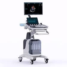
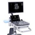
Premium Diagnostic Ultrasound System PU-MT241A
$0.00
Shipped From Abroad
Delivery & Availability:
Typically 10-21 working days – excluding furniture and heavy/bulky equipment. Please contact us for further information.
Typically 10-21 working days – excluding furniture and heavy/bulky equipment. Please contact us for further information.
Description
Ultrasound General imaging Images
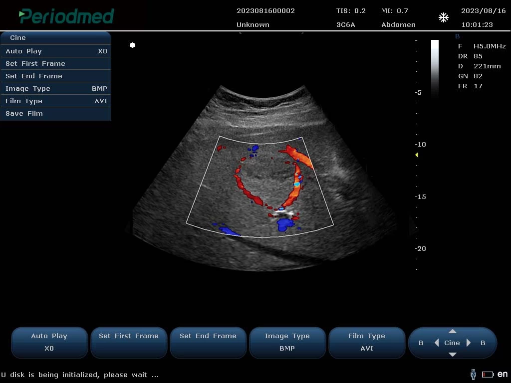
Convex Probe-Color Mode-Liver6
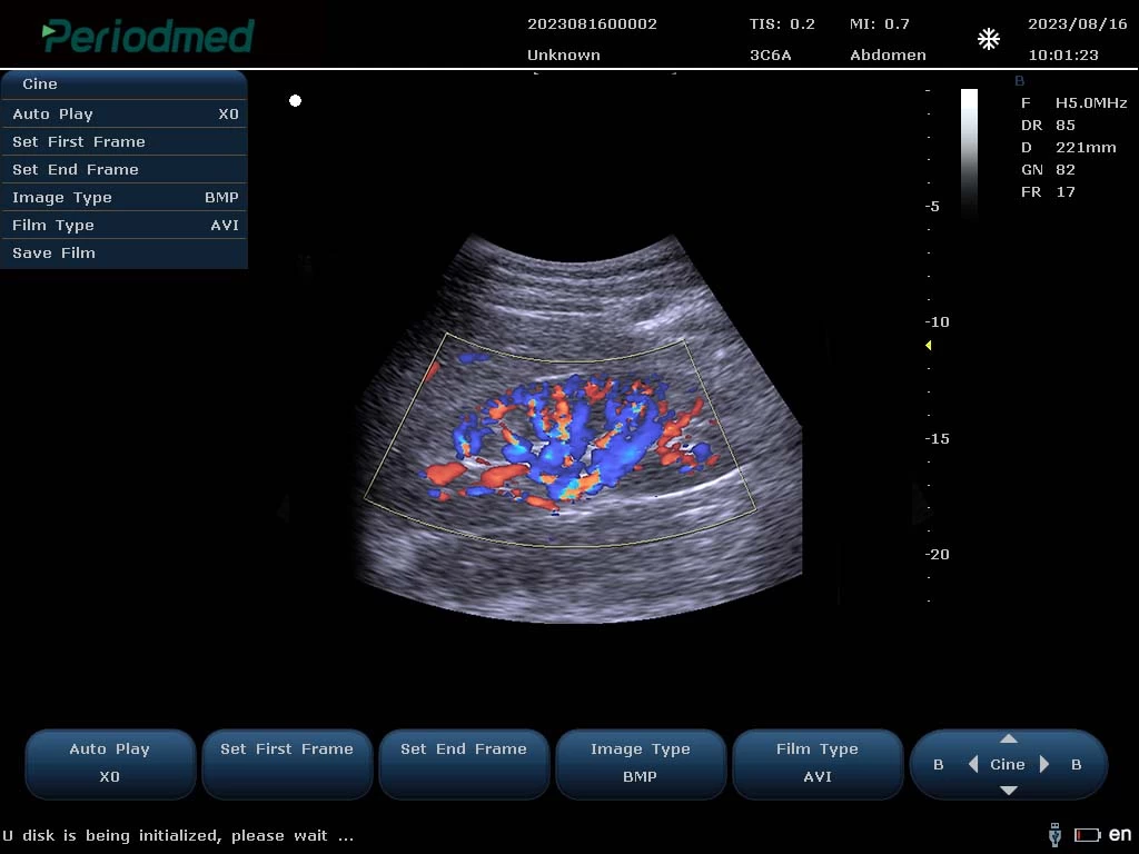
Convex Probe-Color Mode-Kidney 1
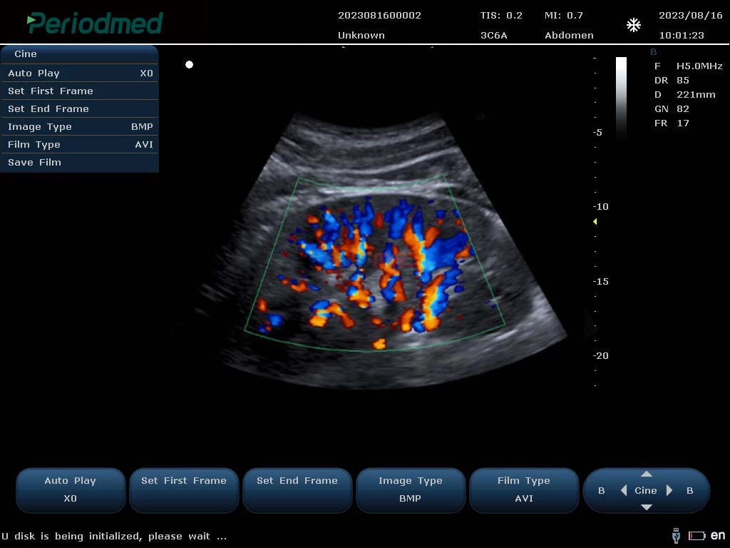
Convex Probe-Color Mode-Kidney 2
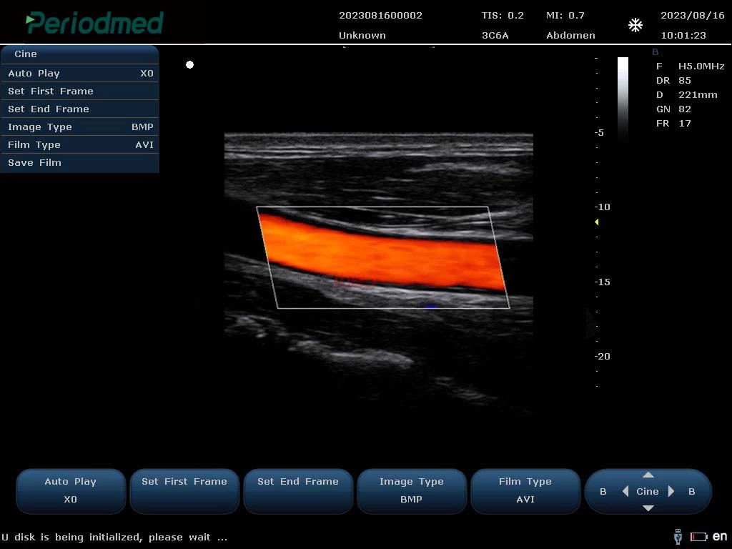
Ultrasound Carotid Images
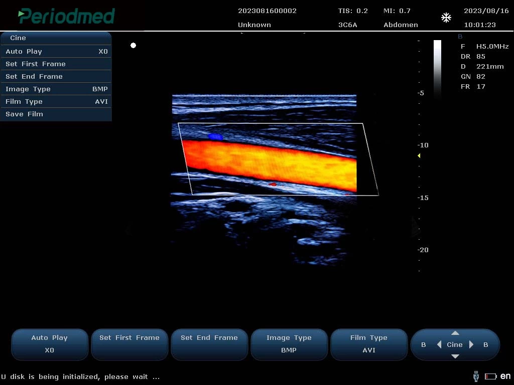
Linear Probe-Color Mode-Carotid1
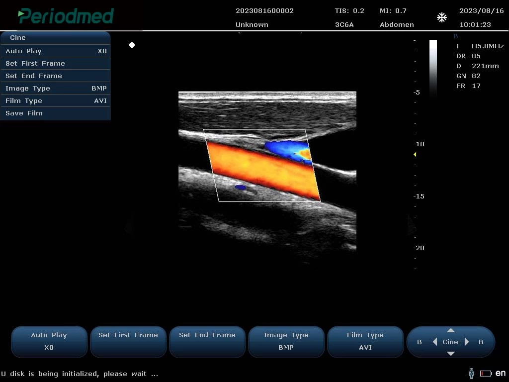
Linear Probe-Color Mode-Carotid2
Linear Probe-Color Mode-Carotid3
Ultrasound Cardiovascular Images
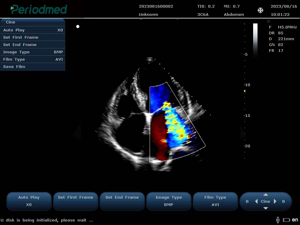
Phased Array Probe-Color Mode-Cardiac1
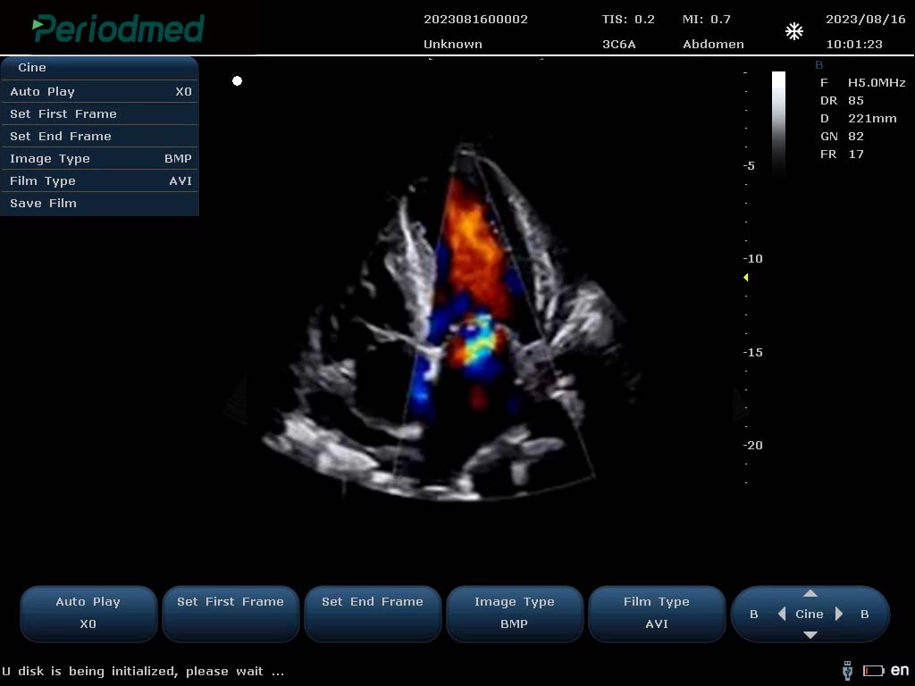
Phased Array Probe-Color Mode-Cardiac2
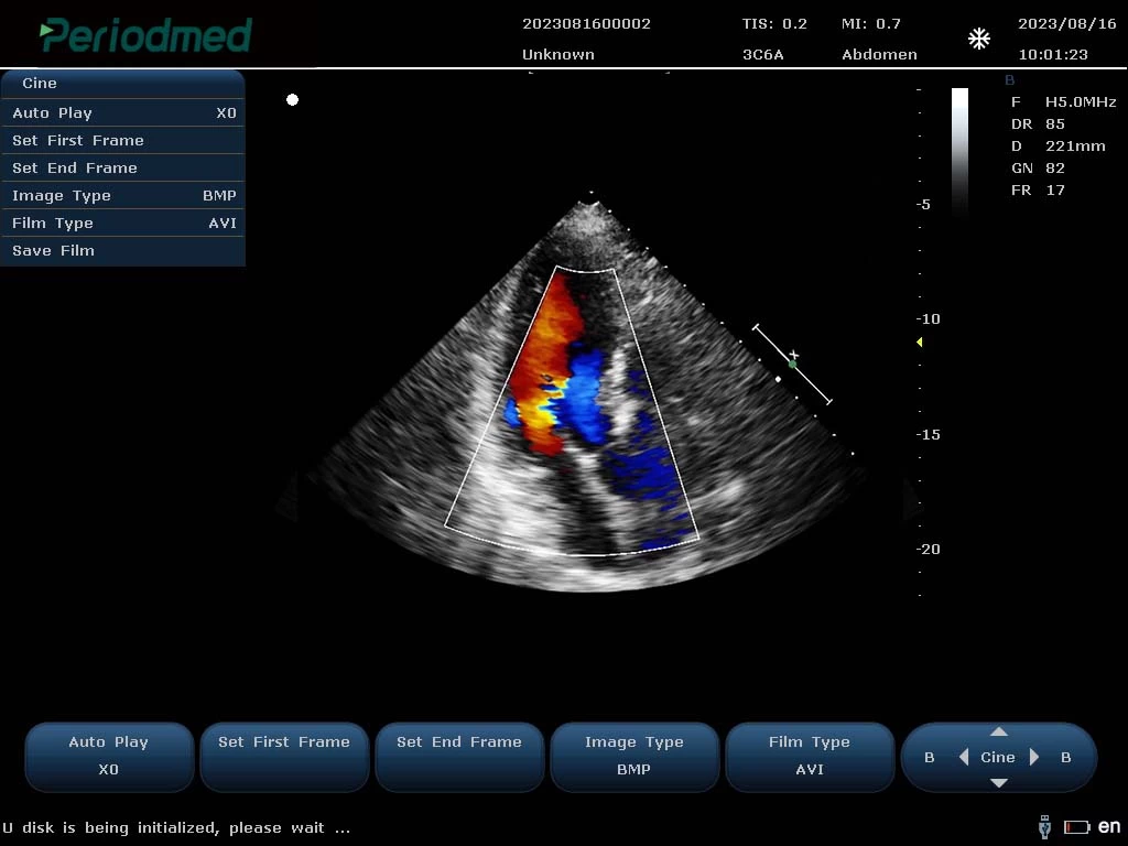
Phased Array Probe-Color Mode-Cardiac3
PU-MT241A Specification
| Multiple Display Modes: | B/2B/4B/BMM/PDI/DPDICF/CW/B+CF/DPDI+PW/B+CF+PW |
| Product Function: | Color Flow(CF) Power Doppler(PDl) Pulsed Doppler(PW) Directional Power Doppler (DPDl) Continuous Doppler (CW) Tissue Doppler (TDI) Color M Type,Anatomical M Type |
| Imaging Technology: | High Pulsed Repetition Frequency(Hprf) Tissue Harmonic Imaging(Thi) Pulsed Reverse Harmonic Imaging(Ithi) Tissue Specific Imaging(Tsi) Spatial Composite Imaging Picture In Picture Imaging Mode |
| Advanced Technology: | 1.Point-By-Point Focusing Application Technology Replaces The Traditional 1-4 Point Multi-Point Transmission Focusing Mode,Making The Transmission Focusing More Accurate 2.The Soft Beamformer Technology Of Gpu Architecture Provides More Powerful Computing Power For The Implementation Of The New Algorithm,And Fundamentally Improves The Quality Of Ultrasound Image 3.192 Elements 128 Channels,With Sound Speed Calibration Technology,16 Beam Compound Algonithm To Provide Dynamic And Accaurate Calculation Parameters,Effectively Improve The Focusing Accuracy 4.The Fast Sound Velocity Calibration Technology Can Automatically Correct The Actual Sound Velocity Of Different Human Bodies And Different Tissues In Real Time,And Provide Dynamic And Accurate Calculation Parameters For Beam Synthesis Algorithm |
| Optional: | Wide Field Imaging Puncture Enhancement Contrast Imaging Elastography Imaging 3D/4D Imaging |
Quick Comparison
| Premium Diagnostic Ultrasound System PU-MT241A remove | ASPEL AsCARD Green B/W ECG Machine remove | Sonoscape S22 Ultrasound Machine remove | Bistos BT-770-12.1" Touchscreen Patient Monitor remove | Littman Stethoscope remove | ASPEL AsCARD Green ECG Machine remove | |||||||||||
|---|---|---|---|---|---|---|---|---|---|---|---|---|---|---|---|---|
| Name | Premium Diagnostic Ultrasound System PU-MT241A remove | ASPEL AsCARD Green B/W ECG Machine remove | Sonoscape S22 Ultrasound Machine remove | Bistos BT-770-12.1" Touchscreen Patient Monitor remove | Littman Stethoscope remove | ASPEL AsCARD Green ECG Machine remove | ||||||||||
| Image | 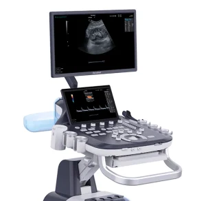 |  |  | 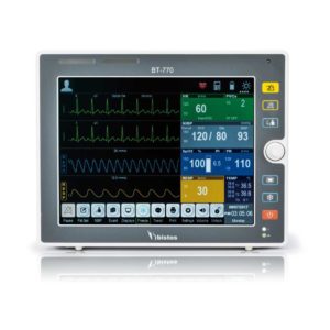 |  | 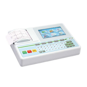 | ||||||||||
| SKU | SF1033560130124-1 | SF1033560075-8 | SF1033560012-3 | SF1033560059-1 | SF1033560084-5 | SF1033560075-9 | ||||||||||
| Rating | ||||||||||||||||
| Price |
|
| $9,350.00 | $902.00 | $13.20 |
| ||||||||||
| Stock | ||||||||||||||||
| Availability | ||||||||||||||||
| Add to cart | ||||||||||||||||
| Description | Shipped From Abroad
Delivery & Availability:
Typically 10-21 working days – excluding furniture and heavy/bulky equipment. Please contact us for further information.
| Shipped from Abroad AsCARD Green electrocardiograph is a 1- and 3-channel ECG unit which enables to make electrocardiogram in full 12 leads. Intended for ECG examinations of adult and paediatric patients aimed at identification of cardiological abnormalities, myocardial ischaemia or infarction. The device is intended for use in healthcare facilities by duly trained personnel. ECG examination may be recorded in manual or automatic mode with the ability to perform the analysis and interpretation. Delivery & Availability: Typically 10 working days – excluding furniture and heavy/bulky equipment. Please contact us for further information. | Shipped from Abroad As SonoScape steps forward to add value and efficiency to ultrasound, the latest S22 was designed in a user-friendly platform to address current and future demanding needs. It represents an excellent mix in performance and price. Delivery & Availability: Typically 5-7 working days – excluding furniture and heavy/bulky equipment. Please contact us for further information. | Shipped from Abroad The Bistos BT-770 patient monitor is equipped with a 12.1" touchscreen display, which allows for an easy operation and readability with a powerful rechargeable battery guaranteeing a continuous operation of 5 hours to monitor ECG, SpO2, NIBP, temperature and respiration Delivery & Availability: Typically 14 working days – excluding furniture and heavy/bulky equipment. Please contact us for further information. | In Stock
Delivery & Availability: Typically 2 working days – excluding furniture and heavy/bulky equipment. Please contact us for further information. | Shipped from Abroad AsCARD Green v.06.101 is a 1-, 3-, 6- and 12-channel ECG unit which enables to make electrocardiogram in full 12 leads. Intended for ECG examinations of adult and paediatric patients aimed at identification of cardiological abnormalities, myocardial ischaemia or infarction. The device is intended for use in healthcare facilities by duly trained personnel. ECG examination may be recorded in manual or automatic mode with the ability to perform the analysis and interpretation. Delivery & Availability: Typically 10 working days – excluding furniture and heavy/bulky equipment. Please contact us for further information. | ||||||||||
| Content | https://youtu.be/K2dzsICPG_s
Ultrasound General imaging Images
Convex Probe-Color Mode-Liver6
Convex Probe-Color Mode-Kidney 1
Convex Probe-Color Mode-Kidney 2 Ultrasound Carotid Images
Linear Probe-Color Mode-Carotid1
Linear Probe-Color Mode-Carotid2
Linear Probe-Color Mode-Carotid3 Ultrasound Cardiovascular Images
Phased Array Probe-Color Mode-Cardiac1
Phased Array Probe-Color Mode-Cardiac2
Phased Array Probe-Color Mode-Cardiac3 PU-MT241A Specification
| AsCARD Green electrocardiograph is a 1- and 3-channel ECG unit which enables to make electrocardiogram in full 12 leads. Intended for ECG examinations of adult and paediatric patients aimed at identification of cardiological abnormalities, myocardial ischaemia or infarction. The device is intended for use in healthcare facilities by duly trained personnel. ECG examination may be recorded in manual or automatic mode with the ability to perform the analysis and interpretation.
Electrocardiograph is based on advanced microprocessor technology. It is equipped with a thermal printer with high-resolution head and graphical LCD display. A hightech membrane keyboard makes the AsCARD Green device operation intuitive, and its menu navigation exceptionally easy. This light-weight, small-footprint and battery powered cause that device can be easily transported to any location. With plastic casing and foil covered keyboard, the device is neat and easy to clean.
Technical Specifications:
Click Here To Download Catalogue | DETAILS
As SonoScape steps forward to add value and efficiency to ultrasound, the latest S22 was designed in a user-friendly platform to address current and future demanding needs. It represents an excellent mix in performance and price.
S22, is a shared service ultrasound system with a slim and elegant package that has combined mobility with utility to fit in specific clinical situations including emergency department, ICU, operating room and so on. Furthermore, its ergonomic design, easy operating and flexible data management will give you a memorable experience.
SPECIFICATION
• Large high-resolution widescreen LED
• Sensitive touch screen
• Four transducer sockets plus one socket for pencil probe
• A comprehensive selection of probes: linear, Convex, Micro-convex, Volumetric, Endocavity, Bi-plane, Phased Array, TEE, Intraoperative, Pencil
• Premium application technology: 4D, μ-scan speckle reduction, compound imaging, Pulse Inversion Harmonic Imaging, Color M-Mode, Steer M-Mode, PDI, TDI, Real-time Panoramic Imaging, Trapezoid Imaging, Auto-IMT…
• Full patient database and image management solutions: DICOM 3.0, AVI/JPG, USB 2.0, HDD, DVD, PDF report
• Multi-Language Input Keyboard
• Built-in battery
Click Here To Download Catalogue |
Bistos BT-770 is a 12.1" touchscreen patient monitor designed for easy operations.
SPECIFICATIONS
Click Here To Download Catalogue | Littman Stethoscope Features:
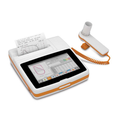
| AsCARD Green v.06.101 is a 1-, 3-, 6- and 12-channel ECG unit which enables to make electrocardiogram in full 12 leads. Intended for ECG examinations of adult and paediatric patients aimed at identification of cardiological abnormalities, myocardial ischaemia or infarction. The device is intended for use in healthcare facilities by duly trained personnel. ECG examination may be recorded in manual or automatic mode with the ability to perform the analysis and interpretation.
Electrocardiograph is based on advanced microprocessor technology .It is equipped with a thermal printer with high-resolution head and 4,3" LCD display. A touch panel and high-tech membrane keyboard makes this device intuitive in usage and its menu navigation exceptionally easy. This light-weight, small-footprint and battery powered cause that device can be easily transported to any location. With plastic casing and foil covered keyboard, the device is neat and easy to clean.
Technical Specifications:
Click Here To Catalogue Download | ||||||||||
| Weight | N/A | N/A | N/A | N/A | N/A | N/A | ||||||||||
| Dimensions | N/A | N/A | N/A | N/A | N/A | N/A | ||||||||||
| Additional information |


Reviews
There are no reviews yet.