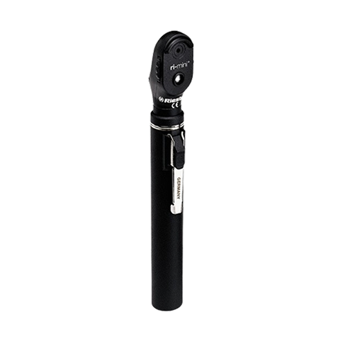
ri-mini® ophthalmoscope
$0.00
Shipped From Abroad
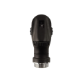
The ri-mini® Ophthalmoscope is a compact, cost-effective instrument featuring 2.5 V Xenon illumination. It includes a focusing wheel with 16 corrective lenses and an aperture hand-wheel with four apertures, housed in an impact-resistant, glass-fibre reinforced plastic casing.
Typically 10-21 working days – excluding furniture and heavy/bulky equipment. Please contact us for further information.
Description
Ideal for clinics and practices, the ri-mini® Ophthalmoscope provides clear fundus visualization with its powerful 2.5 V Xenon (XL) lamp. It features an illuminated dioptre display for precise correction across 16 lenses, an integrated eyeglass protector, and four easy-to-operate apertures. The instrument head is secured by a screw fitting, and the entire unit is made from extremely impact-resistant, glass-fibre reinforced plastic.
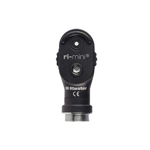
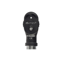
ri-mini® ophthalmoscope details
- Dioptre wheel
- Dioptre display
- Aperture wheel
- On/Off switch
- Battery compartment
Features and benefits of the ri-mini® ophthalmoscope
- ri-mini® with 2.5 V Xenon illumination.
- Screw fitting for quick and secure attachment to the handle.
- Sturdy adjustment ring for positioning the instrument head to the ideal angle on the handle.
- Extremely impact-resistant casing made of glass-fibre reinforced plastic.
- Easy lamp replacement at the base of the instrument head.
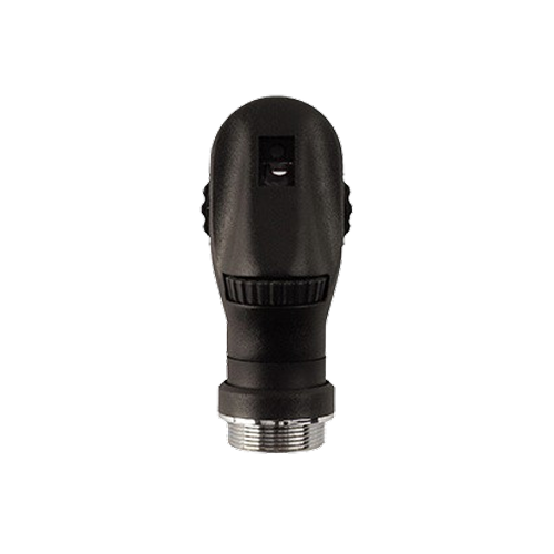
Physician’s side
- Focusing wheel with corrective lenses:
D+ 1 | 2 | 3 | 4 | 6 | 10 | 15 | 20
D- 1 | 2 | 3 | 4 | 6 | 10 | 15 | 20
- Illuminated dioptre display.
- Built-in eyeglass protector.

Patient’s side
- Easy-to-operate aperture hand-wheel.
- Four different apertures:
– Small circle + semi-circle to reduce reflexes in small pupils.
– Large circle for standard examination of the ocular fundus.
– Fixation star for determining central or eccentric fixation.
– Red open filter with a contrast-enhancing effect, for assessing fine vascular changes such as retinal haemorrhages.
Specifications
| Dimensions | .jpg?width=201&height=278&name=Riester%20ri-mini%20ophthalmoscope%20head%20detail%20photo%20(01).jpg) Ophthalmoscope head: 60 mm hight x 26.5 mm wide Ophthalmoscope battery compartment: 107.30 mm height, 20 mm in Ø 23 cm x 14 cm x 4.5 cm in hard case (set) 23 cm x 8.2 cm x 4.5 cm in hard case (single) |
| Magnification lens | The 2.5x magnification lens |
| The lamp for ri-mini® ophthalmoscope | XL, 2.5 V, 750 mA, average life span 16.5 h |
| Battery | 2 Alkaline AA 1.5 V batteries |
| Ambient temperature | 0 °C to 40 °C |
| Relative Humidity | 30% to 70% non-condensing |
| Storage/ Transport Ambient temperature | -10 °C to 55 °C |
| Storage/ Transport relative Humidity | 10% to 95% non-condensing |
Quick Comparison
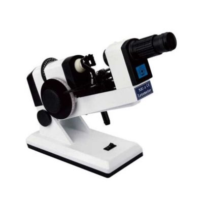
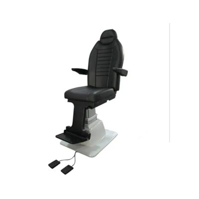
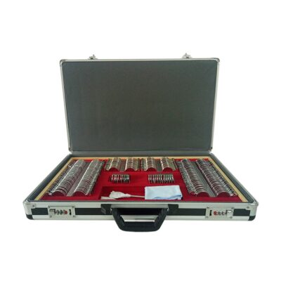
| ri-mini® ophthalmoscope remove | Slit Lamp with Workstation remove | Ophthalmic AB Scan Machine remove | Portable Fundus Camera remove | ENT/Neurosurgery Operating Microscope remove | View Tester (Manual Phoropter) remove | |||||||||||||||||||||||||||||||||||||||||||||||||||||||||||||||||||||||||||||||||||||||||||||||||||||||||||||||||||||||||||||||||||
|---|---|---|---|---|---|---|---|---|---|---|---|---|---|---|---|---|---|---|---|---|---|---|---|---|---|---|---|---|---|---|---|---|---|---|---|---|---|---|---|---|---|---|---|---|---|---|---|---|---|---|---|---|---|---|---|---|---|---|---|---|---|---|---|---|---|---|---|---|---|---|---|---|---|---|---|---|---|---|---|---|---|---|---|---|---|---|---|---|---|---|---|---|---|---|---|---|---|---|---|---|---|---|---|---|---|---|---|---|---|---|---|---|---|---|---|---|---|---|---|---|---|---|---|---|---|---|---|---|---|---|---|---|---|---|---|---|
| Name | ri-mini® ophthalmoscope remove | Slit Lamp with Workstation remove | Ophthalmic AB Scan Machine remove | Portable Fundus Camera remove | ENT/Neurosurgery Operating Microscope remove | View Tester (Manual Phoropter) remove | ||||||||||||||||||||||||||||||||||||||||||||||||||||||||||||||||||||||||||||||||||||||||||||||||||||||||||||||||||||||||||||||||||
| Image | 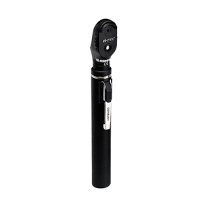 |  | 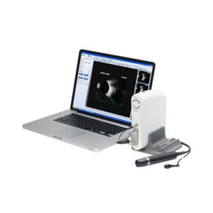 | 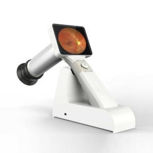 | 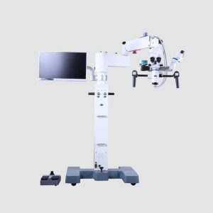 | 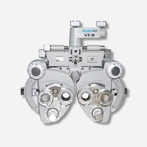 | ||||||||||||||||||||||||||||||||||||||||||||||||||||||||||||||||||||||||||||||||||||||||||||||||||||||||||||||||||||||||||||||||||
| SKU | SF1033560107-7 | SF1033560107-8 | SF1033560107-23 | SF1033560109-1 | SF1033560107-26 | |||||||||||||||||||||||||||||||||||||||||||||||||||||||||||||||||||||||||||||||||||||||||||||||||||||||||||||||||||||||||||||||||||
| Rating | ||||||||||||||||||||||||||||||||||||||||||||||||||||||||||||||||||||||||||||||||||||||||||||||||||||||||||||||||||||||||||||||||||||||||
| Price |
| $3,740.00 | $4,895.00 | $2,310.00 |
| $858.00 | ||||||||||||||||||||||||||||||||||||||||||||||||||||||||||||||||||||||||||||||||||||||||||||||||||||||||||||||||||||||||||||||||||
| Stock | ||||||||||||||||||||||||||||||||||||||||||||||||||||||||||||||||||||||||||||||||||||||||||||||||||||||||||||||||||||||||||||||||||||||||
| Availability | ||||||||||||||||||||||||||||||||||||||||||||||||||||||||||||||||||||||||||||||||||||||||||||||||||||||||||||||||||||||||||||||||||||||||
| Add to cart | ||||||||||||||||||||||||||||||||||||||||||||||||||||||||||||||||||||||||||||||||||||||||||||||||||||||||||||||||||||||||||||||||||||||||
| Description | Shipped From Abroad
The ri-mini® Ophthalmoscope is a compact, cost-effective instrument featuring 2.5 V Xenon illumination. It includes a focusing wheel with 16 corrective lenses and an aperture hand-wheel with four apertures, housed in an impact-resistant, glass-fibre reinforced plastic casing.
Delivery & Availability:
Typically 10-21 working days – excluding furniture and heavy/bulky equipment. Please contact us for further information.
| Shipped from abroad
| Shipped from abroad
| 83Shipped from abroad
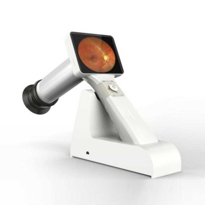
This is a portable medical camera for fundus imaging, diagnosis, and especially for fundus disease screening. It's compact, easy to obtain high definition fundus image. It can be conveniently applied to rapid screening, out diagnosis, bedside diagnosis and remote medical treatment, etc.
| Shipped from abroad
Corder Microscope has Fluid, Responsive and Accurate.Fluid. Responsive. Accurate. These were a few of the principles guiding every phase in the design of the Corder Microscope. With the choicest mechanical machined components, the Corder Microscope has the grace and agility to adjust to every desired position on command. Well designed Apochromatic optics treated with Corder's Mcoatings produce true-to life sharp images with high depth, definition and contrast. | Ship from abroad
| ||||||||||||||||||||||||||||||||||||||||||||||||||||||||||||||||||||||||||||||||||||||||||||||||||||||||||||||||||||||||||||||||||
| Content | Ideal for clinics and practices, the ri-mini® Ophthalmoscope provides clear fundus visualization with its powerful 2.5 V Xenon (XL) lamp. It features an illuminated dioptre display for precise correction across 16 lenses, an integrated eyeglass protector, and four easy-to-operate apertures. The instrument head is secured by a screw fitting, and the entire unit is made from extremely impact-resistant, glass-fibre reinforced plastic.
ri-mini® ophthalmoscope details
Features and benefits of the ri-mini® ophthalmoscope
Physician’s side
Patient’s side
Specifications
| Slit Lamp with Workstation Features:
| Functions of Ophthalmic AB Scan Machine:
| Portable Fundus Camera is a portable medical camera for fundus imaging, diagnosis, and especially for fundus disease screening. It's compact, easy to obtain high definition fundus image. It can be conveniently applied to rapid screening, out diagnosis, bedside diagnosis and remote medical treatment, etc.
Features of Portable Fundus Camera:
| Features:Corder Microscope has Fluid, Responsive and Accurate.Fluid. Responsive. Accurate. These were a few of the principles guiding every phase in the design of the Corder Microscope. With the choicest mechanical machined components, the Corder Microscope has the grace and agility to adjust to every desired position on command. Well designed Apochromatic optics treated with Corder's Mcoatings produce true-to life sharp images with high depth, definition and contrast. More comfortable operation Tiltable binocular tubes available, which can incline more than 60° depending on the posture and physique of the operating surgeon. Movable range: 30° (straight) to 90° (inclined) Corder microscope configured with XYZ motorized movement operated through a comfortable foot /Handle control, a veryeffective co-axial illumnation and 50W halogen light source makes it ideal for Neuro surgeries.Doctor-patient communication is easierTo address digital documentation needs, a host of digital SLR, video camera, and CCD adapters are made available with the ProLine in addition to Corder's proprietary iVu multi-functional imaging solution. 1080P full hd image quality, efficient image management during the operation. Integrate your digital workflow to facilitate case management and facilitate more intuitive patient communication. Technical Permeants: Magnification: motorized zoom system, 1:6 zoom ratio, magnification 3x~16x Focusing range: 50mm Binocular tube: 30°~90° tiltable tube ,(0° ~200° optional) Eyepiece: 12.5x / 10x Objective lens: F 300mm(175mm, 250mm, 350mm optional) pupil distance: 55mm~75mm diopter adjustment: +6D ~ -6D Field of view: Φ74~Φ12mm X-Y translator: Motorized by foot switch or handle controller, ±30mm Assistant tube: 360° Rotating assistant tube Reset functions: YES Illumination System: Coaxial illumination Light source: Halogen lamp Light intensity adjustment: Continuous brightness adjustment 0-100000lux Fiber optic illumination: Dual fiber Field of illumination: Φ50mm Filter: Red free filter, small spot Accessories CCD Camera system: Beam splitter, CCD adapter, CCD, Display XENON LAMP: 150000lux Integrated Video Adapter: SONY / CANON CameraClick Here To Download Catalogue | Features:
| ||||||||||||||||||||||||||||||||||||||||||||||||||||||||||||||||||||||||||||||||||||||||||||||||||||||||||||||||||||||||||||||||||
| Weight | N/A | N/A | N/A | N/A | N/A | N/A | ||||||||||||||||||||||||||||||||||||||||||||||||||||||||||||||||||||||||||||||||||||||||||||||||||||||||||||||||||||||||||||||||||
| Dimensions | N/A | N/A | N/A | N/A | N/A | N/A | ||||||||||||||||||||||||||||||||||||||||||||||||||||||||||||||||||||||||||||||||||||||||||||||||||||||||||||||||||||||||||||||||||
| Additional information | ||||||||||||||||||||||||||||||||||||||||||||||||||||||||||||||||||||||||||||||||||||||||||||||||||||||||||||||||||||||||||||||||||||||||


Reviews
There are no reviews yet.