S-scan-Musculoskeletal MRI System
$0.00
Shipped From Abroad
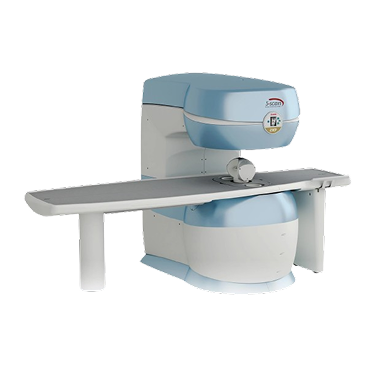
The complete “in-office” musculoskeletal MRI system
S-scan is designed with eXP Technology, an optimised musculoskeletal MRI scanner for any practice with a substantial musculoskeletal workload. S-scan covers all musculoskeletal anatomy, from the foot to the shoulders, including the most important spine segments, including L and C-spine. S-scan’s small footprint makes MRI efficient and cost-effective, whether you perform one or twenty examinations per day.
Typically 10-21 working days – excluding furniture and heavy/bulky equipment. Please contact us for further information.
Description
The next step in office MRI
35% FASTER
- Thanks to the speed-up technique
- Thanks to its smart design, it will fit in a small space.
- during acquisition.
- To maximise sensitivity to the specific anatomies under exam.
Easy patient access
Easy patient access is one of the features that distinguishes the S-scan as an ideal system for more advanced applications such as MRI arthrography. The wide opening and compact magnet provide easy access to the patient. S-scan features a very wide, asymmetric rotating table. This wide table not only makes patient positioning very easy, it also gives maximum stability and comfort to the patient for an easy setup and better images.
Imaging & Visualization
The S-scan is optimised for efficiency, patient comfort, and comprehensive MSK imaging.
-
Comprehensive MSK Coverage: It is capable of imaging all MSK anatomy, encompassing joints and the crucial Cervical (C-spine) and Lumbar (L-spine) segments.
-
eXP Technology: It is designed with eXP Technology, which includes the Speed-Up technique to make acquisitions up to 35% faster.
-
“In-Office” Footprint: The smart design is customer-friendly, requiring only 20 m² of space, making it efficient and cost-effective for private practices.
-
Open Access: The system features a wide, asymmetric rotating table and a compact magnet, providing easy patient access and maximising stability and comfort for better image quality. The wide opening also makes it ideal for more advanced applications like MRI arthrography.
-
Low Energy Use: The average power consumption during acquisition is low (2 kW).
Clinical Applications
The S-scan is tailored for MSK professionals who require a high-quality, efficient scanner capable of covering both large joints and the spine.
-
All Musculoskeletal Anatomy: Primary application is the diagnosis of pathologies in all MSK anatomy:
-
Joints: Foot, ankle, knee, hip, wrist, elbow, and shoulder.
-
Spine: Cervical and Lumbar spine.
-
-
Diagnostic Quality: It provides the high-quality diagnostic information needed to make informed decisions regarding bone, ligament, capsular, soft tissue, and even cartilage lesions.
-
Workflow Integration: It is presented as a complete system that allows practices to efficiently manage a substantial MSK workload.
Coils (Probe Types)
The S-scan uses 14 multi-purpose coils designed to maximise sensitivity across the various anatomies it covers.
| Anatomical Area | Specific Coil Types |
| Spine/Hip | Lumbar, Sacral and Thoracic spine coil (4-channels, large size), Lumbar spine coil (4-channels, medium size), Extra-Large Lumbar Spine coil (2-channels, optional), Cervical spine coil (Linear & DPA, optional), Spine/Hip coil |
| Joints/Extremities | Knee coil DPA, Shoulder coil DPA, Shoulder coil linear, Shoulder coil (3-channels, optional), Ankle-Foot coil DPA, Hand-Wrist coil DPA |
| Head/Other | 4-Channel Head Coil (optional), Flex coil, Flex coil linear (optional), TMJ bilateral coil (2-channels, optional) |
Quick Comparison
| S-scan-Musculoskeletal MRI System remove | Instrument Tray remove | Neck Collar Rigid remove | Agary I.V. Cannula remove | IBIS Neeo R9 Digital Surgical C-Arm remove | DrGem GXR-SD 400mA Floor Mounted Digital X-ray remove | ||||||||||||||||||||||||
|---|---|---|---|---|---|---|---|---|---|---|---|---|---|---|---|---|---|---|---|---|---|---|---|---|---|---|---|---|---|
| Name | S-scan-Musculoskeletal MRI System remove | Instrument Tray remove | Neck Collar Rigid remove | Agary I.V. Cannula remove | IBIS Neeo R9 Digital Surgical C-Arm remove | DrGem GXR-SD 400mA Floor Mounted Digital X-ray remove | |||||||||||||||||||||||
| Image | 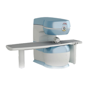 |  |  |  |  |  | |||||||||||||||||||||||
| SKU | SF1033560084-199 | SF1033560084-200 | SF1033560084-221 | SF1033560011-1 | SF1033560074-5 | ||||||||||||||||||||||||
| Rating | |||||||||||||||||||||||||||||
| Price |
| $6.90 | $16.00 | $7.10 |
|
| |||||||||||||||||||||||
| Stock | |||||||||||||||||||||||||||||
| Availability | |||||||||||||||||||||||||||||
| Add to cart | |||||||||||||||||||||||||||||
| Description | Shipped From Abroad
The complete "in-office" musculoskeletal MRI systemS-scan is designed with eXP Technology, an optimised musculoskeletal MRI scanner for any practice with a substantial musculoskeletal workload. S-scan covers all musculoskeletal anatomy, from the foot to the shoulders, including the most important spine segments, including L and C-spine. S-scan’s small footprint makes MRI efficient and cost-effective, whether you perform one or twenty examinations per day. Delivery & Availability:
Typically 10-21 working days – excluding furniture and heavy/bulky equipment. Please contact us for further information.
| In stock
| In stock
Indications:
| In stock
I.V Cannula with wings & injection port 24G/19mm, 0.70mm/20ml/min Injection port with unidirectional valve for facilitating extra medication and preventing back flow. Specially engineered recessed plug with protective ring to avoid risk of contamination. Angle & grooved wings for easy cannulation and to prevent rolling of cannula over patient body. Colour coded cap for easy identification of guage size.
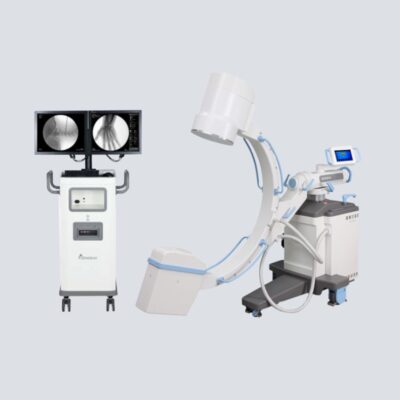
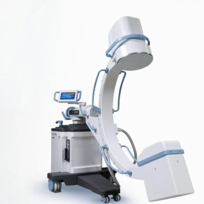
| Shipped from Abroad Our Neeo “C” arms are easy to place, use and are specifically designed to be used in orthopedics, traumatology, abdominal surgery, urology, cardiology and operating rooms. Delivery & Availability: Typically 21 working days – excluding furniture and heavy/bulky equipment. Please contact us for further information. | In Stock The GXR-SD Digital X-ray is a diagnostic digital radiography system that provides reliable high quality digital radiographic images with a reduced dose. The GXR-SD DR systems offer comprehensive digital solutions to all radiography needs, featuring ACQUIDR digital imaging system with stationary or portable digital flat-panel detectors as well as reliable high-frequency x-ray generators that are known worldwide for their excellent performance, lifetime and stability. Patient tables and wall stands are also offered. Delivery & Availability: Typically 21 working days – excluding furniture and heavy/bulky equipment. Please contact us for further information. | |||||||||||||||||||||||
| Content |
https://vimeo.com/963210358?fl=pl&fe=sh
The next step in office MRI
35% FASTER
20 m2-CUSTOMER FRIENDLY
2 Kw AVERAGE CONSUMPTION
14x MULTI-PURPOSE COILS
PATIENT POSITIONING
Easy patient accessEasy patient access is one of the features that distinguishes the S-scan as an ideal system for more advanced applications such as MRI arthrography. The wide opening and compact magnet provide easy access to the patient. S-scan features a very wide, asymmetric rotating table. This wide table not only makes patient positioning very easy, it also gives maximum stability and comfort to the patient for an easy setup and better images. Imaging & VisualizationThe S-scan is optimised for efficiency, patient comfort, and comprehensive MSK imaging.
Clinical ApplicationsThe S-scan is tailored for MSK professionals who require a high-quality, efficient scanner capable of covering both large joints and the spine.
Coils (Probe Types)The S-scan uses 14 multi-purpose coils designed to maximise sensitivity across the various anatomies it covers.
|
| Indications:
| I.V Cannula with wings & injection port 24G/19mm, 0.70mm/20ml/min Injection port with unidirectional valve for facilitating extra medication and preventing back flow. Specially engineered recessed plug with protective ring to avoid risk of contamination. Angle & grooved wings for easy cannulation and to prevent rolling of cannula over patient body. Colour coded cap for easy identification of guage size. | Our Neeo “C” arms are easy to place, use and are specifically designed to be used in orthopedics, traumatology, abdominal surgery, urology, cardiology and operating rooms.
Using Neeo with the RTP (Real Time Processing) option it is possible to perform vascular, urological and cardiological diagnostics. One of the main functions, digital image subtraction, allows to see, as an example, the passage of contrast liquids in a tissue or in a venous or arterial duct; thanks to the possibility of looping, the acquired video can be reproduced several times to monitor more accurately the passage of the fluid within the area in question. Angiographic measurement is another useful function in the vascular field (QA Quantitative Angiography) that allows the measurement of stenoses. Finally, fluoroscopy allows the correct positioning of stents or expanders.
Neeo is used in various interventional and diagnostic procedures in traumatology and orthopedics wards and operating rooms as well. Thanks to low-dose fluoroscopy, it is possible to use the device for positioning bone or subcutaneous grafts, inserting K-wire (Kirschner wire) for stabilization of bone fragments or for the correct positioning of prostheses. The low dose emitted ensures safe use for both the patient and the surgeon or doctor on the operating field.
On the control panel there is a large touch screen display that allows to adjust the basic functions of the equipment. From this display it is possible to select and adjust the fluoroscopic data for the examination, activate or deactivate the laser pointer, select between pulsed, one shot or standard fluoroscopy, rotate the image and perform all operations on collimator. The four side buttons on the display offer the possibility to move the bow vertically thanks to an extremely silent motor.
Neeo has two 19 “medical grade monitors that can be positioned according to the needs of the medical practitioner. Work monitors and feedback monitors are separated to be managed independently. The possible movements are: rotation, revolution, tilting and possibility of height adjustment.
Features:
Click Here To Download Catalogue | DrGem GXR-SD 400mA Floor Mounted Digital X-ray system matches with a radiographic room which perfectly fits your workow and can be easily upgraded to DR system with the help of DR interface and PC interface in GXR generator as well as Bucky suitable to Flat Panel Detector. GXR X-ray system is equipped with a high frequency X-ray generator which consistently produces high quality radiograph in favor of high quality X-ray output with a very small kV ripple and accurate mA and mAs. GXR X-ray system is designed to provide convenience to operator and comfort to patient
Features of DrGem GXR-SD 400mA Floor Mounted Digital X-ray:
Click Here To Download Catalogue | |||||||||||||||||||||||
| Weight | N/A | N/A | N/A | N/A | N/A | N/A | |||||||||||||||||||||||
| Dimensions | N/A | N/A | N/A | N/A | N/A | N/A | |||||||||||||||||||||||
| Additional information |


Reviews
There are no reviews yet.