SonoEye-Echogenic Needle for Ultrasound-Guided Retrobulbar Blocks
$0.00
Shipped From Abroad
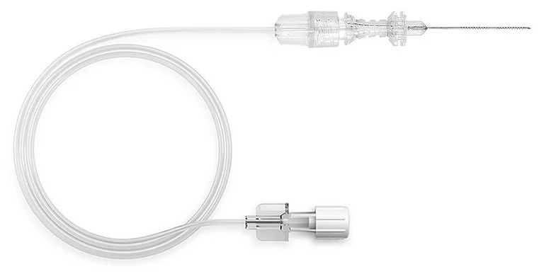
Ultrasound-guided ophthalmic blocks ensure excellent anesthesia with high success rates for eye surgeries. PAJUNK’s SonoEye needle combines the proven Atkinson tip with the innovative Cornerstone Reflectors for optimum needle visibility under ultrasound.
Delivery & Availability:
Typically 10-21 working days – excluding furniture and heavy/bulky equipment. Please contact us for further information.
Typically 10-21 working days – excluding furniture and heavy/bulky equipment. Please contact us for further information.
Description
SonoEye Features & Advantages
Cornerstone Reflectors
360° graduations on the first 20 mm of the needle in two sections:
- Optimized ultrasound visibility of the needle shaft4
- Reliable and optimized needle echogenicity at higher insertion angles.
Echogenic Needle Tip
- Atkinson tip
- Improved needle tip visibility under ultrasound
Quick Comparison
| SonoEye-Echogenic Needle for Ultrasound-Guided Retrobulbar Blocks remove | Ear Irrigation and acumen removal remove | Manual lensmeter remove | Automatic Computer Goldmann (Visual Field Analyzer) remove | Applanation Tonometer remove | Portable Keratometer remove | |||||||||||||||||||||||||||||||||||||||||||||||||||
|---|---|---|---|---|---|---|---|---|---|---|---|---|---|---|---|---|---|---|---|---|---|---|---|---|---|---|---|---|---|---|---|---|---|---|---|---|---|---|---|---|---|---|---|---|---|---|---|---|---|---|---|---|---|---|---|---|
| Name | SonoEye-Echogenic Needle for Ultrasound-Guided Retrobulbar Blocks remove | Ear Irrigation and acumen removal remove | Manual lensmeter remove | Automatic Computer Goldmann (Visual Field Analyzer) remove | Applanation Tonometer remove | Portable Keratometer remove | ||||||||||||||||||||||||||||||||||||||||||||||||||
| Image |  | 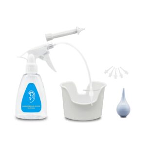 | 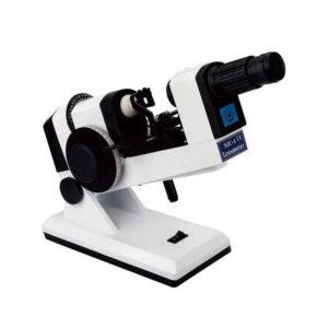 | 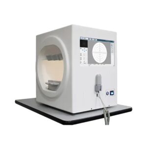 | 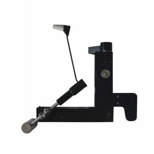 | 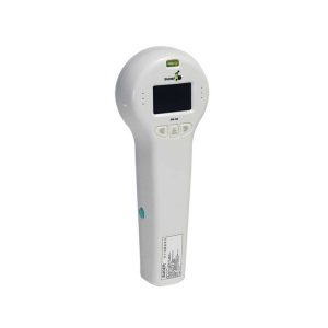 | ||||||||||||||||||||||||||||||||||||||||||||||||||
| SKU | SF1033560130169-9 | SF103356013012 | SF1033560107-22 | SF103356013013 | SF1033560107-1 | SF1033560107-16 | ||||||||||||||||||||||||||||||||||||||||||||||||||
| Rating | ||||||||||||||||||||||||||||||||||||||||||||||||||||||||
| Price |
|
|
| $3,850.00 |
|
| ||||||||||||||||||||||||||||||||||||||||||||||||||
| Stock | ||||||||||||||||||||||||||||||||||||||||||||||||||||||||
| Availability | ||||||||||||||||||||||||||||||||||||||||||||||||||||||||
| Add to cart | ||||||||||||||||||||||||||||||||||||||||||||||||||||||||
| Description | Shipped From Abroad
Ultrasound-guided ophthalmic blocks ensure excellent anesthesia with high success rates for eye surgeries. PAJUNK’s SonoEye needle combines the proven Atkinson tip with the innovative Cornerstone Reflectors for optimum needle visibility under ultrasound.
Delivery & Availability:
Typically 10-21 working days – excluding furniture and heavy/bulky equipment. Please contact us for further information.
| In Stock
Features:
●Professional
Same ear wax removal tool as those used by doctors, you can easily eliminate ear wax buildup at home, really save your money and time on medical visiting. Safe and Environmentally Friendly.
●Quick & Easy
This ear wax removal kit is a quick, effective treatment for excess ear wax buildup. Fill the bottle with solution, Twist on the disposable tip, Use the trigger handle to spray solution into the ear canal. So Easy.
Delivery & Availability:
Typically 7-14 working days – excluding furniture and heavy/bulky equipment. Please contact us for further information.
| Shipped from abroad
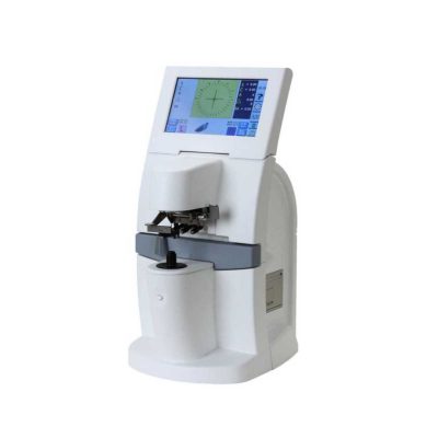
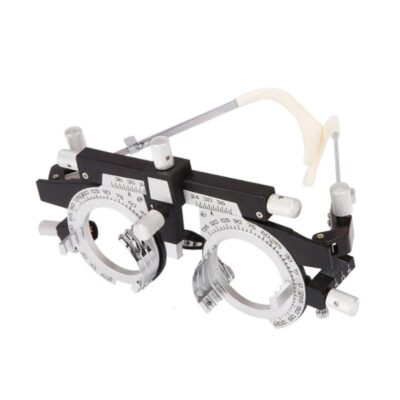
The NJC-4 Lensmeter is used to measure the diopters of spherical lens and cylinder lens, the axis of cylinder lens, the strength and the baseline direction of shuttle lens, it can also stamp the optical center of lens, the axis of cylinder len and the base direction of shuttle lens.
| In Stock
Features:
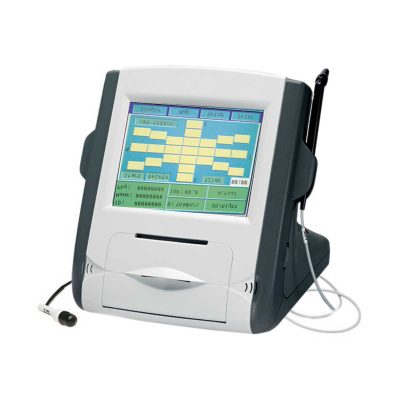
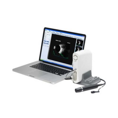
The Bio-1000 automated perimeter absorbs the advantages of international advanced perimetry devices. It comprises the highly integrated computer, optics, machinery and electronics systems.
Delivery & Availability:
Typically 7-14 working days – excluding furniture and heavy/bulky equipment. Please contact us for further information.
| Shipped from abroad
The product is designed on the principle basis of Goldman tonometer. It can be connected with slit lamp(Carl Zeiss type).
| Shipped from abroad
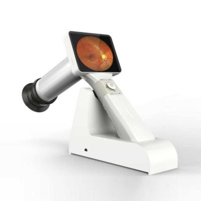
Ultra-small corneal curvature tester, is mainly used to measure the corneal curvature radius and diopter, wireless output print data.
| ||||||||||||||||||||||||||||||||||||||||||||||||||
| Content | SonoEye Features & AdvantagesCornerstone Reflectors
360° graduations on the first 20 mm of the needle in two sections:
Echogenic Needle Tip
| Features: ●Professional Same ear wax removal tool as those used by doctors, you can easily eliminate ear wax buildup at home, really save your money and time on medical visiting. Safe and Environmentally Friendly. ●Quick & Easy This ear wax removal kit is a quick, effective treatment for excess ear wax buildup. Fill the bottle with solution, Twist on the disposable tip, Use the trigger handle to spray solution into the ear canal. So Easy. ●Standard Capacity of the ear cleaner solution bottle is 10.6Oz, it has the most suitable size to hold in hand. Working at condition 32-122℉(0-50℃). Recommend to fill 1/5 of the bottle with OTC hydrogen peroxide, and 4/5 with very warm water. ●Complete Ear Washer System Our earwax removal kit comes with 1× Ear Washer Bottle, 1× Wash Basin, 1× Rubber Bulb, 1× Short Injection Head, 1× Long Hose Injection Head, 5× Disposable Tip, 1× User Manual. | The NJC-4 Lensmeter is used to measure the diopters of the spherical lens and cylinder lens, the axis of cylinder lens, the strength and the baseline direction of shuttle lens, it can also stamp the optical center of the lens, the axis of cylinder lens, and the base direction of shuttle lens. This instrument (Both Ac and Dc are permitted Two cells When Dc) has clear readings and graduations as well as high objective precision and reliabilities except that it can be operated easily and conveniently, AU the lenses and the made glasses can be measured by it, therefore, it is a required and ideal measuring instrument for glasses manufactures, glasses stores and ophthalmological hospitals.
Technical Specifications:
| The Bio-1000 automated perimeter absorbs the advantages of international advanced perimetry devices. It comprises the highly integrated computer, optics, machinery and electronics systems. Incorporated with the advanced configuration, comprehensive software inspection categories, and strictly in accordance with international Goldman standard, it provide scientific means for glaucoma, fundus disease, visual pathway injury and neurological diseases.
Feature:
* Comprehensive real-time monitoring,Heiji-krakau physiological blind spot monitoring,gaze tracking/head position tracking,automatic measurement of pupil diameter, reduce the impact of pupil effect on visual field detection.
* Personalized design,accurate clinical analysis,accurate and repid examination strategy.
* Under international Goldman standard,providing a variety of classic test procedures and report analysis.
Technical Specification:
Click Here To Download Catalogue | Applanation Tonometer is designed on the principle basis of Goldman tonometer. It can be connected with slit lamp(Carl Zeiss type).
Features of Applanation Tonometer:
| Portable Keratometer Features:
Ultra-small corneal curvature tester, is mainly used to measure the corneal curvature radius and diopter, wireless output print data.
Technical Specifications:
| ||||||||||||||||||||||||||||||||||||||||||||||||||
| Weight | N/A | N/A | N/A | N/A | N/A | N/A | ||||||||||||||||||||||||||||||||||||||||||||||||||
| Dimensions | N/A | N/A | N/A | N/A | N/A | N/A | ||||||||||||||||||||||||||||||||||||||||||||||||||
| Additional information | ||||||||||||||||||||||||||||||||||||||||||||||||||||||||


Reviews
There are no reviews yet.