System for measuring hourly diuresis MISURO
$0.00
Shipped From Abroad
Delivery & Availability:
Typically 10-21 working days – excluding furniture and heavy/bulky equipment. Please contact us for further information.
Typically 10-21 working days – excluding furniture and heavy/bulky equipment. Please contact us for further information.
Description
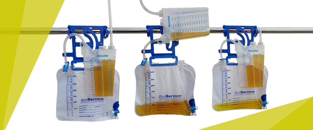
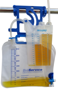
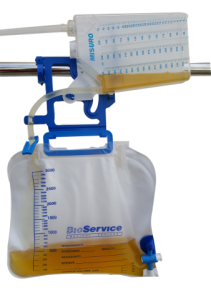
The device is made up of :
- a graduated rigid plastic container ( 500 ml capacity ) for measuring waste fluid , and is equipped with a pre- drip chamber antibacterial filter and a non-return valve.
- drainage tube with wide lumen and smooth internal surface to guarantee high flow.
- graded catheter cone with universal connector with drawing point and protective cap.
- Tube-clamp plate for vesical maneuvers and sheet clip for proper positioning of the tube.
- Collection bag in various capacities with non-return valve , antibacterial filter and drainage tap with rigid storage support.
- System for attachement to bed with two clamps ( optionally the device can be supplied with a hook ) .
| MISURO | |||
|---|---|---|---|
| MODEL | BAG | CODE | CODE (with hook) |
| MISURO 15 | 1500 ml (without drainage tap) | H90033E | H90033E-G |
| MISURO 20 | 2000 ml (with drainage tap) | H90034E | H90034E-G |
| MISURO 30 | 3000 ml (with drainage tap) | H90035E | H90035E-G |
| MISURO 35 | 3500 ml (without drainage tap) | H90037E | H90037E-G |
| MISURO 50 | 5000 ml (without drainage tap) | H90036E | H90036E-G |
All the diveces are available in multi-packs of 10 pieces
MISURO ACCESSORIES
The bag needs to be replaced without having to disconnect the urinometer from the patient , BioService offers a series of spare bags in various capacities.
Collection bag replacement is very practical and fast.
The device is supplied in individual packaging sterilized by EtO
| SPARE BAG | |||
|---|---|---|---|
| CODE | CAPACITY | PACKAGING | |
| H90111E | 1500 ml | 100 pcs | |
Click here to download Catalogue
Quick Comparison
| System for measuring hourly diuresis MISURO remove | Sonoscape HD-500 Gastrointestinal Video Endoscopy Stack remove | Sonoscape E1 Ultrasound Machine With Two Probes remove | Ecleris HD Digital Video Colposcope remove | Sonoscape S11 Ultrasound Machine remove | COMBI 9 Urine Test Kit remove | ||||||||||||||||||||||||||||||||||||||||||||||||||||||||||||||||||||||||||||||||||||||||||||||||||||||||||||
|---|---|---|---|---|---|---|---|---|---|---|---|---|---|---|---|---|---|---|---|---|---|---|---|---|---|---|---|---|---|---|---|---|---|---|---|---|---|---|---|---|---|---|---|---|---|---|---|---|---|---|---|---|---|---|---|---|---|---|---|---|---|---|---|---|---|---|---|---|---|---|---|---|---|---|---|---|---|---|---|---|---|---|---|---|---|---|---|---|---|---|---|---|---|---|---|---|---|---|---|---|---|---|---|---|---|---|---|---|---|---|---|---|---|
| Name | System for measuring hourly diuresis MISURO remove | Sonoscape HD-500 Gastrointestinal Video Endoscopy Stack remove | Sonoscape E1 Ultrasound Machine With Two Probes remove | Ecleris HD Digital Video Colposcope remove | Sonoscape S11 Ultrasound Machine remove | COMBI 9 Urine Test Kit remove | |||||||||||||||||||||||||||||||||||||||||||||||||||||||||||||||||||||||||||||||||||||||||||||||||||||||||||
| Image | 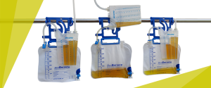 | 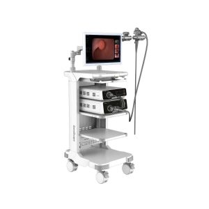 |  | 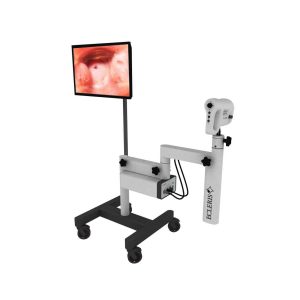 |  | 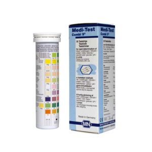 | |||||||||||||||||||||||||||||||||||||||||||||||||||||||||||||||||||||||||||||||||||||||||||||||||||||||||||
| SKU | SF1033560130101-4 | SF1033560012-24 | SF1033560012-20 | SF1033560087-2 | SF1033560012-1 | SF1033560084-247 | |||||||||||||||||||||||||||||||||||||||||||||||||||||||||||||||||||||||||||||||||||||||||||||||||||||||||||
| Rating | |||||||||||||||||||||||||||||||||||||||||||||||||||||||||||||||||||||||||||||||||||||||||||||||||||||||||||||||||
| Price |
|
| $4,620.00 | $3,564.00 | $6,380.00 |
| |||||||||||||||||||||||||||||||||||||||||||||||||||||||||||||||||||||||||||||||||||||||||||||||||||||||||||
| Stock | |||||||||||||||||||||||||||||||||||||||||||||||||||||||||||||||||||||||||||||||||||||||||||||||||||||||||||||||||
| Availability | |||||||||||||||||||||||||||||||||||||||||||||||||||||||||||||||||||||||||||||||||||||||||||||||||||||||||||||||||
| Add to cart | |||||||||||||||||||||||||||||||||||||||||||||||||||||||||||||||||||||||||||||||||||||||||||||||||||||||||||||||||
| Description | Shipped From Abroad
Delivery & Availability:
Typically 10-21 working days – excluding furniture and heavy/bulky equipment. Please contact us for further information.
| Shipped from Abroad Delivery & Availability: Typically 21 working days – excluding furniture and heavy/bulky equipment. Please contact us for further information. | Shipped from Abroad SonoScape has developed a new probe and function for the E1 Exp. With these additions the E1 Exp will bring users a more efficient examination experience with satisfying image quality and a smooth workflow. Delivery & Availability: Typically 5-7 working days – excluding furniture and heavy/bulky equipment. Please contact us for further information. | Shipped from Abroad Content Includes: Floor Mounted with Base with 4 Castors, Video Head, Positioning Arm, LED Light Included on Video Head, 110/220V Power Cable and User´s Guide, Stand For LCD Monitor and Printer, Digital Capturing System for Images, Videos and Sounds, USB 2.0. Includes Main Unit Processor with three Camera Inputs, USB Cable, Software, Hands Free Microphone and Footswitch. Delivery & Availability: Typically 10 working days – excluding furniture and heavy/bulky equipment. Please contact us for further information. | In Stock A Value Choice beyond Your Expectation. SonoScape’s trolley color Doppler system S11 redefines price and performance with practical design. The S11 will go beyond your expectations but not your budget. Delivery & Availability: Typically 2 working days – excluding furniture and heavy/bulky equipment. Please contact us for further information. | In stock
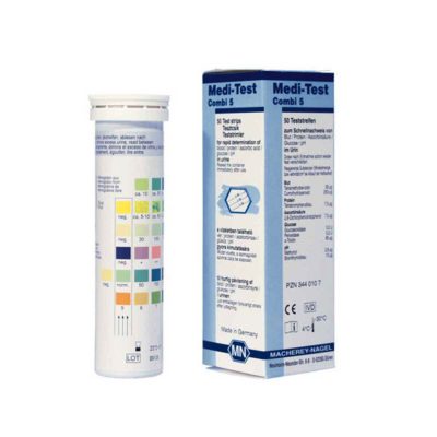
User-friendly urine test strips from Macherey-Nagel for professional urine analysis. Medi-Test Combi 9 test strips are flexible, which enables tests to be carried out even with small samples. This makes them ideal for use in pediatrics.
| |||||||||||||||||||||||||||||||||||||||||||||||||||||||||||||||||||||||||||||||||||||||||||||||||||||||||||
| Content | The device is made up of :
Click here to download Catalogue | HD-500 VIDEO PROCESSOR
Click Here To Download Catalogue | DETAILS
Efficient Diagnosis
μ-Scan, Speckle Reduction & Edge Enhancement
Spatial Compound Imaging
PIH - Pure Inversion Harmonic
Wide Scan - Enlarged Image Area
Tissue-Specific Imaging
SR Flow
Ergonomic Designs
Up to 2 Transducer Ports
Light Weight and Compact
15.6 inch Anti-flickering HD LED Screen
Tilting Monitor Angle Adjustment
Backlit Keyboard and Intelligent Panel
Long-lasting Battery for 90 mins
Ease of Use
Quick Boot Up
Auto-Brightness Adjustment
Auto Image Optimization
Auto IMT
Auto Trace
Equipped Accessories
Wi-Fi and Bluetooth Available
DICOM
500GB Hard Disk
Height Adjustable Trolley
Durable, Carry-on Site Suitcase
Click Here To Download Catalogue | The Ecleris ColpoHD Digital Video Colposcope offers clinicians an illuminated, high definition magnified view of the cervix and tissues of the vagina and vulva. As a screening tool for sexual assault victims or for the early detection of precancerous lesions, the ColpoHD provides insight into relevant pathology changes of tissue shape and color with magnification of up to 50x and a digital zoom up to 128x. The integrated electronic green filter delivers exceptional pathology visualization, with clear, true color imaging and superb depth of field. The swing arm mounted high-definition (HD) camera can be easily maneuvered into place. Optional integrated image capture is available for documentation along with archiving and printing of images.
A powerful tool for cervical cancer screening
The ColpoHD is precise and easy to handle in all examination situations. High quality optics along bright LED illumination and high-tech CMOS image sensors guarantee fantastic quality HD images are displayed for every patient.
The ColpoHD is also ideally suited for the examination of sexual assault victims who may be uncomfortable with a traditional Colposcope examination with the physician working through the standard microscope binoculars. The ColpoHD offers distance and a level of privacy for these patients who may be in a vulnerable frame of mind. The ColpoHD also offers optional image archiving and documentation of the examination in high definition image quality which is vital for sexual assault cases.
Features at a Glance
Click Here To Download Catalogue | DETAILS
SonoScape’s trolley colour Doppler system S11 redefines price and performance with practical design. The S11 will go beyond your expectations but not your budget. As an easy-to-use ultrasound system, the S11 is integrated with a new software platform, especially optimized for a smooth workflow and convenient operation. The system speeds up the exam process and makes file management easier.
SPECIFICATION
- 15-inch high definition LCD monitor with articulating arm
- Compact and agile trolley design
- 3 active transducer sockets available for a wide range of applications
- Duplex, Color Doppler, DPI, PW Doppler, tissue harmonic imaging, μ-scan speckle reduction imaging, compound imaging, trapezoidal imaging
- Customized settings based on your own working style
- Full patient database and image management solutions
Click Here To Download Catalogue | Medi-Test Combi 9
User-friendly urine test strips from Macherey-Nagel for professional urine analysis. Medi-Test Combi 9 test strips are flexible, which enables tests to be carried out even with small samples. This makes them ideal for use in pediatrics.
Product Details for Medi-Test Combi 9
| |||||||||||||||||||||||||||||||||||||||||||||||||||||||||||||||||||||||||||||||||||||||||||||||||||||||||||
| Weight | N/A | N/A | N/A | N/A | N/A | N/A | |||||||||||||||||||||||||||||||||||||||||||||||||||||||||||||||||||||||||||||||||||||||||||||||||||||||||||
| Dimensions | N/A | N/A | N/A | N/A | N/A | N/A | |||||||||||||||||||||||||||||||||||||||||||||||||||||||||||||||||||||||||||||||||||||||||||||||||||||||||||
| Additional information | |||||||||||||||||||||||||||||||||||||||||||||||||||||||||||||||||||||||||||||||||||||||||||||||||||||||||||||||||


Reviews
There are no reviews yet.