
Ophthalmic AB Scan Machine
Ask for Price$0.00
Shipped from abroad
- Software image workstation
- B, B+B, B+A, A modes
- Video review for 100 images
- PDF report output
- Optional 20MHz B Probe: vitreous plus function
Delivery & Availability:
Typically 14 working days – excluding furniture and heavy/bulky equipment. Please contact us for further information.
Description
Functions of Ophthalmic AB Scan Machine:
- Software image workstation
- B, B+B, B+A, A modes
- Video review for 100 images
- PDF report output
- Optional 20MHz B Probe: vitreous plus function
Technical Specifications:
| A scan | 1.Probe: 10MHz frequencies, with LED 2.Depth: 40mm 3.Precision: ±0.05mm 4.Eye mode: Phakic / Aphakic / Dense / Various IOL 5.Measurement: Anterior chamber depth, lens thickness, vitreous body length, total length and average 6.IOL Formula: SRK-II, SRK-T, BINKHORST, HOLLADAY, HOFFER-Q, HAIGIS, Stat. 7.Calculation: Average and standard deviation 8.Store: 10 Scanning results for each eye |
| B scan | 1.Probe: 10MHz/20MHz (optional), Magnetic driven, noiseless 2.Scanning Mode: Sector Scanning 3.Resolution: Lateral ≤0.3mm; Vertical≤0.2mm 4.Geometric Location Precision: Lateral≤10%; Vertical≤5% 5.Depth: 60mm 6.Enhance the part of vitreous body and retina 7.Gain of probe:30dB-105dB 8.Scanning Angle : 53° 9.Gray Scale: 256 10.False Color: Multi colors OTC 11.Measure Mode: distances, perimeter and area 12.Movies: 100 images movie review,AVI ZIP JPG format image output 13.Output: PDF format case report, connect to normal printer |
| Others | 1.Display Mode :B, B+B, B+A, A 2.Hint: preset keyword 3.Case Search: Multi-keywords 4.Working Platform: Windows XP, VISTA, WINDOWS7 5.User-defined report template |
Quick Comparison
| Settings | Ophthalmic AB Scan Machine remove | Sonoscape P20 Ultrasound Machine remove | ASPEL AsCARD Grey ECG Machine remove | DrGem Diamond All-In-One Digital X-ray Machine remove | ASPEL Stress ECG with Treadmill and Software remove | ASPEL AsPEKT 712 Holter Monitor and Software remove | ||||||
|---|---|---|---|---|---|---|---|---|---|---|---|---|
| Name | Ophthalmic AB Scan Machine remove | Sonoscape P20 Ultrasound Machine remove | ASPEL AsCARD Grey ECG Machine remove | DrGem Diamond All-In-One Digital X-ray Machine remove | ASPEL Stress ECG with Treadmill and Software remove | ASPEL AsPEKT 712 Holter Monitor and Software remove | ||||||
| Image | 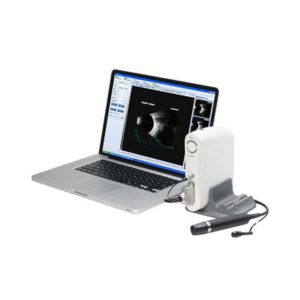 |  |  |  |  |  | ||||||
| SKU | SF1033560107-8 | SF1033560012-9 | SF1033560075-5 | SF1033560074-3 | SF1033560075-2 | SF1033560075-4 | ||||||
| Rating | ||||||||||||
| Price | Ask for Price | Ask for Price | Ask for Price | Ask for Price | Ask for Price | Ask for Price | ||||||
| Stock | ||||||||||||
| Availability | ||||||||||||
| Add to cart | ||||||||||||
| Description | Shipped from abroad
| Shipped from Abroad Incorporating innovative technologies, P20’s user-friendly design with a simple operation panel, intuitive user interface and a variety of intelligent auxiliary scanning tools, will significantly improve your daily examination experience. Besides general imaging applications, P20 has entitled with diagnostic 4D technology which has an extraordinary performance in obstetrics and gynecology applications. Delivery & Availability: Typically 5-7 working days – excluding furniture and heavy/bulky equipment. Please contact us for further information. | Shipped from Abroad Electrocardiograph AsCARD Grey v.07.225 - is a 1, 3, 6, 12 channel ECG unit which enables to make electrocardiogram in full 12 leads. It is intended to conduct ECG examinations of adults and paediatric patients in all types of health care centres. ECG examination may be recorded in manual or automatic mode, with the possibility of analysis and interpretation. The device can be powered from 100 V ÷ 240 V mains supply or by an internal battery. Delivery & Availability: Typically 10 working days – excluding furniture and heavy/bulky equipment. Please contact us for further information. | Shipped from Abroad DrGem Diamond All-In-One Digital X-ray Machine is a fully automatic digital radiography system providing state-of-the-art image quality, image processing and user interface. With a wide selection of anatomical studies on the imaging software, DIAMOND automatically sets up the x-ray generator’s preprogrammed exposure technique settings, motorized radiographic stand positioning, x-ray collimation and post-image processing for the selected study. Specifically designed to increase workflow, this fully digital system offers convenient auto-positioning and advanced image processing to achieve big performance with little effort. Delivery & Availability: Typically 21 working days – excluding furniture and heavy/bulky equipment. Please contact us for further information. | Shipped from Abroad It is a system with professional tool dedicated to exercise and resting ECG examination. Treadmill has 12 lead ECG modules. With ECG Analyzing Software. Delivery & Availability: Typically 21 working days – excluding furniture and heavy/bulky equipment. Please contact us for further information. | Shipped from Abroad The Holta Monitor allows quick analysis of ECG examination and detection, reviewing and editing capability in the qualitative assessment of VE, VT, Single SVE, PSVT, Pauses, Irregular Rhythm, VT, IVR, Brady - and Tachycardia, Couplets, ST-segment elevation and depression, Maximum, Minimum and averaged Heart Rates, artifacts Delivery & Availability: Typically 10 working days – excluding furniture and heavy/bulky equipment. Please contact us for further information. | ||||||
| Content | Functions of Ophthalmic AB Scan Machine:
| DETAILS
Upgraded Images with More Clarity
SonoScape never stops making progress in improving the image quality of its ultrasound products to enhance the confidence of diagnosis for doctors. With extraordinary images provided by P20, the anatomy structures are clearer than ever.
C-Xlasto Imaging
With C-xlasto Imaging, P20 enables comprehensive quantitative elastic analysis. Meanwhile, C-xlasto on P20 is supported by linear, convex and transvaginal probes, to ensure good reproducibility and highly consistent quantitative elastic results.
S-Live
S-Live allows for detailed visualization of subtle anatomical features, thereby enabling intuitive diagnosis with real-time 3D images and enriching patient communication.
Pelvic Floor 4D
Transperineal 4D pelvic floor ultrasound can provide useful clinical values in assessing the vaginal delivery impact on the female anterior compartment, judging whether the pelvic organs are prolapsed or not and the extent, determining if the pelvic muscles were torn accurately.
Anatomic M Mode
Anatomic M Mode helps you observe the myocardial motion at different phases by freely placing sample lines. It accurately measures the myocardial thickness and the heart size of even difficult patients and supports the myocardial function and LV wall-motion assessment.
Tissue Doppler Imaging
P20 is endowed with Tissue Doppler Imaging which provides velocities and other clinical information on myocardial functions, facilitating clinical doctors with the ability to analyze and compare the motions of different parts of the patient's heart.
Click Here To Download Catalogue |
Electrocardiograph AsCARD Grey v.07.225 - is a 1, 3, 6, 12 channel ECG unit which enables to make electrocardiogram in full 12 leads. It is intended to conduct ECG examinations of adults and paediatric patients in all types of health care centres. ECG examination may be recorded in manual or automatic mode, with the possibility of analysis and interpretation. The device can be powered from 100 V ÷ 240 V mains supply or by an internal battery.
Technical Specification:1. Visualisation of 1, 3, 6 or 12 ECG waveforms, analysis results and interpretations, examinations stored in memory.
2. Recording of 12 standard leads.
3. Print out in 1, 3, 6 or 12 ECG waveforms mode. Printing of a selected group:
Click Here To Download Catalogue | DrGem Diamond All-In-One Digital X-ray Machine is a fully automatic digital radiography system providing state-of-the-art image quality, image processing and user interface. With a wide selection of anatomical studies on the imaging software, DIAMOND automatically sets up the x-ray generator’s pre-programmed exposure technique settings, motorized radiographic stand positioning, x-ray collimation and post-image processing for the selected study. Specifically designed to increase workflow, this fully digital system offers convenient auto-positioning and advanced image processing to achieve big performance with little effort.
Features of DrGem Diamond All-In-One Digital X-ray Machine:
Outstanding Image Quality -
Digital radiography via at panel detector improves your workflow, exam speed and comfort with efficiency. Digital at panel detector with Csl screen provides excellent spatial resolution, MTF, DQE and stability based on ne pixel pitch. A 3-field ion-chamber is provided for AEC function.
Automatic Collimation –
Automatic x-ray eld size control of the motorized collimator corresponds to dierent SIDs. Includes user adjustable lamp timer with on/oswitch.
Automatic Positioning –
Click Here To Download Catalogue | It is a system with professional tool dedicated to exercise and resting ECG examination. Treadmill has 12 lead ECG modules. With ECG Analyzing Software.
Technical Specification:
Click Here To Download Catalogue | The Holter Monitor allows quick analysis of ECG examination (arrhythmias and ST segment).
Technical specifications:
HolCARD 24W Software:
Click Here To Download Catalogue | ||||||
| Weight | N/A | N/A | N/A | N/A | N/A | N/A | ||||||
| Dimensions | N/A | N/A | N/A | N/A | N/A | N/A | ||||||
| Additional information |


Reviews
There are no reviews yet.