
Anke Anatom 16 Slice CT Scan
Ask for Price$0.00
Shipped from Abroad
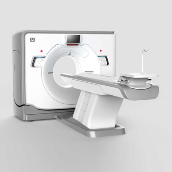
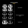
ANATOM16 HD, is a tool of precision medicine in diagnosis imaging. Via the breakthrough designs in precise hardware, software and imaging technologies, ANATOM 16 HD can provide precise diagnosis information and early detection for small lesions.
Delivery & Availability:
Typically 90 working days – excluding furniture and heavy/bulky equipment. Please contact us for further information.
Description
ANATOM16 HD, is a tool of precision medicine in diagnosis imaging. Via the breakthrough designs in precise hardware, software and imaging technologies, ANATOM 16 HD can provide precise diagnosis information and early detection for small lesions.
Features:
- Seamlessly upgrade to meet your future needs: Anke takes full consideration of the increasing clinical requirements of your business in today’s rapidly changing medical environment.
- Precise hardware, Precise technology, Precise imaging: OptiWave detector, High precision gantry control, Dual-mode gantry tilt, Admir3D iterative technology, Dual-energy head imaging, 1024 x1024 matrix imaging technology, High-definition imaging of targeted organs, Low dose platform, 3D enhanced VR
- Precision Technology Platform: ANATOM precision technology platform is equipped with advanced imaging technologies, and adopts OptiWave detector, Ahead dual-energy imaging, Admir3D iterative reconstruction technology and AccuTilt dual-mode tilt gantry technology to provide powerful support for accurate diagnosis
- Admir3D iterative reconstruction technology: Admir applies mathematical and physics models to accurately construct and describe the signal’s quantum characteristics. Iterative operations are performed in the three domains of raw data, projection and image, to greatly reduce the image noise and achieve optimal image quality with low dose
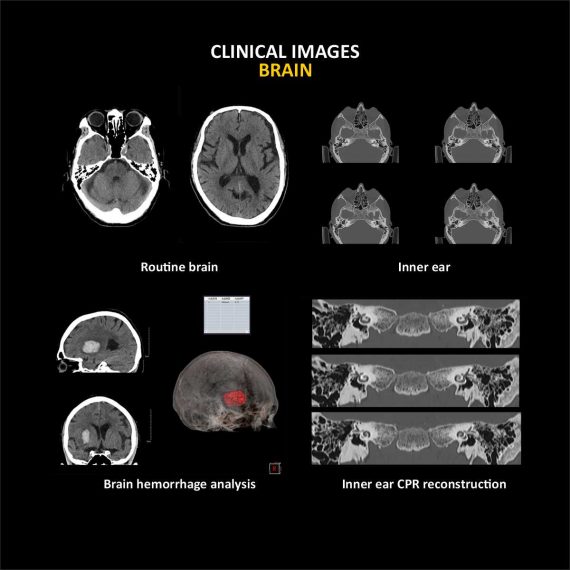
- Ahead-Head dual-energy head imaging technology: Ahead creatively uses 140kV and 80kV dual energy switching scan mode for brain imaging. By careful analyzing the high and low energy characteristics, images can show more valuable information about the brain tissues
- AccuTilt dual-mode gantry tilt technology: The system provides digital and mechanical tilt to accommodate different user habits and clinical needs. Real-time collision preventing system is available for the patients’ safety
- AccuOrgan-Targeted organ imaging: To achieve high precision imaging of each part of human body at low dose and low energy consumption
- AccuDose-Comprehensive low dose imaging: Pediatric Scan Protocol, Individual Dose Monitoring, AccuShape Filter, Efficient Detector, Adose Dose Modulation, Ahead – Head Dual-energy Imaging, Iterative Reconstruction, Amast, Contrast Agent Tracking Technology
- AccuScan-Enjoy ease: Convenient and efficient operation process greatly improve work efficiency to achieve high volume of patients
- Clinical Applications: Fast, precise and low-dose imaging technologies provide a full range of clinical solutions to meet the current and future clinical diagnostic needs
- Service Innovation creating maximum value for customers: Service Support within 24 Hours, Local Service Partners, On-line Service Support, After-sales Maintenance Stations
- AccuSaving Green & Energy-saving: AccuSaving is an innovative energy saving technology. The system will enter the “dormant”, which is a low carbon mode, after a certain idle time or per user’s request. To bring the system back to working status is as easy as pushing a button. The system will also remind the user to perform necessary warm-up and calibration procedures, which are fully automated processes. AccuSaving technology can reduce operation and standby power consumption and save the electricity cost by 30% by adopting different operation modes in working and off hours
Technical Specifications:
| No. | Technical feature | Description |
| 1 | Gantry | |
| 1.01 | Gantry type | Low voltage slip-ring with
AccuSlip-ring technology |
| 1.02 | Gantry driven type | Strap-driven |
| 1.03 | Patient opening | 70cm |
| 1.04 | Gantry tilt mode | Dual-mode gantry tilt |
| 1.05 | Mechanical tilt capability | ±30° |
| 1.06 | Digital tilt capability | ±50° |
| 1.07 | Gantry remote-Control | Provided |
| 1.08 | Detector type | OptiWave rare-earth ceramic detector |
| 1.09 | Numbers of detector rows | 32 |
| 1.10 | Width of Z-axle detector | 20mm |
| 1.11 | Detector columns of channels per row | 912 |
| 1.12 | Numbers of detector columns | 29184 |
| 1.13 | Data-transfer type | RF,optical fiber communication |
| 1.14 | 3D laser orientation | Provided |
| 1.15 | External X-ray enable | Interface for Foot-Pedal Provided |
| 1.16 | Automatic exposure control(mA Modulation) | Provided |
| 1.17 | Auto-voice manager | Breath Graphical Display
Hold Message (Record/Playback) Breath Message(Record/Playback) |
| 1.18 | ANKE energy conservation management | Provided |
| 1.19 | Acquisition mode | 16 × 0.625mm, 16 × 1.25mm |
| 2 | Scan parameter | |
| 2.01 | Shortest 360 degree rotation time | 0.5s |
| 2.02 | Allowed rotation times | 0.5s,0.8s,1.0s,1.5s,2.0s |
| 2.03 | Slice numbers per rotation | 16 |
| 2.04 | Minimum slice thickness of scan | 0.625mm |
| 2.05 | Minimum slice thickness of reconstruction | 0.625mm |
| 2.06 | Maximum slice thickness of scan | 10mm |
| 2.07 | Nominal reconstruction slice thickness | 0.625mm,1.25mm,2.5mm,5.0mm,
7.5mm,10mm |
| 2.08 | Speed of image reconstruction(512×512) | 65 frames/s |
| 2.09 | Scan FOV | 52cm |
| 2.10 | Image reconstruction matrix | 512×512,1024×1024 |
| 2.11 | Image display matrix | 512×512,1024×1024 |
| 2.12 | Maximum continuous scan duration | 120s |
| 2.13 | Maximum continuous scan length | 180cm |
| 2.14 | Direction of TOPO | Front-back,Left-right |
| 2.15 | Max. length of TOPO | 180cm |
| 2.16 | Range of pitch | 0.5~1.5 |
| 2.17 | Scan mode | Scout scan
Axial scan Helical scan Cine scan |
| 3 | HVPS and Tube | |
| 3.01 | Maximum continuous output of HV generator | 50kW |
| 3.02 | Tube kV selections | 80kV,100 kV,120 kV,140 kV |
| 3.03 | Tube mA range | 10~420mA |
| 3.04 | Tube anode heat capacity | 5.0MHU |
| 3.05 | Heat dissipation rate | 815kHU/min |
| 3.06 | Type of cooling | Oil cooling + Air cooling |
| 3.07 | Tube focus | Large:1.0 mm×1.0mm
Small:0.5mm×1.0mm |
| 3.08 | Dynamic flying focal spot technology | Provided |
| 4 | Patient table | |
| 4.01 | Maximum horizontal-movable range | 1850mm |
| 4.02 | Table horizontal-scannable range | 1800mm |
| 4.03 | Table horizontal-position repeatability | ±0.25mm |
| 4.04 | Maximum vertical-movable range | 500mm |
| 4.05 | Maximum speed of vertical movement | 20mm/s |
| 4.06 | Maximum speed of horizontal movement | 150mm/s |
| 4.07 | Maximum patient weight | 205kg |
| 4.08 | Foot pedal of patient table control | Provided |
| 5 | Image Quality | |
| 5.01 | High contrast resolution | 21lp/cm@0%MTF |
| 5.02 | Low contrast resolution | 2.0mm@0.30% |
| 5.03 | Isotropic imaging resolution | 0.625mm |
| 5.04 | Range of CT numbers | -32767~32768 |
| 5.05 | Image noise | ≤0.25@28mGy |
| 6 | Computer subsystem | |
| 6.01 | CPU | 3.5GHz |
| 6.02 | Memory | 16GB×4 |
| 6.03 | Storage of hard-disk | 1T×2 |
| 6.04 | Monitor | 24’’ LCD Monitor |
| 6.05 | Resolution of monitor | 1920×1200 |
| 6.06 | Image-data external storage type | CD/DVD/USB |
| 6.07 | Time of image reconstruction(512×512) | 15.4ms/frame |
| 6.08 | DICOM 3.0 interface | Provided |
| 6.09 | Printer DICOM 3.0 interface | Provided |
| 6.10 | Auto filming | Provided |
| 6.11 | Worklist function | Provided |
| 7 | Advanced application | |
| 7.01 | Multi-Planar Reconstruction(MPR) | Provided |
| 7.02 | Curve Multi-Planar Reconstruction(CPR) | Provided |
| 7.03 | Surface Shaded Display(SSD) | Provided |
| 7.04 | Volume Rendering(VR) | Provided |
| 7.05 | Maximum Intensity Projection(MIP) | Provided |
| 7.06 | Minimum Intensity Projection(MinIP) | Provided |
| 7.07 | Virtual Endoscopy(VE) | Provided |
| 7.08 | CT angiography(CTA) | Provided |
| 7.09 | Tissue segmentation | Provided |
| 7.10 | One click bone remove | Provided |
| 7.11 | One click patient table remove | Provided |
| 7.12 | Bolus-tracking Technology | Provided |
| 7.13 | Spiral auto start | Provided |
| 7.14 | Cine display | Provided |
| 7.15 | AbastTM bone artifact suppression technology | Provided |
| 7.16 | AmastTM metal artifact suppression technology | Provided |
| 7.17 | Admir3D fulll-domain iterative reconstruction | Provided |
| 7.18 | Low-dose pediatric scan technology | Provided |
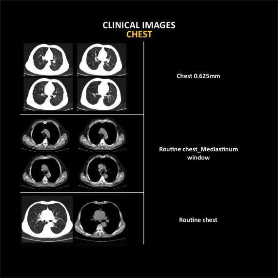
| 7.19 | Low-dose lung scan technology | Provided |
| 7.20 | AccuHead grey-white matter enhanced
technology |
Provided |
| 7.21 | AccuLung high resolution scan technology | Provided |
| 7.22 | AccuOtica inner-ear high resolution scan
technology |
Provided |
| 7.23 | AccuBody high resolution scan technology | Provided |
| 7.24 | AccuBone high resolution scan technology | Provided |
Click Here To Download Catalogue
Additional information
| Model | Advanced, Advanced Plus, Basic, Smart |
|---|
Review(1)
Quick Comparison
| Settings | Anke Anatom 16 Slice CT Scan remove | Sonoscape S8 Exp Portable Ultrasound remove | Sonoscape P10 Ultrasound Machine remove | Jade Mobile X-ray machine (Analogue) remove | ASPEL Stress ECG with Treadmill and Software remove | Topaz Digital X-ray Machine remove | ||||||||||||||||||||||||||||||||||||||||||||||||||||||||||||||||||||||||||||||||||||||||||||||||||||||||||||||||||||||||||||||||||||||||||||||||||||||||||||||||||||||||||||||||||||||||||||||||||||||||||||||||||||||||||||||||||||||||||||||||||||||||||||||||||||||||||||||||||||||||||||||||||||||||||||
|---|---|---|---|---|---|---|---|---|---|---|---|---|---|---|---|---|---|---|---|---|---|---|---|---|---|---|---|---|---|---|---|---|---|---|---|---|---|---|---|---|---|---|---|---|---|---|---|---|---|---|---|---|---|---|---|---|---|---|---|---|---|---|---|---|---|---|---|---|---|---|---|---|---|---|---|---|---|---|---|---|---|---|---|---|---|---|---|---|---|---|---|---|---|---|---|---|---|---|---|---|---|---|---|---|---|---|---|---|---|---|---|---|---|---|---|---|---|---|---|---|---|---|---|---|---|---|---|---|---|---|---|---|---|---|---|---|---|---|---|---|---|---|---|---|---|---|---|---|---|---|---|---|---|---|---|---|---|---|---|---|---|---|---|---|---|---|---|---|---|---|---|---|---|---|---|---|---|---|---|---|---|---|---|---|---|---|---|---|---|---|---|---|---|---|---|---|---|---|---|---|---|---|---|---|---|---|---|---|---|---|---|---|---|---|---|---|---|---|---|---|---|---|---|---|---|---|---|---|---|---|---|---|---|---|---|---|---|---|---|---|---|---|---|---|---|---|---|---|---|---|---|---|---|---|---|---|---|---|---|---|---|---|---|---|---|---|---|---|---|---|---|---|---|---|---|---|---|---|---|---|---|---|---|---|---|---|---|---|---|---|---|---|---|---|---|---|---|---|---|---|---|---|---|---|---|---|
| Name | Anke Anatom 16 Slice CT Scan remove | Sonoscape S8 Exp Portable Ultrasound remove | Sonoscape P10 Ultrasound Machine remove | Jade Mobile X-ray machine (Analogue) remove | ASPEL Stress ECG with Treadmill and Software remove | Topaz Digital X-ray Machine remove | ||||||||||||||||||||||||||||||||||||||||||||||||||||||||||||||||||||||||||||||||||||||||||||||||||||||||||||||||||||||||||||||||||||||||||||||||||||||||||||||||||||||||||||||||||||||||||||||||||||||||||||||||||||||||||||||||||||||||||||||||||||||||||||||||||||||||||||||||||||||||||||||||||||||||||||
| Image | 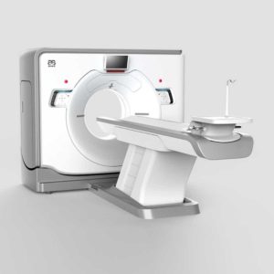 |  |  |  |  |  | ||||||||||||||||||||||||||||||||||||||||||||||||||||||||||||||||||||||||||||||||||||||||||||||||||||||||||||||||||||||||||||||||||||||||||||||||||||||||||||||||||||||||||||||||||||||||||||||||||||||||||||||||||||||||||||||||||||||||||||||||||||||||||||||||||||||||||||||||||||||||||||||||||||||||||||
| SKU | SF1033560092-5 | SF1033560012-15 | SF1033560012-7 | SF1033560074-2 | SF1033560075-2 | SF1033560074-1 | ||||||||||||||||||||||||||||||||||||||||||||||||||||||||||||||||||||||||||||||||||||||||||||||||||||||||||||||||||||||||||||||||||||||||||||||||||||||||||||||||||||||||||||||||||||||||||||||||||||||||||||||||||||||||||||||||||||||||||||||||||||||||||||||||||||||||||||||||||||||||||||||||||||||||||||
| Rating | ||||||||||||||||||||||||||||||||||||||||||||||||||||||||||||||||||||||||||||||||||||||||||||||||||||||||||||||||||||||||||||||||||||||||||||||||||||||||||||||||||||||||||||||||||||||||||||||||||||||||||||||||||||||||||||||||||||||||||||||||||||||||||||||||||||||||||||||||||||||||||||||||||||||||||||||||||
| Price | Ask for Price | $7,700.00 | Ask for Price | $6,930.00 | Ask for Price | $52,000.00 | ||||||||||||||||||||||||||||||||||||||||||||||||||||||||||||||||||||||||||||||||||||||||||||||||||||||||||||||||||||||||||||||||||||||||||||||||||||||||||||||||||||||||||||||||||||||||||||||||||||||||||||||||||||||||||||||||||||||||||||||||||||||||||||||||||||||||||||||||||||||||||||||||||||||||||||
| Stock | ||||||||||||||||||||||||||||||||||||||||||||||||||||||||||||||||||||||||||||||||||||||||||||||||||||||||||||||||||||||||||||||||||||||||||||||||||||||||||||||||||||||||||||||||||||||||||||||||||||||||||||||||||||||||||||||||||||||||||||||||||||||||||||||||||||||||||||||||||||||||||||||||||||||||||||||||||
| Availability | ||||||||||||||||||||||||||||||||||||||||||||||||||||||||||||||||||||||||||||||||||||||||||||||||||||||||||||||||||||||||||||||||||||||||||||||||||||||||||||||||||||||||||||||||||||||||||||||||||||||||||||||||||||||||||||||||||||||||||||||||||||||||||||||||||||||||||||||||||||||||||||||||||||||||||||||||||
| Add to cart | ||||||||||||||||||||||||||||||||||||||||||||||||||||||||||||||||||||||||||||||||||||||||||||||||||||||||||||||||||||||||||||||||||||||||||||||||||||||||||||||||||||||||||||||||||||||||||||||||||||||||||||||||||||||||||||||||||||||||||||||||||||||||||||||||||||||||||||||||||||||||||||||||||||||||||||||||||
| Description | Shipped from Abroad
ANATOM16 HD, is a tool of precision medicine in diagnosis imaging. Via the breakthrough designs in precise hardware, software and imaging technologies, ANATOM 16 HD can provide precise diagnosis information and early detection for small lesions.
Delivery & Availability: Typically 90 working days – excluding furniture and heavy/bulky equipment. Please contact us for further information. | Shipped from Abroad With ultra-modern innovative design and the clinically-proven technologies, S8 Exp is portable ultrasound scanner well equipped as a low-physical-effort and enhanced-image-quality ultrasound scanner, which not only provides optimized solutions for versatile applications, but does help to improve the user-experience for both routine and non-traditional challenges. Delivery & Availability: Typically 5-7 working days – excluding furniture and heavy/bulky equipment. Please contact us for further information. | Shipped from Abroad The P10 color Doppler ultrasound system is a new generation product from SonoScape. It is designed to give high quality images, rich probe configurations, various clinical tools and automatic analysis software to provide you with comprehensive solutions for your growing demand for clinical applications. Delivery & Availability: Typically 5-7 working days – excluding furniture and heavy/bulky equipment. Please contact us for further information. | In Stock JADE is one of the lightest portable X-ray systems on the market, allowing it to be used in any imaginable way including bedside, operating rooms, intensive care units and in veterinary fields. With a simple, easy-to-use operator console, three-way control, two-step foldable stand and auto lock system, JADE is a user-friendly portable X-ray system. Delivery & Availability: Typically 21 working days – excluding furniture and heavy/bulky equipment. Please contact us for further information. | Shipped from Abroad It is a system with professional tool dedicated to exercise and resting ECG examination. Treadmill has 12 lead ECG modules. With ECG Analyzing Software. Delivery & Availability: Typically 21 working days – excluding furniture and heavy/bulky equipment. Please contact us for further information. | In Stock DRGEM’s TOPAZ X-ray machine is a state-of-the-art mobile digital radiography system, designed with maximum comfort for patients and users in mind. From its user-friendly software to smooth movements, TOPAZ is made to improve your workflow and provide you with high-quality images. Delivery & Availability: Typically 21 working days – excluding furniture and heavy/bulky equipment. Please contact us for further information. | ||||||||||||||||||||||||||||||||||||||||||||||||||||||||||||||||||||||||||||||||||||||||||||||||||||||||||||||||||||||||||||||||||||||||||||||||||||||||||||||||||||||||||||||||||||||||||||||||||||||||||||||||||||||||||||||||||||||||||||||||||||||||||||||||||||||||||||||||||||||||||||||||||||||||||||
| Content | ANATOM16 HD, is a tool of precision medicine in diagnosis imaging. Via the breakthrough designs in precise hardware, software and imaging technologies, ANATOM 16 HD can provide precise diagnosis information and early detection for small lesions.
Features:
Click Here To Download Catalogue | Sonoscape S8 Exp Portable Ultrasound scannerDETAILS Agile and Versatile With ultra-modern innovative design and the clinically-proven technologies, S8 Exp Portable Ultrasound scanner is well equipped as a low-physical-effort and enhanced-image-quality ultrasound scanner, which not only provides optimized solutions for versatile applications but does help to improve the user experience for both routine and non-traditional challenges. Working with S8 Exp, it will trigger your unlimited reverie and endow you with endless charm. Carrying forward the classical design of SonoScape's portable ultrasound products, S8 Exp successfully combines the best ergonomics, attractive design and ease of use. This charismatic identity is also enhanced by a sophisticated color palette—with sedate grey as its interior paint color and pearl white as exterior cover, S8 Exp reveals a style of aristocrat and strong character among SonoScape's ultrasound systems. Workflow The S8 Exp is a portable ultrasound scanner that adapts to your workflow, whether you are in the consulting room, at the bedside, or at a remote location. With easy-to-use new platform designed for sonographers' needs and full connection interfaces for easy connectivity and data sharing, S8 Exp leads to improved user comfort and clinical outcome as well as patient throughput and working efficiency. Powerful Platform Embedded with SonoScape's core imaging technologies such as μ-scan, PHI and Spatial Compound, S8 Exp boasts exceptional 2D image, sensitive spectral, Color and Power Doppler, displaying well-defined anatomy and pathology and facilitating a highly optimized diagnostic user environment for conclusive diagnoses. Besides, S8 Exp offers a comprehensive selection of electronic probes to maximally extend its capabilities to meet a wide range of applications including the abdomen, pediatric, OB/GYN, cardiovascular, musculoskeletal, etc. The advanced probe technologies also effectively enhance the image quality and confidence in reaching clinical diagnoses even in difficult patients.Click Here To Download Catalogue | DETAILS
B + Compound
B + Compound utilizes several lines of sight for optimal contrast resolution, speckle reduction and border detection, with which P10 is ideal for superficial and abdominal imaging with better clarity and improved continuity of structures.
μ-Scan
The new generation μ-Scan imaging technology gives you better image quality by reducing noise, improving signal strength and improving visualization.
P10 offers a comprehensive selection of electronic probes to maximize its capabilities to meet a wide range of applications including abdomen, pediatric, OB/GYN, cardiovascular, musculoskeletal, etc. The advanced probe technologies also effectively enhance the image quality and confidence in reaching clinical diagnoses, even in difficult patients.
Convex Probe 3C-A
Ideal for an abundant of application such as abdomen, gynecology, obstetrics, urology and even abdomen biopsy.
Linear Probe L741
This linear probe is designed to satisfy vascular, breast, thyroid, and other small parts diagnosis, and its adjustable parameters could also present users a clear view of MSK and deep vessels.
Phase Array Probe 3P-A
For the purpose of adult and pediatric cardiology and emergency, the phase array probe provides elaborate presets for different exam modes, even for difficult patients.
Intracavitary Probe 6V1
Intracavitary probe could face application of gynecology, urology, prostate, and its temperature detection technology not only protects the patient but also extends the service life.
Click Here To Download Catalogue | JADE Mobile X-ray machine is one of the lightest portable X-ray systems on the market, allowing it to be used in any imaginable way including bedside, operating rooms, intensive care units and veterinary fields. With a simple, easy-to-use operator console, three-way control, two-step foldable stand and auto-lock system, the JADE Mobile X-ray machine is a user-friendly portable X-ray system.
Convenient & Intuitive Operation:
JADE is one of the lightest portable X-ray systems on the market, allowing it to be used in any imaginable way including bedside, operating rooms, intensive care units and in veterinary fields. With a simple, easy-to-use operator console, three-way control, two-step foldable stand and auto-lock system, JADE is a user-friendly portable X-ray system.
Compact & Powerful Design:
JADE Mobile X-ray machine is an innovative, highly versatile portable X-ray system suitable for a variety of clinical uses. Utilizing the unique technology used in DRGEM’s universally recognized X-ray generators, JADE is a compact but powerful unit with a 4kW output and thoughtfully designed components to increase efficiency and maximize workflow. The core part of X-ray source adopts high-quality tube assembly, X-ray collimator and high frequency X-ray generator with excellent performance, lifetime and stability.
Features:
Click Here To Download Catalogue | It is a system with professional tool dedicated to exercise and resting ECG examination. Treadmill has 12 lead ECG modules. With ECG Analyzing Software.
Technical Specification:
Click Here To Download Catalogue | TOPAZ X-ray machine is among the high end X-ray machine manufactured by DRGEM, a digital X-ray system that provides quality images with little or no effort.
It begins with Advanced Technology
Integrating high technology and over a decade of experience in conventional and digital radiography systems, DRGEM’s TOPAZ X-ray machine is a state-of-the-art mobile digital radiography system, designed with maximum comfort for patients and users. From its user-friendly software to smooth movements, TOPAZ X-ray machine is made to improve your workflow and provide you with high-quality images.
Full Featured Imaging Software & Excellent Digital Image Processing
With a high-performance, built-in touchscreen, TOPAZ X-ray machine offers a user-friendly interface and powerful software for easy operation and increased workflow. The anatomical view-based digital image processing, automatically optimizes and enhances the quality of the image. it also comes with automatic image storage and print with DICOM 3.0 networking capability. additionally, the system offers increasing exam throughput while decreasing examination time.
Click Here To Download Catalogue | ||||||||||||||||||||||||||||||||||||||||||||||||||||||||||||||||||||||||||||||||||||||||||||||||||||||||||||||||||||||||||||||||||||||||||||||||||||||||||||||||||||||||||||||||||||||||||||||||||||||||||||||||||||||||||||||||||||||||||||||||||||||||||||||||||||||||||||||||||||||||||||||||||||||||||||
| Weight | N/A | N/A | N/A | N/A | N/A | N/A | ||||||||||||||||||||||||||||||||||||||||||||||||||||||||||||||||||||||||||||||||||||||||||||||||||||||||||||||||||||||||||||||||||||||||||||||||||||||||||||||||||||||||||||||||||||||||||||||||||||||||||||||||||||||||||||||||||||||||||||||||||||||||||||||||||||||||||||||||||||||||||||||||||||||||||||
| Dimensions | N/A | N/A | N/A | N/A | N/A | N/A | ||||||||||||||||||||||||||||||||||||||||||||||||||||||||||||||||||||||||||||||||||||||||||||||||||||||||||||||||||||||||||||||||||||||||||||||||||||||||||||||||||||||||||||||||||||||||||||||||||||||||||||||||||||||||||||||||||||||||||||||||||||||||||||||||||||||||||||||||||||||||||||||||||||||||||||
| Additional information |
|


Jorge Meléndez
Please send me price and more information
samson faluro
please send a request email to biz.development@halomedicals.com