- Sorry, this product cannot be purchased.


3B Scientific 3B MICROanatomy™ Human Eye Model – 3B Smart Anatomy
Ask for Price$0.00
Ship from abroad
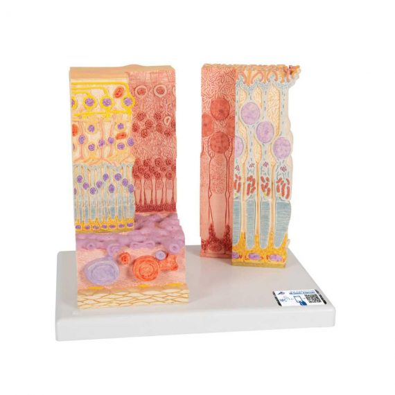
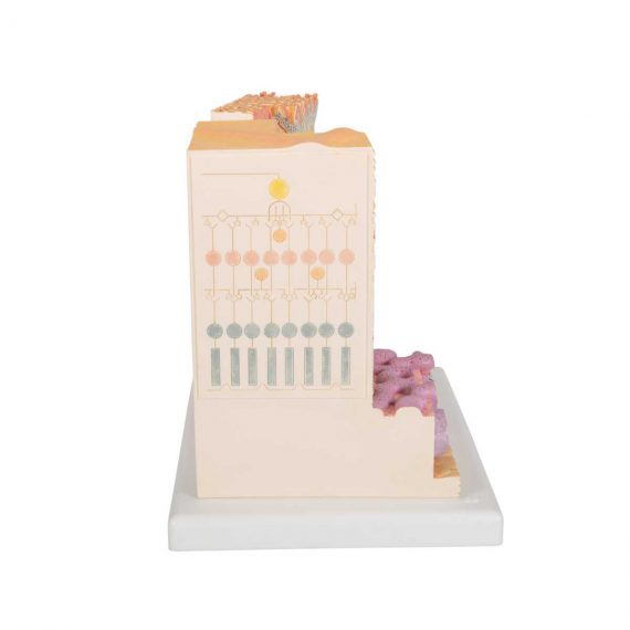
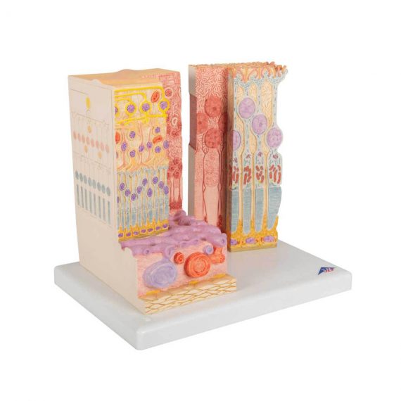
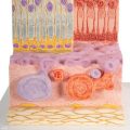
The MICROanatomy™ Eye model illustrates the microscopic anatomical structure of the retina with choroid and sclera. The left block-like, layered side of the eye model shows the complete structure of the retina including the supplying vascular layer and parts of the sclera from a light microscopic view.
Delivery & Availability:
Typically 5-7 working days – excluding furniture and heavy/bulky equipment. Please contact us for further information.
Description
The MICROanatomy™ Eye model illustrates the microscopic anatomical structure of the retina with choroid and sclera. The left block-like, layered side of the eye model shows the complete structure of the retina including the supplying vascular layer and parts of the sclera from a light microscopic view.
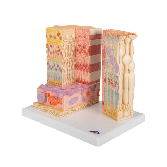
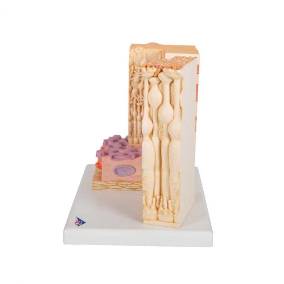
The right part of the eye model is a sectional enlargement. MICROanatomy™ Eye shows the microscopic structure of the photoreceptors and the cells of the pigmented layer.
Left part of MICROanatomy™ Eye 850-times enlarged – right part 3800-times enlarged. You’ve never seen the human eye like this before!
Quick Comparison
| Settings | 3B Scientific 3B MICROanatomy™ Human Eye Model - 3B Smart Anatomy remove | Chemical Weighing Balance remove | Petri Dish Per Pack remove | Measuring Beaker 500ml remove | Glass Measuring Cylinder 500ml remove | OPTIKA B-382PL-ALC Binocular Brightfeld Microscope remove | ||||||||||||||
|---|---|---|---|---|---|---|---|---|---|---|---|---|---|---|---|---|---|---|---|---|
| Name | 3B Scientific 3B MICROanatomy™ Human Eye Model - 3B Smart Anatomy remove | Chemical Weighing Balance remove | Petri Dish Per Pack remove | Measuring Beaker 500ml remove | Glass Measuring Cylinder 500ml remove | OPTIKA B-382PL-ALC Binocular Brightfeld Microscope remove | ||||||||||||||
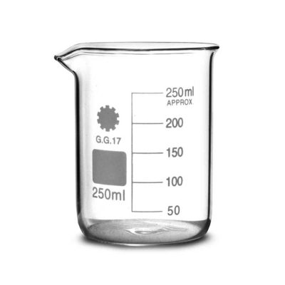
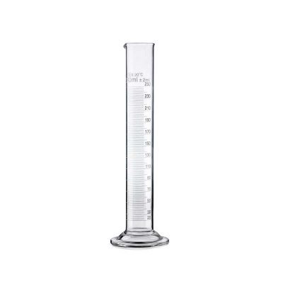
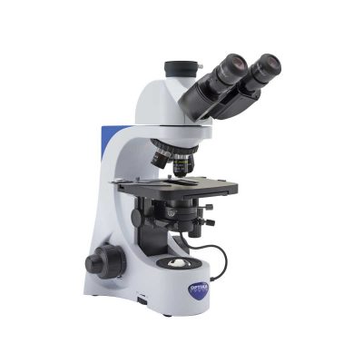
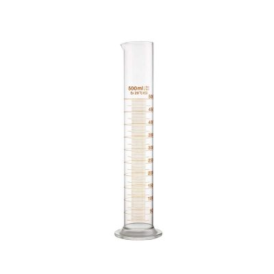
| Image |  | 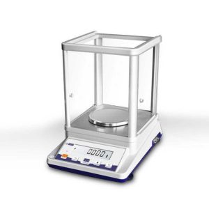 | 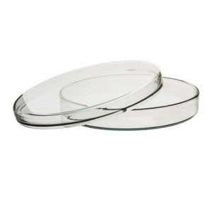 | 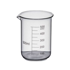 | 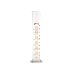 | 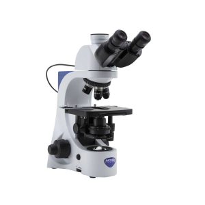 | ||||||||||||||
| SKU | SF1033560099-34 | SF1033560084-190 | SF1033560084-172 | SF1033560084-154 | SF1033560084-163 | SF1033560098-13 | ||||||||||||||
| Rating | ||||||||||||||||||||
| Price | Ask for Price | $658.00 | Ask for Price | $4.95 | $7.00 | Ask for Price | ||||||||||||||
| Stock | ||||||||||||||||||||
| Availability | ||||||||||||||||||||
| Add to cart | ||||||||||||||||||||
| Description | Ship from abroad
The MICROanatomy™ Eye model illustrates the microscopic anatomical structure of the retina with choroid and sclera. The left block-like, layered side of the eye model shows the complete structure of the retina including the supplying vascular layer and parts of the sclera from a light microscopic view.
| In stock
This is a 210g/0.001g Analytical electronic chemical balance for industrial and scientific field.
| In stock
Premium petri dishes made to withstand wet or dry repeated sterilisation. The heavy borosilicate glass and can handle direct flames.
| In stock
| In stock
| Shipped from Abroad The OPTIKA B-380 is an educational and laboratory microscope for routine applications. Dye-cast frame, with high stability and ergonomy, for transmitted light observation. Delivery & Availability: Typically 21 working days – excluding furniture and heavy/bulky equipment. Please contact us for further information. | ||||||||||||||
| Content | The MICROanatomy™ Eye model illustrates the microscopic anatomical structure of the retina with choroid and sclera. The left block-like, layered side of the eye model shows the complete structure of the retina including the supplying vascular layer and parts of the sclera from a light microscopic view. The right part of the eye model is a sectional enlargement. MICROanatomy™ Eye shows the microscopic structure of the photoreceptors and the cells of the pigmented layer. Left part of MICROanatomy™ Eye 850-times enlarged - right part 3800-times enlarged. You've never seen the human eye like this before! | This is a 210g/0.001g Analytical electronic chemical balance for industrial and scientific field.
| Premium petri dishes made to withstand wet or dry repeated sterilisation. The heavy borosilicate glass and can handle direct flames.
Features:
|
|
| Observation mode: Brightfield.
Head: Binocular, 30° inclined, 360° rotating (when ALC cable is unplugged).
Interpupillary distance: Adjustable between 48 and 75 mm.
Dioptric adjustment: On the left eyepiece tube.
Eyepieces: WF10x/20 mm, high eye-point and secured by screw.
Nosepiece: Quintuple revolving nosepiece, rotation on ball bearings.
Objectives:
N-PLAN 4x/0.10
N-PLAN 10x/0.25
N-PLAN 40x/0.65
N-PLAN 100x/1.25 (Oil/Water)
All with anti-fungus treatment.
Specimen stage: Double layer rackless mechanical stage, 150×139 mm, 75×33 mm X-Y range.
Focusing: Coaxial coarse (adjustable tension) and fine focusing mechanism with limit stop to prevent the contact between objective and specimen. Condenser: Abbe N.A. 1.25, with objective-coded iris diaphragm, focusable and centerable.
Illumination (Fixed Koehler type): X-LED3 with white 3.6 W LED (6,300K) and brightness control. ALC system. Multi-plug 100-240Vac/6Vdc external power supply.
Click Here To Download Catalogue | ||||||||||||||
| Weight | N/A | N/A | N/A | N/A | N/A | N/A | ||||||||||||||
| Dimensions | N/A | N/A | N/A | N/A | N/A | N/A | ||||||||||||||
| Additional information |


Reviews
There are no reviews yet.