

Anke Anatom 32 Fit Multi-Slice Spiral CT Scan
$0.00
Shipped from Abroad
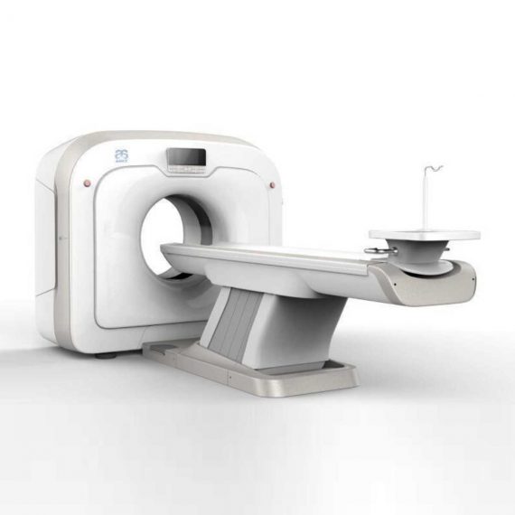
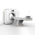
This Machine gives a possibility to perform computed tomography without any problems and on high quality level. This device is used to conduct exams of internal organs and their functioning. With its help, a physician has a possibility to assess the condition of the human body as a whole.
Delivery & Availability:
Typically 90 working days – excluding furniture and heavy/bulky equipment. Please contact us for further information.
Description
This Machine gives a possibility to perform computed tomography without any problems and on high quality level. This device is used to conduct exams of internal organs and their functioning. With its help, a physician has a possibility to assess the condition of the human body as a whole.
Features:
- It is easy to use;
- Convenience;
- Multi functionality;
- Obtained images are of high definition;
- High-definition 3D images of the area under study;
- The procedure is pain-free;
- The data is processed fast;
- The image can be stored in the computer memory;
- The diagnostics does not take a lot of time;
- Acceptable radiation dose.
Technical Specifications:
| No. | Technical Features | Descriptions |
| 1 | Gantry | |
| 1.01 | Gantry type | Low voltage slip-ring |
| 1.02 | Gantry driven type | Strap-driven |
| 1.03 | Patient opening | 70cm |
| 1.04 | Gantry tilt mode | Digital gantry tilt |
| 1.05 | Digital tilt capability | ±50° |
| 1.06 | Detector type | OptiWave rare-earth ceramic detector |
| 1.07 | Numbers of detector rows | 16 |
| 1.08 | Width of Z-axle detector | 20mm |
| 1.09 | Detector columns of channels per row | 848 |
| 1.10 | Numbers of detector columns | 13568 |
| 1.11 | Data-transfer type | RF, optical fiber communication |
| 1.12 | Distance of focus-ISO-center | 53cm |
| 1.13 | Distance of focus-detector | 94cm |
| 1.14 | 3D laser orientation | Provided |
| 1.15 | 13″ integrated display panel | Provided |
| 1.16 | Adose automatic exposure control (mA
Modulation) |
Provided |
| 1.17 | Auto-voice manager | Breath Graphical Display
Hold Message (Record/Playback) Breath Message (Record/Playback) |
| 1.18 | AccuSaving energy conservation management | Provided |
| 2 | HVPS and X-ray tube | |
| 2.01 | Maximum continuous output of HVgenerator | 42kW |
| 2.02 | Tube kV selections | 70kV, 80kV, 100 kV, 120 kV, 140 kV |
| 2.03 | Tube mA range | 10~350mA |
| 2.04 | Tube anode heat capacity | 3.5MHU |
| 2.05 | Max. anode cooling rate | 735kHU/min |
| 2.06 | Type of cooling | Oil cooling + Air cooling |
| 2.07 | Tube focus | Large: 1.2mm×1.4mm
Small: 0.7mm×0.8mm |
| 2.08 | Collimator width selection | 4-level election |
| 2.09 | Focus spot tracking technology | Provided |
| 3 | Patient table | |
| 3.01 | Maximum horizontal-movable range | 1850mm |
| 3.02 | Table horizontal-scannablerange | 1800mm |
| 3.03 | Table horizontal-position repeatability | ±0.25mm |
| 3.04 | Minimum height above floor | 430mm |
| 3.05 | Maximum vertical-movable range | 500mm |
| 3.06 | Maximum speed of vertical movement | 35mm |
| 3.07 | Maximum speed of horizontal movement | 150mm/s |
| 3.08 | Maximum patient weight | 205kg |
| 3.09 | Foot pedal of patient table control | Provided |
| 4 | Computer | |
| 4.01 | CPU | 3.5GHz |
| 4.02 | Memory | 32GB |
| 4.03 | Storage of hard-disk | 1TB×2 |
| 4.04 | Monitor | 24’’ LCD Monitor |
| 4.05 | Resolution of monitor | 1920×1200 |
| 4.06 | Image-data external storage type | CD/DVD/USB |
| 4.07 | Time of image reconstruction (512×512) | 33.3ms/image |
| 4.08 | Speed of image reconstruction (512×12) | 30fps |
| 4.09 | DICOM 3.0 interface | Provided |
| 4.10 | Printer DICOM 3.0 interface | Provided |
| 4.11 | Auto filming | Provided |
| 4.12 | Worklist function | Provided |
| 5 | Scan parameters | |
| 5.01 | Shortest 360 degree rotation time | 0.75s |
| 5.02 | Allowed rotation times | 0.75s, 1.0s, 1.5s, 2.0s, 3.0s, 4.0s |
| 5.03 | Maximum slice numbers per rotation | 32 |
| 5.04 | Minimum slice thickness of scan | 1.25mm |
| 5.05 | Minimum slice thickness of reconstruction | 0.625mm |
| 5.06 | Maximum slice thickness of scan | 20mm |
| 5.07 | Nominal reconstruction slice thickness | 0.625mm, 1.25mm, 2.5mm, 5.0mm, 7.5mm,
10mm, 20mm |
| 5.08 | Speed of image reconstruction (512×512) | 30 frames/s |
| 5.09 | Scan FOV | 50cm |
| 5.10 | Image reconstruction matrix | 512×512, 1024×1024 (Optional) |
| 5.11 | Image reconstruction matrix | 512×512, 1024×1024 (Optional) |
| 5.12 | Image display matrix | 512×512, 1024×1024 (Optional) |
| 5.13 | Maximum continuous scan duration | 120s |
| 5.14 | Maximum continuous scan length | 180cm |
| 5.15 | Direction of TOPO | Front-back, Left-right |
| 5.16 | Max. length of TOPO | 180cm |
| 5.17 | Range of pitch | 0.5~1.5 |
| 5.18 | Scan mode | Scout scan
Axial scan Helical scan Cine scan |
| 6 | Image Quality | |
| 6.01 | High contrast resolution | 21lp/cm@0%MTF |
| 6.02 | Low contrast resolution | 2.0mm@0.30% |
| 6.03 | Isotropic imaging resolution | 0.24mm |
| 6.04 | Range of CT numbers | -32767~32768 |
| 6.05 | Image noise | ≤0.29@28mGy |
| 7 | Advanced application | |
| 7.01 | Multi-Planar Reconstruction (MPR) | Provided |
| 7.02 | Curve Multi-Planar Reconstruction (CPR) | Provided |
| 7.03 | Surface Shaded Display (SSD) | Provided |
| 7.04 | Volume Rendering (VR) | Provided |
| 7.05 | Maximum Intensity Projection (MIP) | Provided |
| 7.06 | Minimum Intensity Projection (MinIP) | Provided |
| 7.07 | Virtual Endoscopy (VE) | Provided |
| 7.08 | CT angiography (CTA) | Provided |
| 7.09 | Tissue segmentation | Provided |
| 7.10 | One click bone remove | Provided |
| 7.11 | One click patient table remove | Provided |
| 7.12 | Bolus-tracking Technology | Provided |
| 7.13 | Spiral auto start | Provided |
| 7.14 | Cine display | Provided |
| 7.15 | AbastTM bone artifact suppression technology | Provided |
| 7.16 | AmastTM metal artifact suppression technology | Provided |
| 7.17 | Admir3D all-domain iterative reconstruction | Provided |
| 7.18 | Low-dose pediatric scan technology | Provided |
| 7.19 | Low-dose lung scan technology | Provided |
| 7.20 | AccuHead grey-white matter enhanced
technology |
Provided |
| 7.21 | AccuOrgan lung high resolution scan technology | Provided |
| 7.22 | AccuOrgan inner-ear high resolution scan
technology |
Provided |
| 7.23 | AccuOrgan body high resolution scan technology | Provided |
| 7.24 | AccuOrgan bone high resolution scan technology | Provided |
| 7.25 | AccuMatter dual-energy with Admir3D for new
application |
Provided |
Click Here To Download Catalogue
Additional information
| Model | Advanced, Advanced Plus, Basic, Smart |
|---|
Quick Comparison
| Anke Anatom 32 Fit Multi-Slice Spiral CT Scan remove | Bistos BT-770-12.1" Touchscreen Patient Monitor remove | ASPEL AsCARD Green ECG Machine remove | Sonoscape S8 Exp Portable Ultrasound remove | ASPEL AsCARD Grey ECG Machine remove | DrGem Diamond All-In-One Digital X-ray Machine remove | |||||||||||||||||||||||||||||||||||||||||||||||||||||||||||||||||||||||||||||||||||||||||||||||||||||||||||||||||||||||||||||||||||||||||||||||||||||||||||||||||||||||||||||||||||||||||||||||||||||||||||||||||||||||||||||||||||||||||||||||||||||||||||||||||||||||||||||||||||||||||||||||||||||||||||||||||||||||||
|---|---|---|---|---|---|---|---|---|---|---|---|---|---|---|---|---|---|---|---|---|---|---|---|---|---|---|---|---|---|---|---|---|---|---|---|---|---|---|---|---|---|---|---|---|---|---|---|---|---|---|---|---|---|---|---|---|---|---|---|---|---|---|---|---|---|---|---|---|---|---|---|---|---|---|---|---|---|---|---|---|---|---|---|---|---|---|---|---|---|---|---|---|---|---|---|---|---|---|---|---|---|---|---|---|---|---|---|---|---|---|---|---|---|---|---|---|---|---|---|---|---|---|---|---|---|---|---|---|---|---|---|---|---|---|---|---|---|---|---|---|---|---|---|---|---|---|---|---|---|---|---|---|---|---|---|---|---|---|---|---|---|---|---|---|---|---|---|---|---|---|---|---|---|---|---|---|---|---|---|---|---|---|---|---|---|---|---|---|---|---|---|---|---|---|---|---|---|---|---|---|---|---|---|---|---|---|---|---|---|---|---|---|---|---|---|---|---|---|---|---|---|---|---|---|---|---|---|---|---|---|---|---|---|---|---|---|---|---|---|---|---|---|---|---|---|---|---|---|---|---|---|---|---|---|---|---|---|---|---|---|---|---|---|---|---|---|---|---|---|---|---|---|---|---|---|---|---|---|---|---|---|---|---|---|---|---|---|---|---|---|---|---|---|---|---|---|---|---|---|---|---|---|---|---|---|---|---|---|---|---|---|---|---|---|---|---|---|---|
| Name | Anke Anatom 32 Fit Multi-Slice Spiral CT Scan remove | Bistos BT-770-12.1" Touchscreen Patient Monitor remove | ASPEL AsCARD Green ECG Machine remove | Sonoscape S8 Exp Portable Ultrasound remove | ASPEL AsCARD Grey ECG Machine remove | DrGem Diamond All-In-One Digital X-ray Machine remove | ||||||||||||||||||||||||||||||||||||||||||||||||||||||||||||||||||||||||||||||||||||||||||||||||||||||||||||||||||||||||||||||||||||||||||||||||||||||||||||||||||||||||||||||||||||||||||||||||||||||||||||||||||||||||||||||||||||||||||||||||||||||||||||||||||||||||||||||||||||||||||||||||||||||||||||||||||||||||
| Image |  | 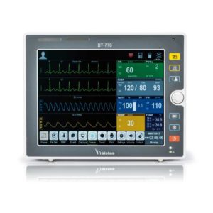 |  |  |  |  | ||||||||||||||||||||||||||||||||||||||||||||||||||||||||||||||||||||||||||||||||||||||||||||||||||||||||||||||||||||||||||||||||||||||||||||||||||||||||||||||||||||||||||||||||||||||||||||||||||||||||||||||||||||||||||||||||||||||||||||||||||||||||||||||||||||||||||||||||||||||||||||||||||||||||||||||||||||||||
| SKU | SF1033560092-1 | SF1033560059-1 | SF1033560075-9 | SF1033560012-15 | SF1033560075-5 | SF1033560074-3 | ||||||||||||||||||||||||||||||||||||||||||||||||||||||||||||||||||||||||||||||||||||||||||||||||||||||||||||||||||||||||||||||||||||||||||||||||||||||||||||||||||||||||||||||||||||||||||||||||||||||||||||||||||||||||||||||||||||||||||||||||||||||||||||||||||||||||||||||||||||||||||||||||||||||||||||||||||||||||
| Rating | ||||||||||||||||||||||||||||||||||||||||||||||||||||||||||||||||||||||||||||||||||||||||||||||||||||||||||||||||||||||||||||||||||||||||||||||||||||||||||||||||||||||||||||||||||||||||||||||||||||||||||||||||||||||||||||||||||||||||||||||||||||||||||||||||||||||||||||||||||||||||||||||||||||||||||||||||||||||||||||||
| Price |
| $902.00 |
| $9,350.00 | $1,166.00 |
| ||||||||||||||||||||||||||||||||||||||||||||||||||||||||||||||||||||||||||||||||||||||||||||||||||||||||||||||||||||||||||||||||||||||||||||||||||||||||||||||||||||||||||||||||||||||||||||||||||||||||||||||||||||||||||||||||||||||||||||||||||||||||||||||||||||||||||||||||||||||||||||||||||||||||||||||||||||||||
| Stock | ||||||||||||||||||||||||||||||||||||||||||||||||||||||||||||||||||||||||||||||||||||||||||||||||||||||||||||||||||||||||||||||||||||||||||||||||||||||||||||||||||||||||||||||||||||||||||||||||||||||||||||||||||||||||||||||||||||||||||||||||||||||||||||||||||||||||||||||||||||||||||||||||||||||||||||||||||||||||||||||
| Availability | ||||||||||||||||||||||||||||||||||||||||||||||||||||||||||||||||||||||||||||||||||||||||||||||||||||||||||||||||||||||||||||||||||||||||||||||||||||||||||||||||||||||||||||||||||||||||||||||||||||||||||||||||||||||||||||||||||||||||||||||||||||||||||||||||||||||||||||||||||||||||||||||||||||||||||||||||||||||||||||||
| Add to cart | ||||||||||||||||||||||||||||||||||||||||||||||||||||||||||||||||||||||||||||||||||||||||||||||||||||||||||||||||||||||||||||||||||||||||||||||||||||||||||||||||||||||||||||||||||||||||||||||||||||||||||||||||||||||||||||||||||||||||||||||||||||||||||||||||||||||||||||||||||||||||||||||||||||||||||||||||||||||||||||||
| Description | Shipped from Abroad
This Machine gives a possibility to perform computed tomography without any problems and on high quality level. This device is used to conduct exams of internal organs and their functioning. With its help, a physician has a possibility to assess the condition of the human body as a whole.
Delivery & Availability: Typically 90 working days – excluding furniture and heavy/bulky equipment. Please contact us for further information. | Shipped from Abroad The Bistos BT-770 patient monitor is equipped with a 12.1" touchscreen display, which allows for an easy operation and readability with a powerful rechargeable battery guaranteeing a continuous operation of 5 hours to monitor ECG, SpO2, NIBP, temperature and respiration Delivery & Availability: Typically 14 working days – excluding furniture and heavy/bulky equipment. Please contact us for further information. | Shipped from Abroad AsCARD Green v.06.101 is a 1-, 3-, 6- and 12-channel ECG unit which enables to make electrocardiogram in full 12 leads. Intended for ECG examinations of adult and paediatric patients aimed at identification of cardiological abnormalities, myocardial ischaemia or infarction. The device is intended for use in healthcare facilities by duly trained personnel. ECG examination may be recorded in manual or automatic mode with the ability to perform the analysis and interpretation. Delivery & Availability: Typically 10 working days – excluding furniture and heavy/bulky equipment. Please contact us for further information. | Shipped from Abroad With ultra-modern innovative design and the clinically-proven technologies, S8 Exp is portable ultrasound scanner well equipped as a low-physical-effort and enhanced-image-quality ultrasound scanner, which not only provides optimized solutions for versatile applications, but does help to improve the user-experience for both routine and non-traditional challenges. Delivery & Availability: Typically 5-7 working days – excluding furniture and heavy/bulky equipment. Please contact us for further information. | Shipped from Abroad Electrocardiograph AsCARD Grey v.07.225 - is a 1, 3, 6, 12 channel ECG unit which enables to make electrocardiogram in full 12 leads. It is intended to conduct ECG examinations of adults and paediatric patients in all types of health care centres. ECG examination may be recorded in manual or automatic mode, with the possibility of analysis and interpretation. The device can be powered from 100 V ÷ 240 V mains supply or by an internal battery. Delivery & Availability: Typically 10 working days – excluding furniture and heavy/bulky equipment. Please contact us for further information. | Shipped from Abroad DrGem Diamond All-In-One Digital X-ray Machine is a fully automatic digital radiography system providing state-of-the-art image quality, image processing and user interface. With a wide selection of anatomical studies on the imaging software, DIAMOND automatically sets up the x-ray generator’s preprogrammed exposure technique settings, motorized radiographic stand positioning, x-ray collimation and post-image processing for the selected study. Specifically designed to increase workflow, this fully digital system offers convenient auto-positioning and advanced image processing to achieve big performance with little effort. Delivery & Availability: Typically 21 working days – excluding furniture and heavy/bulky equipment. Please contact us for further information. | ||||||||||||||||||||||||||||||||||||||||||||||||||||||||||||||||||||||||||||||||||||||||||||||||||||||||||||||||||||||||||||||||||||||||||||||||||||||||||||||||||||||||||||||||||||||||||||||||||||||||||||||||||||||||||||||||||||||||||||||||||||||||||||||||||||||||||||||||||||||||||||||||||||||||||||||||||||||||
| Content | This Machine gives a possibility to perform computed tomography without any problems and on high quality level. This device is used to conduct exams of internal organs and their functioning. With its help, a physician has a possibility to assess the condition of the human body as a whole.
Features:
Click Here To Download Catalogue |
Bistos BT-770 is a 12.1" touchscreen patient monitor designed for easy operations.
SPECIFICATIONS
Click Here To Download Catalogue | AsCARD Green v.06.101 is a 1-, 3-, 6- and 12-channel ECG unit which enables to make electrocardiogram in full 12 leads. Intended for ECG examinations of adult and paediatric patients aimed at identification of cardiological abnormalities, myocardial ischaemia or infarction. The device is intended for use in healthcare facilities by duly trained personnel. ECG examination may be recorded in manual or automatic mode with the ability to perform the analysis and interpretation.
Electrocardiograph is based on advanced microprocessor technology .It is equipped with a thermal printer with high-resolution head and 4,3" LCD display. A touch panel and high-tech membrane keyboard makes this device intuitive in usage and its menu navigation exceptionally easy. This light-weight, small-footprint and battery powered cause that device can be easily transported to any location. With plastic casing and foil covered keyboard, the device is neat and easy to clean.
Technical Specifications:
Click Here To Catalogue Download | Sonoscape S8 Exp Portable Ultrasound scannerDETAILS Agile and Versatile With ultra-modern innovative design and the clinically-proven technologies, S8 Exp Portable Ultrasound scanner is well equipped as a low-physical-effort and enhanced-image-quality ultrasound scanner, which not only provides optimized solutions for versatile applications but does help to improve the user experience for both routine and non-traditional challenges. Working with S8 Exp, it will trigger your unlimited reverie and endow you with endless charm. Carrying forward the classical design of SonoScape's portable ultrasound products, S8 Exp successfully combines the best ergonomics, attractive design and ease of use. This charismatic identity is also enhanced by a sophisticated color palette—with sedate grey as its interior paint color and pearl white as exterior cover, S8 Exp reveals a style of aristocrat and strong character among SonoScape's ultrasound systems. Workflow The S8 Exp is a portable ultrasound scanner that adapts to your workflow, whether you are in the consulting room, at the bedside, or at a remote location. With easy-to-use new platform designed for sonographers' needs and full connection interfaces for easy connectivity and data sharing, S8 Exp leads to improved user comfort and clinical outcome as well as patient throughput and working efficiency. Powerful Platform Embedded with SonoScape's core imaging technologies such as μ-scan, PHI and Spatial Compound, S8 Exp boasts exceptional 2D image, sensitive spectral, Color and Power Doppler, displaying well-defined anatomy and pathology and facilitating a highly optimized diagnostic user environment for conclusive diagnoses. Besides, S8 Exp offers a comprehensive selection of electronic probes to maximally extend its capabilities to meet a wide range of applications including the abdomen, pediatric, OB/GYN, cardiovascular, musculoskeletal, etc. The advanced probe technologies also effectively enhance the image quality and confidence in reaching clinical diagnoses even in difficult patients.Click Here To Download Catalogue |
Electrocardiograph AsCARD Grey v.07.225 - is a 1, 3, 6, 12 channel ECG unit which enables to make electrocardiogram in full 12 leads. It is intended to conduct ECG examinations of adults and paediatric patients in all types of health care centres. ECG examination may be recorded in manual or automatic mode, with the possibility of analysis and interpretation. The device can be powered from 100 V ÷ 240 V mains supply or by an internal battery.
Technical Specification:1. Visualisation of 1, 3, 6 or 12 ECG waveforms, analysis results and interpretations, examinations stored in memory.
2. Recording of 12 standard leads.
3. Print out in 1, 3, 6 or 12 ECG waveforms mode. Printing of a selected group:
Click Here To Download Catalogue | DrGem Diamond All-In-One Digital X-ray Machine is a fully automatic digital radiography system providing state-of-the-art image quality, image processing and user interface. With a wide selection of anatomical studies on the imaging software, DIAMOND automatically sets up the x-ray generator’s pre-programmed exposure technique settings, motorized radiographic stand positioning, x-ray collimation and post-image processing for the selected study. Specifically designed to increase workflow, this fully digital system offers convenient auto-positioning and advanced image processing to achieve big performance with little effort.
Features of DrGem Diamond All-In-One Digital X-ray Machine:
Outstanding Image Quality -
Digital radiography via at panel detector improves your workflow, exam speed and comfort with efficiency. Digital at panel detector with Csl screen provides excellent spatial resolution, MTF, DQE and stability based on ne pixel pitch. A 3-field ion-chamber is provided for AEC function.
Automatic Collimation –
Automatic x-ray eld size control of the motorized collimator corresponds to dierent SIDs. Includes user adjustable lamp timer with on/oswitch.
Automatic Positioning –
Click Here To Download Catalogue | ||||||||||||||||||||||||||||||||||||||||||||||||||||||||||||||||||||||||||||||||||||||||||||||||||||||||||||||||||||||||||||||||||||||||||||||||||||||||||||||||||||||||||||||||||||||||||||||||||||||||||||||||||||||||||||||||||||||||||||||||||||||||||||||||||||||||||||||||||||||||||||||||||||||||||||||||||||||||
| Weight | N/A | N/A | N/A | N/A | N/A | N/A | ||||||||||||||||||||||||||||||||||||||||||||||||||||||||||||||||||||||||||||||||||||||||||||||||||||||||||||||||||||||||||||||||||||||||||||||||||||||||||||||||||||||||||||||||||||||||||||||||||||||||||||||||||||||||||||||||||||||||||||||||||||||||||||||||||||||||||||||||||||||||||||||||||||||||||||||||||||||||
| Dimensions | N/A | N/A | N/A | N/A | N/A | N/A | ||||||||||||||||||||||||||||||||||||||||||||||||||||||||||||||||||||||||||||||||||||||||||||||||||||||||||||||||||||||||||||||||||||||||||||||||||||||||||||||||||||||||||||||||||||||||||||||||||||||||||||||||||||||||||||||||||||||||||||||||||||||||||||||||||||||||||||||||||||||||||||||||||||||||||||||||||||||||
| Additional information |
|


Reviews
There are no reviews yet.