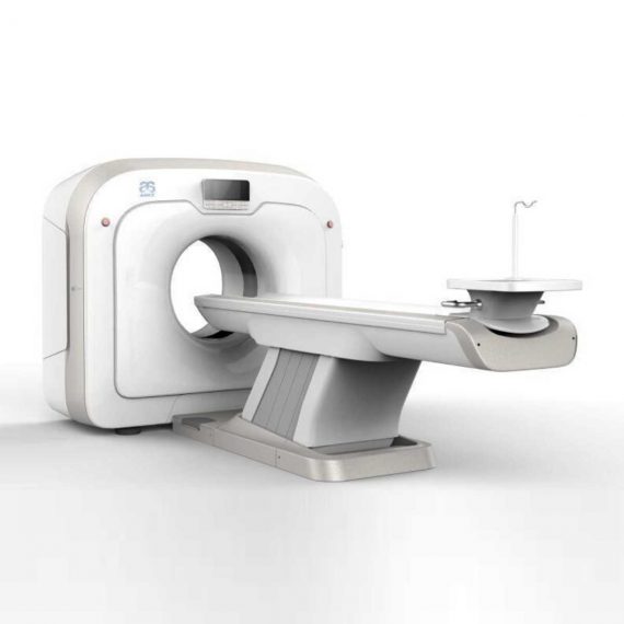

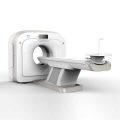

Anke Anatom 32 Fit Multi-Slice Spiral CT Scan
$0.00
Shipped from Abroad
This Machine gives a possibility to perform computed tomography without any problems and on high quality level. This device is used to conduct exams of internal organs and their functioning. With its help, a physician has a possibility to assess the condition of the human body as a whole.
Delivery & Availability:
Typically 90 working days – excluding furniture and heavy/bulky equipment. Please contact us for further information.
Description
This Machine gives a possibility to perform computed tomography without any problems and on high quality level. This device is used to conduct exams of internal organs and their functioning. With its help, a physician has a possibility to assess the condition of the human body as a whole.
Features:
- It is easy to use;
- Convenience;
- Multi functionality;
- Obtained images are of high definition;
- High-definition 3D images of the area under study;
- The procedure is pain-free;
- The data is processed fast;
- The image can be stored in the computer memory;
- The diagnostics does not take a lot of time;
- Acceptable radiation dose.
Technical Specifications:
| No. | Technical Features | Descriptions |
| 1 | Gantry | |
| 1.01 | Gantry type | Low voltage slip-ring |
| 1.02 | Gantry driven type | Strap-driven |
| 1.03 | Patient opening | 70cm |
| 1.04 | Gantry tilt mode | Digital gantry tilt |
| 1.05 | Digital tilt capability | ±50° |
| 1.06 | Detector type | OptiWave rare-earth ceramic detector |
| 1.07 | Numbers of detector rows | 16 |
| 1.08 | Width of Z-axle detector | 20mm |
| 1.09 | Detector columns of channels per row | 848 |
| 1.10 | Numbers of detector columns | 13568 |
| 1.11 | Data-transfer type | RF, optical fiber communication |
| 1.12 | Distance of focus-ISO-center | 53cm |
| 1.13 | Distance of focus-detector | 94cm |
| 1.14 | 3D laser orientation | Provided |
| 1.15 | 13″ integrated display panel | Provided |
| 1.16 | Adose automatic exposure control (mA
Modulation) |
Provided |
| 1.17 | Auto-voice manager | Breath Graphical Display
Hold Message (Record/Playback) Breath Message (Record/Playback) |
| 1.18 | AccuSaving energy conservation management | Provided |
| 2 | HVPS and X-ray tube | |
| 2.01 | Maximum continuous output of HVgenerator | 42kW |
| 2.02 | Tube kV selections | 70kV, 80kV, 100 kV, 120 kV, 140 kV |
| 2.03 | Tube mA range | 10~350mA |
| 2.04 | Tube anode heat capacity | 3.5MHU |
| 2.05 | Max. anode cooling rate | 735kHU/min |
| 2.06 | Type of cooling | Oil cooling + Air cooling |
| 2.07 | Tube focus | Large: 1.2mm×1.4mm
Small: 0.7mm×0.8mm |
| 2.08 | Collimator width selection | 4-level election |
| 2.09 | Focus spot tracking technology | Provided |
| 3 | Patient table | |
| 3.01 | Maximum horizontal-movable range | 1850mm |
| 3.02 | Table horizontal-scannablerange | 1800mm |
| 3.03 | Table horizontal-position repeatability | ±0.25mm |
| 3.04 | Minimum height above floor | 430mm |
| 3.05 | Maximum vertical-movable range | 500mm |
| 3.06 | Maximum speed of vertical movement | 35mm |
| 3.07 | Maximum speed of horizontal movement | 150mm/s |
| 3.08 | Maximum patient weight | 205kg |
| 3.09 | Foot pedal of patient table control | Provided |
| 4 | Computer | |
| 4.01 | CPU | 3.5GHz |
| 4.02 | Memory | 32GB |
| 4.03 | Storage of hard-disk | 1TB×2 |
| 4.04 | Monitor | 24’’ LCD Monitor |
| 4.05 | Resolution of monitor | 1920×1200 |
| 4.06 | Image-data external storage type | CD/DVD/USB |
| 4.07 | Time of image reconstruction (512×512) | 33.3ms/image |
| 4.08 | Speed of image reconstruction (512×12) | 30fps |
| 4.09 | DICOM 3.0 interface | Provided |
| 4.10 | Printer DICOM 3.0 interface | Provided |
| 4.11 | Auto filming | Provided |
| 4.12 | Worklist function | Provided |
| 5 | Scan parameters | |
| 5.01 | Shortest 360 degree rotation time | 0.75s |
| 5.02 | Allowed rotation times | 0.75s, 1.0s, 1.5s, 2.0s, 3.0s, 4.0s |
| 5.03 | Maximum slice numbers per rotation | 32 |
| 5.04 | Minimum slice thickness of scan | 1.25mm |
| 5.05 | Minimum slice thickness of reconstruction | 0.625mm |
| 5.06 | Maximum slice thickness of scan | 20mm |
| 5.07 | Nominal reconstruction slice thickness | 0.625mm, 1.25mm, 2.5mm, 5.0mm, 7.5mm,
10mm, 20mm |
| 5.08 | Speed of image reconstruction (512×512) | 30 frames/s |
| 5.09 | Scan FOV | 50cm |
| 5.10 | Image reconstruction matrix | 512×512, 1024×1024 (Optional) |
| 5.11 | Image reconstruction matrix | 512×512, 1024×1024 (Optional) |
| 5.12 | Image display matrix | 512×512, 1024×1024 (Optional) |
| 5.13 | Maximum continuous scan duration | 120s |
| 5.14 | Maximum continuous scan length | 180cm |
| 5.15 | Direction of TOPO | Front-back, Left-right |
| 5.16 | Max. length of TOPO | 180cm |
| 5.17 | Range of pitch | 0.5~1.5 |
| 5.18 | Scan mode | Scout scan
Axial scan Helical scan Cine scan |
| 6 | Image Quality | |
| 6.01 | High contrast resolution | 21lp/cm@0%MTF |
| 6.02 | Low contrast resolution | 2.0mm@0.30% |
| 6.03 | Isotropic imaging resolution | 0.24mm |
| 6.04 | Range of CT numbers | -32767~32768 |
| 6.05 | Image noise | ≤0.29@28mGy |
| 7 | Advanced application | |
| 7.01 | Multi-Planar Reconstruction (MPR) | Provided |
| 7.02 | Curve Multi-Planar Reconstruction (CPR) | Provided |
| 7.03 | Surface Shaded Display (SSD) | Provided |
| 7.04 | Volume Rendering (VR) | Provided |
| 7.05 | Maximum Intensity Projection (MIP) | Provided |
| 7.06 | Minimum Intensity Projection (MinIP) | Provided |
| 7.07 | Virtual Endoscopy (VE) | Provided |
| 7.08 | CT angiography (CTA) | Provided |
| 7.09 | Tissue segmentation | Provided |
| 7.10 | One click bone remove | Provided |
| 7.11 | One click patient table remove | Provided |
| 7.12 | Bolus-tracking Technology | Provided |
| 7.13 | Spiral auto start | Provided |
| 7.14 | Cine display | Provided |
| 7.15 | AbastTM bone artifact suppression technology | Provided |
| 7.16 | AmastTM metal artifact suppression technology | Provided |
| 7.17 | Admir3D all-domain iterative reconstruction | Provided |
| 7.18 | Low-dose pediatric scan technology | Provided |
| 7.19 | Low-dose lung scan technology | Provided |
| 7.20 | AccuHead grey-white matter enhanced
technology |
Provided |
| 7.21 | AccuOrgan lung high resolution scan technology | Provided |
| 7.22 | AccuOrgan inner-ear high resolution scan
technology |
Provided |
| 7.23 | AccuOrgan body high resolution scan technology | Provided |
| 7.24 | AccuOrgan bone high resolution scan technology | Provided |
| 7.25 | AccuMatter dual-energy with Admir3D for new
application |
Provided |
Click Here To Download Catalogue
Additional information
| Model | Advanced, Advanced Plus, Basic, Smart |
|---|
Quick Comparison
| Settings | Anke Anatom 32 Fit Multi-Slice Spiral CT Scan remove | Jade Mobile X-ray machine (Analogue) remove | ASPEL Stress ECG with Ergometer and Software remove | Sonoscape E2 Ultrasound Machine remove | Sonoscape P20 Ultrasound Machine remove | Sonoscape P15 Ultrasound Machine With Four Probes remove | ||||||||||||||||||||||||||||||||||||||||||||||||||||||||||||||||||||||||||||||||||||||||||||||||||||||||||||||||||||||||||||||||||||||||||||||||||||||||||||||||||||||||||||||||||||||||||||||||||||||||||||||||||||||||||||||||||||||||||||||||||||||||||||||||||||||||||||||||||||||||||||||||||||||||||||||||||||||||
|---|---|---|---|---|---|---|---|---|---|---|---|---|---|---|---|---|---|---|---|---|---|---|---|---|---|---|---|---|---|---|---|---|---|---|---|---|---|---|---|---|---|---|---|---|---|---|---|---|---|---|---|---|---|---|---|---|---|---|---|---|---|---|---|---|---|---|---|---|---|---|---|---|---|---|---|---|---|---|---|---|---|---|---|---|---|---|---|---|---|---|---|---|---|---|---|---|---|---|---|---|---|---|---|---|---|---|---|---|---|---|---|---|---|---|---|---|---|---|---|---|---|---|---|---|---|---|---|---|---|---|---|---|---|---|---|---|---|---|---|---|---|---|---|---|---|---|---|---|---|---|---|---|---|---|---|---|---|---|---|---|---|---|---|---|---|---|---|---|---|---|---|---|---|---|---|---|---|---|---|---|---|---|---|---|---|---|---|---|---|---|---|---|---|---|---|---|---|---|---|---|---|---|---|---|---|---|---|---|---|---|---|---|---|---|---|---|---|---|---|---|---|---|---|---|---|---|---|---|---|---|---|---|---|---|---|---|---|---|---|---|---|---|---|---|---|---|---|---|---|---|---|---|---|---|---|---|---|---|---|---|---|---|---|---|---|---|---|---|---|---|---|---|---|---|---|---|---|---|---|---|---|---|---|---|---|---|---|---|---|---|---|---|---|---|---|---|---|---|---|---|---|---|---|---|---|---|---|---|---|---|---|---|---|---|---|---|---|---|
| Name | Anke Anatom 32 Fit Multi-Slice Spiral CT Scan remove | Jade Mobile X-ray machine (Analogue) remove | ASPEL Stress ECG with Ergometer and Software remove | Sonoscape E2 Ultrasound Machine remove | Sonoscape P20 Ultrasound Machine remove | Sonoscape P15 Ultrasound Machine With Four Probes remove | ||||||||||||||||||||||||||||||||||||||||||||||||||||||||||||||||||||||||||||||||||||||||||||||||||||||||||||||||||||||||||||||||||||||||||||||||||||||||||||||||||||||||||||||||||||||||||||||||||||||||||||||||||||||||||||||||||||||||||||||||||||||||||||||||||||||||||||||||||||||||||||||||||||||||||||||||||||||||
| Image |  |  | 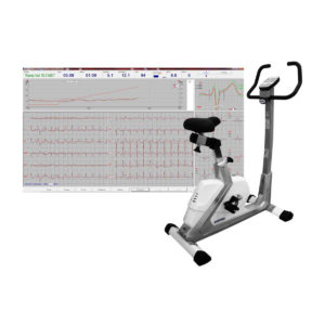 |  |  |  | ||||||||||||||||||||||||||||||||||||||||||||||||||||||||||||||||||||||||||||||||||||||||||||||||||||||||||||||||||||||||||||||||||||||||||||||||||||||||||||||||||||||||||||||||||||||||||||||||||||||||||||||||||||||||||||||||||||||||||||||||||||||||||||||||||||||||||||||||||||||||||||||||||||||||||||||||||||||||
| SKU | SF1033560092-1 | SF1033560074-2 | SF1033560075-1 | SF1033560012-17 | SF1033560012-9 | SF1033560012-8 | ||||||||||||||||||||||||||||||||||||||||||||||||||||||||||||||||||||||||||||||||||||||||||||||||||||||||||||||||||||||||||||||||||||||||||||||||||||||||||||||||||||||||||||||||||||||||||||||||||||||||||||||||||||||||||||||||||||||||||||||||||||||||||||||||||||||||||||||||||||||||||||||||||||||||||||||||||||||||
| Rating | ||||||||||||||||||||||||||||||||||||||||||||||||||||||||||||||||||||||||||||||||||||||||||||||||||||||||||||||||||||||||||||||||||||||||||||||||||||||||||||||||||||||||||||||||||||||||||||||||||||||||||||||||||||||||||||||||||||||||||||||||||||||||||||||||||||||||||||||||||||||||||||||||||||||||||||||||||||||||||||||
| Price |
|
| $4,202.00 | $5,500.00 |
| $13,900.00 | ||||||||||||||||||||||||||||||||||||||||||||||||||||||||||||||||||||||||||||||||||||||||||||||||||||||||||||||||||||||||||||||||||||||||||||||||||||||||||||||||||||||||||||||||||||||||||||||||||||||||||||||||||||||||||||||||||||||||||||||||||||||||||||||||||||||||||||||||||||||||||||||||||||||||||||||||||||||||
| Stock | ||||||||||||||||||||||||||||||||||||||||||||||||||||||||||||||||||||||||||||||||||||||||||||||||||||||||||||||||||||||||||||||||||||||||||||||||||||||||||||||||||||||||||||||||||||||||||||||||||||||||||||||||||||||||||||||||||||||||||||||||||||||||||||||||||||||||||||||||||||||||||||||||||||||||||||||||||||||||||||||
| Availability | ||||||||||||||||||||||||||||||||||||||||||||||||||||||||||||||||||||||||||||||||||||||||||||||||||||||||||||||||||||||||||||||||||||||||||||||||||||||||||||||||||||||||||||||||||||||||||||||||||||||||||||||||||||||||||||||||||||||||||||||||||||||||||||||||||||||||||||||||||||||||||||||||||||||||||||||||||||||||||||||
| Add to cart | ||||||||||||||||||||||||||||||||||||||||||||||||||||||||||||||||||||||||||||||||||||||||||||||||||||||||||||||||||||||||||||||||||||||||||||||||||||||||||||||||||||||||||||||||||||||||||||||||||||||||||||||||||||||||||||||||||||||||||||||||||||||||||||||||||||||||||||||||||||||||||||||||||||||||||||||||||||||||||||||
| Description | Shipped from Abroad
This Machine gives a possibility to perform computed tomography without any problems and on high quality level. This device is used to conduct exams of internal organs and their functioning. With its help, a physician has a possibility to assess the condition of the human body as a whole.
Delivery & Availability: Typically 90 working days – excluding furniture and heavy/bulky equipment. Please contact us for further information. | In Stock JADE is one of the lightest portable X-ray systems on the market, allowing it to be used in any imaginable way including bedside, operating rooms, intensive care units and in veterinary fields. With a simple, easy-to-use operator console, three-way control, two-step foldable stand and auto lock system, JADE is a user-friendly portable X-ray system. Delivery & Availability: Typically 21 working days – excluding furniture and heavy/bulky equipment. Please contact us for further information. | Shipped from Abroad Ergometer CRG 200 is dedicated for Exercise Stress Tests System CardioTEST. The Ergometer has been designed according to modern technologies. It is controlled from PC equipped in CardioTEST software. Load level is controlled by a microprocessor, therefore it does not depend on speed, which in turn can be adjusted according to patient’s individual needs. Ergometer is equipped with ECG mode recording 12 standard leads. Delivery & Availability: Typically 21 working days – excluding furniture and heavy/bulky equipment. Please contact us for further information. | Shipped from Abroad Sonoscape E2 portable ultrasound machine is a color Doppler ultrasound system that reaches beyond your expectations due to its compact and fashionable appearance. It fulfills GI, OB/GYN, Cardiac and POC applications to fit your routine scanning needs while its color mode will help you for more accurate and efficient diagnosis of lesions. E2 provides a wide range of applications to assist users with routine scanning. E2 provides automatic calculations to enhance your diagnostic confidence and save you time for patient communication. Delivery & Availability: Typically 14 working days – excluding furniture and heavy/bulky equipment. Please contact us for further information. | Shipped from Abroad Incorporating innovative technologies, P20’s user-friendly design with a simple operation panel, intuitive user interface and a variety of intelligent auxiliary scanning tools, will significantly improve your daily examination experience. Besides general imaging applications, P20 has entitled with diagnostic 4D technology which has an extraordinary performance in obstetrics and gynecology applications. Delivery & Availability: Typically 5-7 working days – excluding furniture and heavy/bulky equipment. Please contact us for further information. | In Stock A feature-rich system inheriting the Wi-Sono high-end platform, the P15 uses an array of advanced tools to help enhance the image quality. It's a cost-effective, simplified console with an intuitive user interface and multiple intelligent functions. Delivery & Availability: Typically 2 working days – excluding furniture and heavy/bulky equipment. Please contact us for further information. | ||||||||||||||||||||||||||||||||||||||||||||||||||||||||||||||||||||||||||||||||||||||||||||||||||||||||||||||||||||||||||||||||||||||||||||||||||||||||||||||||||||||||||||||||||||||||||||||||||||||||||||||||||||||||||||||||||||||||||||||||||||||||||||||||||||||||||||||||||||||||||||||||||||||||||||||||||||||||
| Content | This Machine gives a possibility to perform computed tomography without any problems and on high quality level. This device is used to conduct exams of internal organs and their functioning. With its help, a physician has a possibility to assess the condition of the human body as a whole.
Features:
Click Here To Download Catalogue | JADE Mobile X-ray machine is one of the lightest portable X-ray systems on the market, allowing it to be used in any imaginable way including bedside, operating rooms, intensive care units and veterinary fields. With a simple, easy-to-use operator console, three-way control, two-step foldable stand and auto-lock system, the JADE Mobile X-ray machine is a user-friendly portable X-ray system.
Convenient & Intuitive Operation:
JADE is one of the lightest portable X-ray systems on the market, allowing it to be used in any imaginable way including bedside, operating rooms, intensive care units and in veterinary fields. With a simple, easy-to-use operator console, three-way control, two-step foldable stand and auto-lock system, JADE is a user-friendly portable X-ray system.
Compact & Powerful Design:
JADE Mobile X-ray machine is an innovative, highly versatile portable X-ray system suitable for a variety of clinical uses. Utilizing the unique technology used in DRGEM’s universally recognized X-ray generators, JADE is a compact but powerful unit with a 4kW output and thoughtfully designed components to increase efficiency and maximize workflow. The core part of X-ray source adopts high-quality tube assembly, X-ray collimator and high frequency X-ray generator with excellent performance, lifetime and stability.
Features:
Click Here To Download Catalogue | Ergometer CRG 200 is dedicated for Exercise Stress Tests System CardioTEST. The Ergometer has been designed according to modern technologies. It is controlled from PC equipped in CardioTEST software. Load level is controlled by a microprocessor, therefore it does not depend on speed, which in turn can be adjusted according to patient’s individual needs. Ergometer is equipped with ECG mode recording 12 standard leads.
Technical Specifications:
Click Here To Download Catalogue | SONOSCAPE E2 DETAILS
Auto Image Optimization
A portable ultrasound machine with the press of a button, the image is automatically adjusted and optimized, saving you time with parameter adjustments. Additionally, with Auto Focus on, the focus area follows the depth of the ROI box as it is moved in the scanning field, providing users with excellent image quality in the desired area of interest.
Automated Calculation
Auto IMT is used when determining the level of vascular sclerosis present in the patient by automatically tracing the thickness of the carotid vessels.
Auto trace provides users sensitive and accurate wave tracing, avoiding the error of manual trace and giving out calculation result in no time
In-Build Battery pack
This portable ultrasound machine was equipped with an in-build battery pack which enable the user to perform image scanning when AC power is not available.
Click Here To Download Catalogue | DETAILS
Upgraded Images with More Clarity
SonoScape never stops making progress in improving the image quality of its ultrasound products to enhance the confidence of diagnosis for doctors. With extraordinary images provided by P20, the anatomy structures are clearer than ever.
C-Xlasto Imaging
With C-xlasto Imaging, P20 enables comprehensive quantitative elastic analysis. Meanwhile, C-xlasto on P20 is supported by linear, convex and transvaginal probes, to ensure good reproducibility and highly consistent quantitative elastic results.
S-Live
S-Live allows for detailed visualization of subtle anatomical features, thereby enabling intuitive diagnosis with real-time 3D images and enriching patient communication.
Pelvic Floor 4D
Transperineal 4D pelvic floor ultrasound can provide useful clinical values in assessing the vaginal delivery impact on the female anterior compartment, judging whether the pelvic organs are prolapsed or not and the extent, determining if the pelvic muscles were torn accurately.
Anatomic M Mode
Anatomic M Mode helps you observe the myocardial motion at different phases by freely placing sample lines. It accurately measures the myocardial thickness and the heart size of even difficult patients and supports the myocardial function and LV wall-motion assessment.
Tissue Doppler Imaging
P20 is endowed with Tissue Doppler Imaging which provides velocities and other clinical information on myocardial functions, facilitating clinical doctors with the ability to analyze and compare the motions of different parts of the patient's heart.
Click Here To Download Catalogue | DETAILS
Super Wide-bandwidth Platform
Inheriting Wi-sono's ultra-wide system platform and with the advanced probe technology, high-resolution and deep penetration images are provided for precision medicine.
Spatial Compound Imaging
Spatial Compound Imaging utilizes several lines of sight for optimal contrast resolution, speckle reduction and border detection, with which P15 is ideal for superficial and abdominal imaging with better clarity and improved continuity of structures.
μ-Scan+
The new generation μ-Scan imaging technology gives you better image quality by reducing noise, improving signal strength and improving visualization.
Dynamic Color
Dynamic color improves upon already existing color Doppler technologies for a clearer capture of color flow and detailed visualization of even tiny veins with lower velocities.
Real-time Panoramic
With real-time panoramic, you can acquire an extended field of view for large organs or long vessels for easy measurement and diagnostic efficiency. Accomplished in real-time for the convenience of the sonographers, any mistake can also be easily back tracked and corrected without interrupting the scan.
3D/4D
Outstanding volume performance with speed and convenience makes P15 outshine others on volume imaging.
Tissue Doppler Imaging
Tissue Doppler Imaging allows clinical doctors to quantitatively evaluate local myocardial movements and functions, facilitating them with the ability to analyze and compare the motions of the different parts of the patient's heart.
Auto IMT
Quick measurement of intra-media vessel thickness ensures good reproducibility and high diagnostic efficiency.
Click Here To Download Catalogue | ||||||||||||||||||||||||||||||||||||||||||||||||||||||||||||||||||||||||||||||||||||||||||||||||||||||||||||||||||||||||||||||||||||||||||||||||||||||||||||||||||||||||||||||||||||||||||||||||||||||||||||||||||||||||||||||||||||||||||||||||||||||||||||||||||||||||||||||||||||||||||||||||||||||||||||||||||||||||
| Weight | N/A | N/A | N/A | N/A | N/A | N/A | ||||||||||||||||||||||||||||||||||||||||||||||||||||||||||||||||||||||||||||||||||||||||||||||||||||||||||||||||||||||||||||||||||||||||||||||||||||||||||||||||||||||||||||||||||||||||||||||||||||||||||||||||||||||||||||||||||||||||||||||||||||||||||||||||||||||||||||||||||||||||||||||||||||||||||||||||||||||||
| Dimensions | N/A | N/A | N/A | N/A | N/A | N/A | ||||||||||||||||||||||||||||||||||||||||||||||||||||||||||||||||||||||||||||||||||||||||||||||||||||||||||||||||||||||||||||||||||||||||||||||||||||||||||||||||||||||||||||||||||||||||||||||||||||||||||||||||||||||||||||||||||||||||||||||||||||||||||||||||||||||||||||||||||||||||||||||||||||||||||||||||||||||||
| Additional information |
|


Reviews
There are no reviews yet.