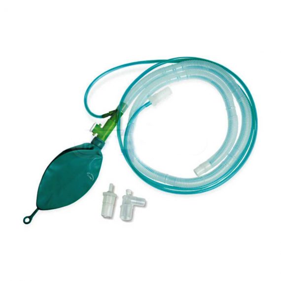
MediPlus Bain Circuit (Adult)
$28.00
In Stock
- Provided with 22mm Corrugated tube of length 1.6m
- APL Valve
- 2 L Antistatic Rebreathing bag
- 6mm Green PVC tube of length 1.6m
Delivery & Availability:
Typically 2 working days – excluding furniture and heavy/bulky equipment. Please contact us for further information.
Description
- Provided with 22mm Corrugated tube of length 1.6m
- APL Valve
- 2 L Antistatic Rebreathing bag
- 6mm Green PVC tube of length 1.6m
Quick Comparison
| MediPlus Bain Circuit (Adult) remove | Sonoscape P10 Ultrasound Machine remove | Sonoscape E2 Ultrasound Machine remove | ASPEL AsCARD Coral PC Based ECG Machine remove | ASPEL AsPEKT 712 Holter Monitor and Software remove | DrGem Ceiling Analogue X-ray Machine remove | |
|---|---|---|---|---|---|---|
| Name | MediPlus Bain Circuit (Adult) remove | Sonoscape P10 Ultrasound Machine remove | Sonoscape E2 Ultrasound Machine remove | ASPEL AsCARD Coral PC Based ECG Machine remove | ASPEL AsPEKT 712 Holter Monitor and Software remove | DrGem Ceiling Analogue X-ray Machine remove |
| Image | 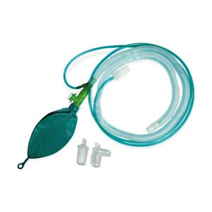 |  |  | 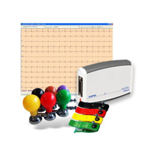 |  |  |
| SKU | SF1033560084-21 | SF1033560012-7 | SF1033560012-17 | SF1033560075-11 | SF1033560075-4 | SF1033560074-7 |
| Rating | ||||||
| Price | $28.00 | $9,350.00 | $5,500.00 | $486.00 | $1,991.00 |
|
| Stock | ||||||
| Availability | ||||||
| Add to cart | ||||||
| Description | In Stock
| Shipped from Abroad The P10 color Doppler ultrasound system is a new generation product from SonoScape. It is designed to give high quality images, rich probe configurations, various clinical tools and automatic analysis software to provide you with comprehensive solutions for your growing demand for clinical applications. Delivery & Availability: Typically 5-7 working days – excluding furniture and heavy/bulky equipment. Please contact us for further information. | Shipped from Abroad Sonoscape E2 portable ultrasound machine is a color Doppler ultrasound system that reaches beyond your expectations due to its compact and fashionable appearance. It fulfills GI, OB/GYN, Cardiac and POC applications to fit your routine scanning needs while its color mode will help you for more accurate and efficient diagnosis of lesions. E2 provides a wide range of applications to assist users with routine scanning. E2 provides automatic calculations to enhance your diagnostic confidence and save you time for patient communication. Delivery & Availability: Typically 14 working days – excluding furniture and heavy/bulky equipment. Please contact us for further information. | Shipped from Abroad AsCARD Coral electrocardiograph is a 3-, 6-, 12-channel ECG equipped with CardioTEKA software allows transmission of full 12 ECG leads to the user PC through USB interface. It is intended for carrying out ECG examinations in adults and pediatric patients in all types of health care centres. ECG procedures can be performed by qualified personnel only. AsCARD Coral can cooperate also with CardioTEST system as 12-channel ECG device allows transmission of full 12 ECG leads to the user PC through USB interface. Delivery & Availability: Typically 10 working days – excluding furniture and heavy/bulky equipment. Please contact us for further information. | Shipped from Abroad The Holta Monitor allows quick analysis of ECG examination and detection, reviewing and editing capability in the qualitative assessment of VE, VT, Single SVE, PSVT, Pauses, Irregular Rhythm, VT, IVR, Brady - and Tachycardia, Couplets, ST-segment elevation and depression, Maximum, Minimum and averaged Heart Rates, artifacts Delivery & Availability: Typically 10 working days – excluding furniture and heavy/bulky equipment. Please contact us for further information. | Shipped from abroad The DrGem Ceiling Analogue X-ray Machine is a diagnostic radiography system that provides reliable high quality radiographic images with a reduced dose. The reliable high-frequency x-ray generators that are known worldwide for their excellent performance, lifetime and stability. Patient tables and wall stands are also offered. Delivery & Availability: Typically 21 working days – excluding furniture and heavy/bulky equipment. Please contact us for further information. |
| Content |
| DETAILS
B + Compound
B + Compound utilizes several lines of sight for optimal contrast resolution, speckle reduction and border detection, with which P10 is ideal for superficial and abdominal imaging with better clarity and improved continuity of structures.
μ-Scan
The new generation μ-Scan imaging technology gives you better image quality by reducing noise, improving signal strength and improving visualization.
P10 offers a comprehensive selection of electronic probes to maximize its capabilities to meet a wide range of applications including abdomen, pediatric, OB/GYN, cardiovascular, musculoskeletal, etc. The advanced probe technologies also effectively enhance the image quality and confidence in reaching clinical diagnoses, even in difficult patients.
Convex Probe 3C-A
Ideal for an abundant of application such as abdomen, gynecology, obstetrics, urology and even abdomen biopsy.
Linear Probe L741
This linear probe is designed to satisfy vascular, breast, thyroid, and other small parts diagnosis, and its adjustable parameters could also present users a clear view of MSK and deep vessels.
Phase Array Probe 3P-A
For the purpose of adult and pediatric cardiology and emergency, the phase array probe provides elaborate presets for different exam modes, even for difficult patients.
Intracavitary Probe 6V1
Intracavitary probe could face application of gynecology, urology, prostate, and its temperature detection technology not only protects the patient but also extends the service life.
Click Here To Download Catalogue | SONOSCAPE E2 DETAILS
Auto Image Optimization
A portable ultrasound machine with the press of a button, the image is automatically adjusted and optimized, saving you time with parameter adjustments. Additionally, with Auto Focus on, the focus area follows the depth of the ROI box as it is moved in the scanning field, providing users with excellent image quality in the desired area of interest.
Automated Calculation
Auto IMT is used when determining the level of vascular sclerosis present in the patient by automatically tracing the thickness of the carotid vessels.
Auto trace provides users sensitive and accurate wave tracing, avoiding the error of manual trace and giving out calculation result in no time
In-Build Battery pack
This portable ultrasound machine was equipped with an in-build battery pack which enable the user to perform image scanning when AC power is not available.
Click Here To Download Catalogue |
AsCARD Coral electrocardiograph is a 3-, 6-, 12-channel ECG equipped with CardioTEKA software allows transmission of full 12 ECG leads to the user PC through USB interface. It is intended for carrying out ECG examinations in adults and pediatric patients in all types of health care centres. ECG procedures can be performed by qualified personnel only. AsCARD Coral can cooperate also with CardioTEST system as 12-channel ECG device allows transmission of full 12 ECG leads to the user PC through USB interface.
Technical Specification:
Click Here To Download Catalogue | The Holter Monitor allows quick analysis of ECG examination (arrhythmias and ST segment).
Technical specifications:
HolCARD 24W Software:
Click Here To Download Catalogue | DrGem Ceiling Analogue X-ray Machine is a diagnostic radiography system X-ray Machine that provides reliable high quality radiographic images with a reduced dose. The reliable high-frequency x-ray generators that are known worldwide for their excellent performance, lifetime and stability. Patient tables and wall stands are also offered.
Features of DrGem Ceiling Analogue X-ray Machine
Click Here To Download Catalogue |
| Weight | N/A | N/A | N/A | N/A | N/A | N/A |
| Dimensions | N/A | N/A | N/A | N/A | N/A | N/A |
| Additional information |


Reviews
There are no reviews yet.