Bistos BT-350 Fetal Monitor
$825.00
In Stock
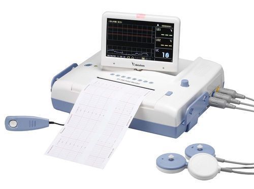
The BT-350 is a microprocessor-based fetal monitor, providing continuous monitoring, display and recording of fetal heart rate(FHR) and uterine contraction(UC) for antepartum testing and monitoring.
Delivery & Availability:
Typically 2 working days – excluding furniture and heavy/bulky equipment. Please contact us for further information.
Brands:: Bistos

Description
Product Details:
| Minimum Order Quantity | 1 Piece |
| Model | BT-350 |
| Dimension | 80mm (H) X 330mm (L) X 280mm (D) |
| Application | Hospital |
| Brand | Bistos |
| Screen rotating | 180 Degree |
| LCD display | 7″ wide TFT color LCD display |
Product Description:
- Up to 150 patients date saving
- Fetal heart sound play and record in PC
- Multiple language support
- Quick guide display
- Various installation method
Dual Pulsed Doppler:
- Ultrasound Frequency: 0.985MHz
- Intensity: 10mW/cm2 or less
- FHR Range: 30~240bpm FHR
- Accuracy: ±2% of Range
- Auto-detection of Dual Fetal Movement
- External Type
- Frequency Response: DC~0.5HZ
- Reference(Zero) Control
- Measurement Range: 0~99 units
Printer:
- Thermal Array Type
- Print Speed: 1,2,3, cm/min, high speed
- Auto print: Off,10,20,30,40,50,60 min
- Paper Feeding Function
Display:
- 3 Channels (FHR I, FHR II, UC)
- 7″ Wide TFT-Color LCD:800(W) X 480(H) BT-350LCD
- Large 7-segment volume lever LED : BT-350 LED
Function:
- Mark Function
- FHR II Offset Function
- Auto Print function
- Fetal movement print function
- Multi-language quick guide function: BT-350 LCD
Trend:
Data Saving for 450 hours(3hr/person): BT-350 LCD
Accessory Standard:
- Ultrasound Doppler Probe 2ea
- Event Marker Jack 1ea
- Power Adaptor Cord 1ea
- Probe Belt 3ea
Click Here To Download Catalogue
Quick Comparison
| Settings | Bistos BT-350 Fetal Monitor remove | ASPEL AsPEKT 712 Holter Monitor and Software remove | Sonoscape E2 Ultrasound Machine remove | DrGem Ceiling Mounted Digital X-ray remove | Sonoscape E1 Ultrasound Machine With Two Probes remove | Sonoscape P20 Ultrasound Machine remove | ||||||||||||||
|---|---|---|---|---|---|---|---|---|---|---|---|---|---|---|---|---|---|---|---|---|
| Name | Bistos BT-350 Fetal Monitor remove | ASPEL AsPEKT 712 Holter Monitor and Software remove | Sonoscape E2 Ultrasound Machine remove | DrGem Ceiling Mounted Digital X-ray remove | Sonoscape E1 Ultrasound Machine With Two Probes remove | Sonoscape P20 Ultrasound Machine remove | ||||||||||||||
| Image | 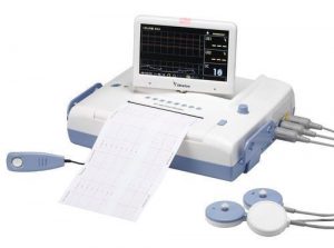 |  |  |  |  |  | ||||||||||||||
| SKU | SF1033560059-15 | SF1033560075-4 | SF1033560012-17 | SF1033560074-4 | SF1033560012-20 | SF1033560012-9 | ||||||||||||||
| Rating | ||||||||||||||||||||
| Price | $825.00 | $1,991.00 | $5,500.00 |
| $4,620.00 |
| ||||||||||||||
| Stock | ||||||||||||||||||||
| Availability | ||||||||||||||||||||
| Add to cart | ||||||||||||||||||||
| Description | In Stock
The BT-350 is a microprocessor-based fetal monitor, providing continuous monitoring, display and recording of fetal heart rate(FHR) and uterine contraction(UC) for antepartum testing and monitoring.
| Shipped from Abroad The Holta Monitor allows quick analysis of ECG examination and detection, reviewing and editing capability in the qualitative assessment of VE, VT, Single SVE, PSVT, Pauses, Irregular Rhythm, VT, IVR, Brady - and Tachycardia, Couplets, ST-segment elevation and depression, Maximum, Minimum and averaged Heart Rates, artifacts Delivery & Availability: Typically 10 working days – excluding furniture and heavy/bulky equipment. Please contact us for further information. | Shipped from Abroad Sonoscape E2 portable ultrasound machine is a color Doppler ultrasound system that reaches beyond your expectations due to its compact and fashionable appearance. It fulfills GI, OB/GYN, Cardiac and POC applications to fit your routine scanning needs while its color mode will help you for more accurate and efficient diagnosis of lesions. E2 provides a wide range of applications to assist users with routine scanning. E2 provides automatic calculations to enhance your diagnostic confidence and save you time for patient communication. Delivery & Availability: Typically 14 working days – excluding furniture and heavy/bulky equipment. Please contact us for further information. | In Stock The GXR-SD is a diagnostic digital radiography system that provides reliable high quality digital radiographic images with a reduced dose. The GXR-SD DR systems offer comprehensive digital solutions to all radiography needs, featuring ACQUIDR digital imaging system with stationary or portable digital flat-panel detectors as well as reliable high-frequency x-ray generators that are known worldwide for their excellent performance, lifetime and stability. Patient tables and wall stands are also offered. Delivery & Availability: Typically 21 working days – excluding furniture and heavy/bulky equipment. Please contact us for further information. | Shipped from Abroad SonoScape has developed a new probe and function for the E1 Exp. With these additions the E1 Exp will bring users a more efficient examination experience with satisfying image quality and a smooth workflow. Delivery & Availability: Typically 5-7 working days – excluding furniture and heavy/bulky equipment. Please contact us for further information. | Shipped from Abroad Incorporating innovative technologies, P20’s user-friendly design with a simple operation panel, intuitive user interface and a variety of intelligent auxiliary scanning tools, will significantly improve your daily examination experience. Besides general imaging applications, P20 has entitled with diagnostic 4D technology which has an extraordinary performance in obstetrics and gynecology applications. Delivery & Availability: Typically 5-7 working days – excluding furniture and heavy/bulky equipment. Please contact us for further information. | ||||||||||||||
| Content | Product Details:
Click Here To Download Catalogue | The Holter Monitor allows quick analysis of ECG examination (arrhythmias and ST segment).
Technical specifications:
HolCARD 24W Software:
Click Here To Download Catalogue | SONOSCAPE E2 DETAILS
Auto Image Optimization
A portable ultrasound machine with the press of a button, the image is automatically adjusted and optimized, saving you time with parameter adjustments. Additionally, with Auto Focus on, the focus area follows the depth of the ROI box as it is moved in the scanning field, providing users with excellent image quality in the desired area of interest.
Automated Calculation
Auto IMT is used when determining the level of vascular sclerosis present in the patient by automatically tracing the thickness of the carotid vessels.
Auto trace provides users sensitive and accurate wave tracing, avoiding the error of manual trace and giving out calculation result in no time
In-Build Battery pack
This portable ultrasound machine was equipped with an in-build battery pack which enable the user to perform image scanning when AC power is not available.
Click Here To Download Catalogue | DrGem Ceiling Mounted Digital X-ray is a diagnostic digital radiography system that provides reliable high quality digital radiographic images with a reduced dose. The GXR-SD DR systems offer comprehensive digital solutions to all radiography needs, featuring ACQUIDR digital imaging system with stationary or portable digital flat-panel detectors as well as reliable high-frequency x-ray generators that are known worldwide for their excellent performance, lifetime and stability. Patient tables and wall stands are also offered.
Features:
Click Here To Download Catalogue | DETAILS
Efficient Diagnosis
μ-Scan, Speckle Reduction & Edge Enhancement
Spatial Compound Imaging
PIH - Pure Inversion Harmonic
Wide Scan - Enlarged Image Area
Tissue-Specific Imaging
SR Flow
Ergonomic Designs
Up to 2 Transducer Ports
Light Weight and Compact
15.6 inch Anti-flickering HD LED Screen
Tilting Monitor Angle Adjustment
Backlit Keyboard and Intelligent Panel
Long-lasting Battery for 90 mins
Ease of Use
Quick Boot Up
Auto-Brightness Adjustment
Auto Image Optimization
Auto IMT
Auto Trace
Equipped Accessories
Wi-Fi and Bluetooth Available
DICOM
500GB Hard Disk
Height Adjustable Trolley
Durable, Carry-on Site Suitcase
Click Here To Download Catalogue | DETAILS
Upgraded Images with More Clarity
SonoScape never stops making progress in improving the image quality of its ultrasound products to enhance the confidence of diagnosis for doctors. With extraordinary images provided by P20, the anatomy structures are clearer than ever.
C-Xlasto Imaging
With C-xlasto Imaging, P20 enables comprehensive quantitative elastic analysis. Meanwhile, C-xlasto on P20 is supported by linear, convex and transvaginal probes, to ensure good reproducibility and highly consistent quantitative elastic results.
S-Live
S-Live allows for detailed visualization of subtle anatomical features, thereby enabling intuitive diagnosis with real-time 3D images and enriching patient communication.
Pelvic Floor 4D
Transperineal 4D pelvic floor ultrasound can provide useful clinical values in assessing the vaginal delivery impact on the female anterior compartment, judging whether the pelvic organs are prolapsed or not and the extent, determining if the pelvic muscles were torn accurately.
Anatomic M Mode
Anatomic M Mode helps you observe the myocardial motion at different phases by freely placing sample lines. It accurately measures the myocardial thickness and the heart size of even difficult patients and supports the myocardial function and LV wall-motion assessment.
Tissue Doppler Imaging
P20 is endowed with Tissue Doppler Imaging which provides velocities and other clinical information on myocardial functions, facilitating clinical doctors with the ability to analyze and compare the motions of different parts of the patient's heart.
Click Here To Download Catalogue | ||||||||||||||
| Weight | N/A | N/A | N/A | N/A | N/A | N/A | ||||||||||||||
| Dimensions | N/A | N/A | N/A | N/A | N/A | N/A | ||||||||||||||
| Additional information |


Reviews
There are no reviews yet.