
BONE DENSITOMETER (EXCELLUS)
$0.00
Shipped From Abroad
EXCELLUS is a half body fan beam Dual X-ray Absorptiometry(DXA) device, which is designed under the new concept of a body composition analyzer capable of measuring the BMD(Bone Mineral Density) and analyzing a half body composition.
Typically 10-21 working days – excluding furniture and heavy/bulky equipment. Please contact us for further information.
Description
Description
8 Channel Half Body DXA Analyzer
The Most Accurate & Precise DXA for Half Body Fat, Lean and Bone Mass
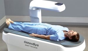
The new body analyzer, EXCELLUS, can quickly and easily analyze fat mass, lean mass and bone mass with the utmost accuracy and its special function makes it possible to analyze Gynoid and Android. And also, you can scope fat and lean mass in a specific site of a body with OsteoSys’s exclusive function of B-Scope.
Features
- Smart Fan Beam DXA
A Smart Fan Beam DXA of a compact concept, EXCELLUS is introduced as a new category for DXA market by maximizing the high-end functions and the strong points of our two previous models, Central DXA ‘DEXXUM T’ and Whole Body DXA ‘PRIMUS’
- Half Body Analysis with Multi-channel Detector
Based on DXA technology, EXCELLUS scopes the half body (from shoulder to knee) and effectively performs an examination using its Fan beam technology and multi-channel detector.
- From BMD to Visceral Fat Analysis
From the basic measurement of the BMD (Bone Mineral Density) of AP spine, Dual Femur, Forearm, and Lateral spine, EXCELLUS supports the various analysis such as VAT(Visceral fat Adipose Tissue) or orthopedics.
- Combination of Beauty and Cutting Edge
Technology
We enhanced both the beauty and the convenience by combining modern, contemporary design and cutting edge technology.
- Swing Arm
You can install EXCELLUS as a normal table for ultrasound scanner or use it in a chest X-ray room by utilizing its swing arm function.
- Compact Design
We are very proud of the compact, sophisticated and refined design of EXCELLUS
Specifications
| Measurement Type | Half body DXA (Half body Composition and assessment) |
| Measurement Method | Narrow fan beam |
| Scan site | Half body, AP spine, Femur(Dual femur), Forearm, Lateral spine, LVA(VFA) |
| Scan area | 800 ×480mm |
| Scan time | AP spine – 23Sec. (± 2Sec.) Femur – 19Sec. (± 2Sec.) Forearm – 18Sec. (± 2Sec.) Half body – 3Min. 30Sec. (± 2Sec.) |
| Special feature | Automatic real one-scan Swing arm |
| Reproducibility | ≤ 1.0% CV |
| Measured parameter | BMD, BMC, BMI, T-score, Z-score, Area, Half body BMD, Body Composition(Fat/Lean/BMC), HA(Hip Analysis), Dual femur Orthopedics / Pediatrics / B-Scope(body-Scope) / FRAX / Color mapping / Trend report / DICOM & PACS |
| Dimension | (W)1900mm × (D)800mm × (H)1230mm |
| Table height | 650mm |
| Weight | 160kg |
| Power consumption | 110VAC / 220VAC(+/- 10%) |
Click here to download Catalogue
Quick Comparison
| BONE DENSITOMETER (EXCELLUS) remove | Topaz Digital X-ray Machine remove | DrGem Ceiling Analogue X-ray Machine remove | Sonoscape P10 Ultrasound Machine remove | Sonoscape E2 Ultrasound Machine remove | DrGem GXR-SD 400mA Floor Mounted Digital X-ray remove | |||||||||||||||||||||||||
|---|---|---|---|---|---|---|---|---|---|---|---|---|---|---|---|---|---|---|---|---|---|---|---|---|---|---|---|---|---|---|
| Name | BONE DENSITOMETER (EXCELLUS) remove | Topaz Digital X-ray Machine remove | DrGem Ceiling Analogue X-ray Machine remove | Sonoscape P10 Ultrasound Machine remove | Sonoscape E2 Ultrasound Machine remove | DrGem GXR-SD 400mA Floor Mounted Digital X-ray remove | ||||||||||||||||||||||||
| Image | 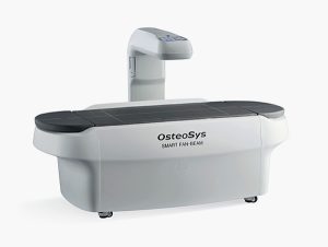 |  |  |  |  |  | ||||||||||||||||||||||||
| SKU | SF1033560130103-1 | SF1033560074-1 | SF1033560074-7 | SF1033560012-7 | SF1033560012-17 | SF1033560074-5 | ||||||||||||||||||||||||
| Rating | ||||||||||||||||||||||||||||||
| Price |
|
|
| $9,350.00 | $5,500.00 |
| ||||||||||||||||||||||||
| Stock | ||||||||||||||||||||||||||||||
| Availability | ||||||||||||||||||||||||||||||
| Add to cart | ||||||||||||||||||||||||||||||
| Description | Shipped From Abroad
EXCELLUS is a half body fan beam Dual X-ray Absorptiometry(DXA) device, which is designed under the new concept of a body composition analyzer capable of measuring the BMD(Bone Mineral Density) and analyzing a half body composition.
Delivery & Availability:
Typically 10-21 working days – excluding furniture and heavy/bulky equipment. Please contact us for further information.
| In Stock DRGEM’s TOPAZ X-ray machine is a state-of-the-art mobile digital radiography system, designed with maximum comfort for patients and users in mind. From its user-friendly software to smooth movements, TOPAZ is made to improve your workflow and provide you with high-quality images. Delivery & Availability: Typically 21 working days – excluding furniture and heavy/bulky equipment. Please contact us for further information. | Shipped from abroad The DrGem Ceiling Analogue X-ray Machine is a diagnostic radiography system that provides reliable high quality radiographic images with a reduced dose. The reliable high-frequency x-ray generators that are known worldwide for their excellent performance, lifetime and stability. Patient tables and wall stands are also offered. Delivery & Availability: Typically 21 working days – excluding furniture and heavy/bulky equipment. Please contact us for further information. | Shipped from Abroad The P10 color Doppler ultrasound system is a new generation product from SonoScape. It is designed to give high quality images, rich probe configurations, various clinical tools and automatic analysis software to provide you with comprehensive solutions for your growing demand for clinical applications. Delivery & Availability: Typically 5-7 working days – excluding furniture and heavy/bulky equipment. Please contact us for further information. | Shipped from Abroad Sonoscape E2 portable ultrasound machine is a color Doppler ultrasound system that reaches beyond your expectations due to its compact and fashionable appearance. It fulfills GI, OB/GYN, Cardiac and POC applications to fit your routine scanning needs while its color mode will help you for more accurate and efficient diagnosis of lesions. E2 provides a wide range of applications to assist users with routine scanning. E2 provides automatic calculations to enhance your diagnostic confidence and save you time for patient communication. Delivery & Availability: Typically 14 working days – excluding furniture and heavy/bulky equipment. Please contact us for further information. | In Stock The GXR-SD Digital X-ray is a diagnostic digital radiography system that provides reliable high quality digital radiographic images with a reduced dose. The GXR-SD DR systems offer comprehensive digital solutions to all radiography needs, featuring ACQUIDR digital imaging system with stationary or portable digital flat-panel detectors as well as reliable high-frequency x-ray generators that are known worldwide for their excellent performance, lifetime and stability. Patient tables and wall stands are also offered. Delivery & Availability: Typically 21 working days – excluding furniture and heavy/bulky equipment. Please contact us for further information. | ||||||||||||||||||||||||
| Content | Descriptionhttps://youtu.be/ofOxoH4CQLw?si=HjWSYzof9z7u5b5h8 Channel Half Body DXA AnalyzerThe Most Accurate & Precise DXA for Half Body Fat, Lean and Bone Mass
The new body analyzer, EXCELLUS, can quickly and easily analyze fat mass, lean mass and bone mass with the utmost accuracy and its special function makes it possible to analyze Gynoid and Android. And also, you can scope fat and lean mass in a specific site of a body with OsteoSys’s exclusive function of B-Scope.
Features
Specifications
Click here to download Catalogue | TOPAZ X-ray machine is among the high end X-ray machine manufactured by DRGEM, a digital X-ray system that provides quality images with little or no effort.
It begins with Advanced Technology
Integrating high technology and over a decade of experience in conventional and digital radiography systems, DRGEM’s TOPAZ X-ray machine is a state-of-the-art mobile digital radiography system, designed with maximum comfort for patients and users. From its user-friendly software to smooth movements, TOPAZ X-ray machine is made to improve your workflow and provide you with high-quality images.
Full Featured Imaging Software & Excellent Digital Image Processing
With a high-performance, built-in touchscreen, TOPAZ X-ray machine offers a user-friendly interface and powerful software for easy operation and increased workflow. The anatomical view-based digital image processing, automatically optimizes and enhances the quality of the image. it also comes with automatic image storage and print with DICOM 3.0 networking capability. additionally, the system offers increasing exam throughput while decreasing examination time.
Click Here To Download Catalogue | DrGem Ceiling Analogue X-ray Machine is a diagnostic radiography system X-ray Machine that provides reliable high quality radiographic images with a reduced dose. The reliable high-frequency x-ray generators that are known worldwide for their excellent performance, lifetime and stability. Patient tables and wall stands are also offered.
Features of DrGem Ceiling Analogue X-ray Machine
Click Here To Download Catalogue | DETAILS
B + Compound
B + Compound utilizes several lines of sight for optimal contrast resolution, speckle reduction and border detection, with which P10 is ideal for superficial and abdominal imaging with better clarity and improved continuity of structures.
μ-Scan
The new generation μ-Scan imaging technology gives you better image quality by reducing noise, improving signal strength and improving visualization.
P10 offers a comprehensive selection of electronic probes to maximize its capabilities to meet a wide range of applications including abdomen, pediatric, OB/GYN, cardiovascular, musculoskeletal, etc. The advanced probe technologies also effectively enhance the image quality and confidence in reaching clinical diagnoses, even in difficult patients.
Convex Probe 3C-A
Ideal for an abundant of application such as abdomen, gynecology, obstetrics, urology and even abdomen biopsy.
Linear Probe L741
This linear probe is designed to satisfy vascular, breast, thyroid, and other small parts diagnosis, and its adjustable parameters could also present users a clear view of MSK and deep vessels.
Phase Array Probe 3P-A
For the purpose of adult and pediatric cardiology and emergency, the phase array probe provides elaborate presets for different exam modes, even for difficult patients.
Intracavitary Probe 6V1
Intracavitary probe could face application of gynecology, urology, prostate, and its temperature detection technology not only protects the patient but also extends the service life.
Click Here To Download Catalogue | SONOSCAPE E2 DETAILS
Auto Image Optimization
A portable ultrasound machine with the press of a button, the image is automatically adjusted and optimized, saving you time with parameter adjustments. Additionally, with Auto Focus on, the focus area follows the depth of the ROI box as it is moved in the scanning field, providing users with excellent image quality in the desired area of interest.
Automated Calculation
Auto IMT is used when determining the level of vascular sclerosis present in the patient by automatically tracing the thickness of the carotid vessels.
Auto trace provides users sensitive and accurate wave tracing, avoiding the error of manual trace and giving out calculation result in no time
In-Build Battery pack
This portable ultrasound machine was equipped with an in-build battery pack which enable the user to perform image scanning when AC power is not available.
Click Here To Download Catalogue | DrGem GXR-SD 400mA Floor Mounted Digital X-ray system matches with a radiographic room which perfectly fits your workow and can be easily upgraded to DR system with the help of DR interface and PC interface in GXR generator as well as Bucky suitable to Flat Panel Detector. GXR X-ray system is equipped with a high frequency X-ray generator which consistently produces high quality radiograph in favor of high quality X-ray output with a very small kV ripple and accurate mA and mAs. GXR X-ray system is designed to provide convenience to operator and comfort to patient
Features of DrGem GXR-SD 400mA Floor Mounted Digital X-ray:
Click Here To Download Catalogue | ||||||||||||||||||||||||
| Weight | N/A | N/A | N/A | N/A | N/A | N/A | ||||||||||||||||||||||||
| Dimensions | N/A | N/A | N/A | N/A | N/A | N/A | ||||||||||||||||||||||||
| Additional information |


Reviews
There are no reviews yet.