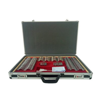
| Description | Shipped From Abroad
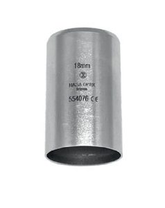
18mm
Delivery & Availability:
Typically 10-21 working days – excluding furniture and heavy/bulky equipment. Please contact us for further information.
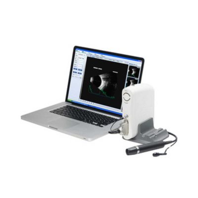
| Shipped from abroad
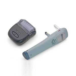
Tonometer SW-500 with vertical and horizontal two working modes, wireless output print data.
Delivery & Availability:
Typically 14 working days – excluding furniture and heavy/bulky equipment. Please contact us for further information.
| In Stock
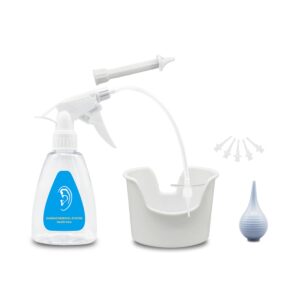
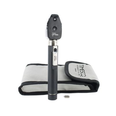
Features:
●Professional
Same ear wax removal tool as those used by doctors, you can easily eliminate ear wax buildup at home, really save your money and time on medical visiting. Safe and Environmentally Friendly.
●Quick & Easy
This ear wax removal kit is a quick, effective treatment for excess ear wax buildup. Fill the bottle with solution, Twist on the disposable tip, Use the trigger handle to spray solution into the ear canal. So Easy.
Delivery & Availability:
Typically 7-14 working days – excluding furniture and heavy/bulky equipment. Please contact us for further information.
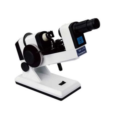
| Ship from abroad
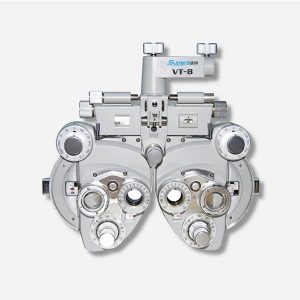
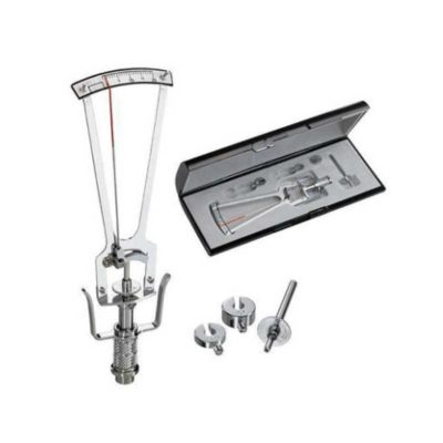
- Equipped with comprehensive measuring functions, it provides SPH, CYL, AXIS and pupil distance optometry
- Durable and easy to operate
- Easily and intuitively read the sphere focal scale value
- High eco-friendly materials
- Design fitting the face curve and no stimulation
Delivery & Availability:
Typically 14 working days – excluding furniture and heavy/bulky equipment. Please contact us for further information.
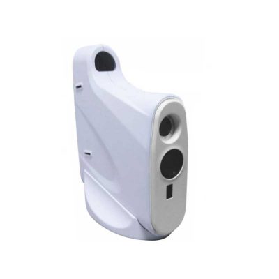
| Shipped from abroad
The product is designed on the principle basis of Goldman tonometer. It can be connected with slit lamp(Carl Zeiss type).
Delivery & Availability:
Typically 14 working days – excluding furniture and heavy/bulky equipment. Please contact us for further information.
| Ship from abroad
- Easy installation and maintenance: It is quite easy to replace light bulks, and unnecessary to adjust its focus and position.
- Customer Programmable: Icons can be displayed in predefined sequence by programming.
- Display Single Icon: Display single icon by multi-function mask plate.
- Tilted Placement: Perfect image position can be obtained by adjusting the horizontal axis of projector.
- Standards parts: Metal plate with polarized coating (410x 410mm)
Delivery & Availability:
Typically 14 working days – excluding furniture and heavy/bulky equipment. Please contact us for further information.
|
| Content | | Tonometer SW-500 with vertical and horizontal two working modes, wireless output print data. The equipment is used to measure intraocular pressure, using the principle of: the probe hits the surfaces of different hardness at a certain speed, has a different reaction when the probe rebounds. Be of advantages of high accuracy, portable, without anesthesia, without the cross-infection, etc.
Features:
- Measurement can go along both in standing and lying posture
- Wireless data printing
- Easy to learn, easy to use
- Lightweight and handy
- No anesthesia, avoiding discomfort reaction
Technical Specifications:
| Performance Index: |
| Measuring Range : |
3mmHg~70mmHg |
| Measuring Error: |
±1.5mmHg(3mmHg≤intraocular pressure≤25mmHg) |
| ±2.5mmHg(25mmHg<intraocular pressure≤70mmHg) |
| Environment Requirement: |
| Transport and Storage: |
Ambient temperature:-20℃~+55℃ |
| Relative moisture:≦95% |
| Atmosphere pressure:500hPa~1060hPa |
| Running: |
Ambient temperature:+5℃~+40℃ |
| Relative moisture:≦80% |
| Atmosphere pressure:700hPa~1060hPa |
| Rating voltage: DC3V(2 AA Batteries) |
| Rating input power:1VA |
| Features:
●Professional
Same ear wax removal tool as those used by doctors, you can easily eliminate ear wax buildup at home, really save your money and time on medical visiting. Safe and Environmentally Friendly.
●Quick & Easy
This ear wax removal kit is a quick, effective treatment for excess ear wax buildup. Fill the bottle with solution, Twist on the disposable tip, Use the trigger handle to spray solution into the ear canal. So Easy.
●Standard
Capacity of the ear cleaner solution bottle is 10.6Oz, it has the most suitable size to hold in hand. Working at condition 32-122℉(0-50℃). Recommend to fill 1/5 of the bottle with OTC hydrogen peroxide, and 4/5 with very warm water.
●Complete Ear Washer System
Our earwax removal kit comes with 1× Ear Washer Bottle, 1× Wash Basin, 1× Rubber Bulb, 1× Short Injection Head, 1× Long Hose Injection Head, 5× Disposable Tip, 1× User Manual. | Features:
- Equipped with comprehensive measuring functions, it provides SPH, CYL, AXIS and pupil distance optometry
- Durable and easy to operate
- Easily and intuitively read the sphere focal scale value
- High eco-friendly materials
- Design fitting the face curve and no stimulation
- Easy to take and clean
- Free switch between the cross-cylindrical lens and the rotary prism
- When the rotating risk is turning by the sphere, it can make sphere power adjust 3.00D for big scope.
- It is designed expediently and smartly for a particular cross cylinder. Supporting supplementary lens could increase scope of measurement.
Technical Specifications:
|
Sphere |
Range:-19.00~+16.75m-1 Step: 0.25m-1, 3.00m-1 |
|
Cylinder |
Range: 0.00~-6.00m-1(Measuring Range With Accessories0.00~-8.00m-1) Step: 0.25m-1 |
|
Cylinder Axis |
Range: 0~180°, Step:5° |
|
Distance of Optical center (also known as Pupil) |
Range: 50~75mmStep: 1mm |
|
Sight Switch |
Range:∞~380mm (distance of Optical center is64mm) |
|
Front Chin Test |
Range: 0~16mm |
|
Distance (from cornea vertex to the lens surface) |
16mm |
|
Standard Accessories Lens |
two pieces of Auxiliary Cylinder -2.00m-1 and -0.12m-1 respectively |
|
Standard Accessories |
one piece of M2 Hexagon wrench , one piece of a Myopia Standard Card, two piece of Myopia Standard Card , one piece of standard card holder , a dust cover |
|
Auxiliary Lens |
“O”:Open aperture
“R”:Retinoscope lens
“R”:Retinoscope lens
“R”:Retinoscope lens
*Lens of +1.50m-1 ,It is suit for the distance of 67 centimeters
“P”:Polaroid
* it is used for examining the dioptric balance of eyes , Implicit strabismus and stereo vision
“RMV”:Red Vertical maddox
*Be used to examine Implicit strabismus
“RMH”:Red horizontal maddox
*Be used to examine Implicit strabismus
“WMV”:Plane Vertical Maddox
*Be used to examine Implicit strabismus
“WMH”:Plane horizontal maddox
*Be used to examine Implicit strabismus
“RL”:Red lens
*Be used to examine eye function, Blending function and Implicit strabismus
“GL”:Green lens
*Be used to examine eye function, Blending function and Implicit strabismus
“+”:Test mark of optical center adjustment
“+.12”:Dioptric of the Spherical Lens is +0.12m-1
*Be used for the semi-adjustment of sphere lens, 0.25m-1
“PH”:1mmPinhole lens
*Be used to exclude visual non-refractive errors of the tested eye
“6ΔU”:6ΔBottom-up prism
*Be used to examine the rotating prism with the detection of nearly horizontal squint
“10ΔI”:10ΔBottom-up prism
*Be used to examine the rotating prism with the detection of nearly horizontal squint
“±0.50”:Cross-cylindrical lens
*Be used to examine the corrected dioptric of the Presbyopia and spherical lens
“OC”:Black lens
|
|
size |
338(L)×99(W)×292(H)mm |
|
NW |
about 5kg |
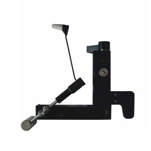
| Applanation Tonometer is designed on the principle basis of Goldman tonometer. It can be connected with slit lamp(Carl Zeiss type).
Features of Applanation Tonometer:
- When used with slit lamp, it can be used to examine the eyes and measure ocular pressure.
- Accurate measurement and total tolerance are less than 0.066KPa(0.55Hg).
- Directly get the ocular pressure and do not need to look up the conversion table.
- The measured ocular pressure is not affected by hardness of eye.
- The affected?ocular volume is just 0.56 cubic millimeter.
- Adjustable measuring pressure ensures the long-term stability and reliability.
Technical Specifications Applanation Tonometer:
- Measuring Range: 0~1064 Kpa
- Light Ring Displacement: 1.53×2=3.06mm
- Diameter of Prism Head: 7mm
- Moving Range of Prism Head: 3mm
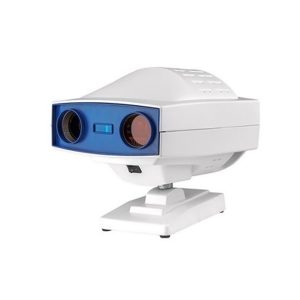
| Digital Chart Projector-Features:
- Easy installation and maintenance: It is quite easy to replace light bulks, and unnecessary to adjust its focus and position.
- Customer Programmable: Icons can be displayed in predefined sequence by programming.
- Display Single Icon: Display single icon by multi-function mask plate.
- Tilted Placement: Perfect image position can be obtained by adjusting the horizontal axis of projector.
- Standards parts: Metal plate with polarized coating (410x 410mm)
Technical Specifications of Digital Chart Projector:
|
Projection Distance |
1.5m~6m |
|
Projection Magnification |
30x (at 5m) |
|
Projection Size |
330x270mm (at 5 m) |
|
Chart Switching Speed |
One chart per 0.03s |
|
Mask Switching Speed |
1 open, 5 horizontal different lines, 8 vertical lines, 21 single letters, 1 red/green |
|
Program |
2 sets programs, each program contains up to 30 steps |
|
Speed of mask conversion |
One mask per 0.03s |
|
Lamp |
LED lamp |
|
Auto-off function |
After 10 minutes idle time |
|
Power Source |
AC 220V, 50Hz or 110V, 60Hz |
|
Power consumption |
40W |
|
Accessories |
Remote control, polarized metal screen, halogen lamp, polarized glasses, fuses (2), batteries (2) |
|


Reviews
There are no reviews yet.