| Description | Shipped From Abroad
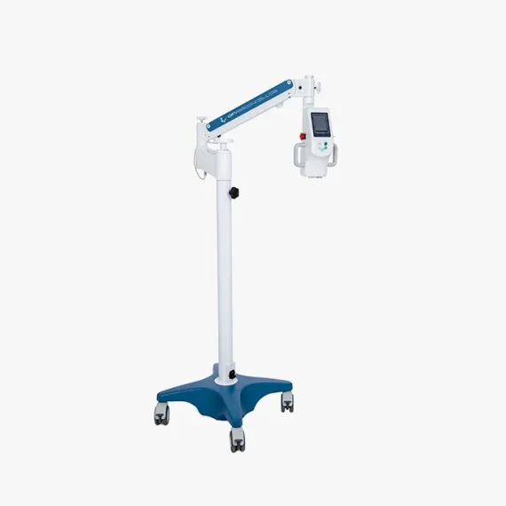
DFV Cerathos was designed with state-of-the-art technology to provide safe and effective treatment. Its optical system was developed to provide a safe dosage of energy homogeneously over the cornea to be treated. It has a dedicated microprocessor system that controls all functions and monitors the amount of energy emitted by the equipment. It's versatile and accurate!
Delivery & Availability:
Typically 10-21 working days – excluding furniture and heavy/bulky equipment. Please contact us for further information.
| Shipped from abroad
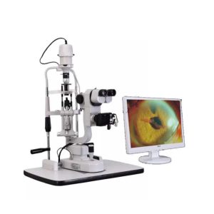
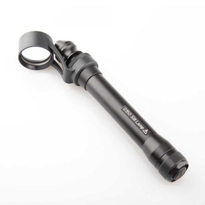
- Digital Slit lamp system,Galilean optical system,Five magnifications.
- Independent image process and management software system
- More than 10 million pixel image resolution
Delivery & Availability:
Typically 14 working days – excluding furniture and heavy/bulky equipment. Please contact us for further information.
| Shipped from abroad
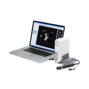
- Software image workstation
- B, B+B, B+A, A modes
- Video review for 100 images
- PDF report output
- Optional 20MHz B Probe: vitreous plus function
Delivery & Availability:
Typically 14 working days – excluding furniture and heavy/bulky equipment. Please contact us for further information.
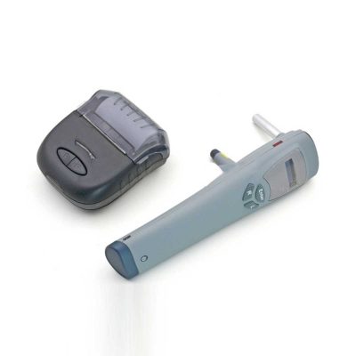
| 83Shipped from abroad
This is a portable medical camera for fundus imaging, diagnosis, and especially for fundus disease screening. It's compact, easy to obtain high definition fundus image. It can be conveniently applied to rapid screening, out diagnosis, bedside diagnosis and remote medical treatment, etc.
Delivery & Availability:
Typically 14 working days – excluding furniture and heavy/bulky equipment. Please contact us for further information.
| Ship from abroad
- Easy installation and maintenance: It is quite easy to replace light bulks, and unnecessary to adjust its focus and position.
- Customer Programmable: Icons can be displayed in predefined sequence by programming.
- Display Single Icon: Display single icon by multi-function mask plate.
- Tilted Placement: Perfect image position can be obtained by adjusting the horizontal axis of projector.
- Standards parts: Metal plate with polarized coating (410x 410mm)
Delivery & Availability:
Typically 14 working days – excluding furniture and heavy/bulky equipment. Please contact us for further information.
| Shipped from abroad
- 7.0-inch color LCD touch panel.
- Hartman sensor with 108 multiple measure points.
- Green measurement light beam.
- Dyeing lens, sunglasses measured easily.
Delivery & Availability:
Typically 14 working days – excluding furniture and heavy/bulky equipment. Please contact us for further information.
|
| Content |
Description
RELIABLE!
Power control with automatic calibration and reliability certified according to medical safety standards evaluated by Anvisa.
VERSATILE AND MODERN!
Dedicated microprocessor system; Commands integrated into the console; Agility in adjusting parameters on the touch screen;
Independent aiming system; Integrated color camera;
Treatment evolution bar.
FRIENDLY INTERFACE!
° Touch screen display that facilitates treatment configuration and monitoring.
° Integrated high sensitivity Watec color camera (0.3lx. F2.0).
° Color touchscreen display with high contrast.
° All controls integrated into a single-volume cabinet close to the patient.
° Clear identification of important treatment parameters.
° Agility in adjusting parameters on the same display screen.
° Progressive bar that indicates the temporal evolution of the treatment and the correct times for riboflavin application.
° Three treatment profile options record the desired parameters.
| Slit Lamp with Workstation Features:
- Digital Slit lamp system, Galilean optical system, Five magnifications
- Independent image process and management software system
- More than 10 million pixel image resolution
Technical Specifications:
| Microscope Type |
Galileo Parallel |
| Digital System |
External Digital Slit Lamp with 10 Mega pixels |
| Magnification Change Way |
Drum Five Magnifications |
| Eyepiece Magnification |
12.5x |
| Total Magnifications |
6x, 10x, 16x, 25x,40x |
| Diopter Adjustment |
-5D ~+5D |
| Slit Width |
0-14MM Continuous |
| Slit Height |
1-14MM Continuous |
| Slit Angle |
0°- 180° Adjustable |
| Slit Inclination Angle |
5°,10°,15°, 20° |
| Light Spot Diameter |
0.2mm, 2mm, 3mm, 5mm, 10mm, 14mm |
| Filter |
Heat Absorption; Grey; Red free; Cobalt Blue |
| Fixation |
Red LED |
| Illumination Bulb |
12V/50W German OSRAM Halogen Tungsten Lamp |
| Functions of Ophthalmic AB Scan Machine:
- Software image workstation
- B, B+B, B+A, A modes
- Video review for 100 images
- PDF report output
- Optional 20MHz B Probe: vitreous plus function
Technical Specifications:
| A scan |
1.Probe: 10MHz frequencies, with LED
2.Depth: 40mm
3.Precision: ±0.05mm
4.Eye mode: Phakic / Aphakic / Dense / Various IOL
5.Measurement: Anterior chamber depth, lens thickness, vitreous body length, total length and average
6.IOL Formula: SRK-II, SRK-T, BINKHORST, HOLLADAY, HOFFER-Q, HAIGIS, Stat.
7.Calculation: Average and standard deviation
8.Store: 10 Scanning results for each eye |
| B scan |
1.Probe: 10MHz/20MHz (optional), Magnetic driven, noiseless
2.Scanning Mode: Sector Scanning
3.Resolution: Lateral ≤0.3mm; Vertical≤0.2mm
4.Geometric Location Precision: Lateral≤10%; Vertical≤5%
5.Depth: 60mm
6.Enhance the part of vitreous body and retina
7.Gain of probe:30dB-105dB
8.Scanning Angle : 53°
9.Gray Scale: 256
10.False Color: Multi colors OTC
11.Measure Mode: distances, perimeter and area
12.Movies: 100 images movie review,AVI ZIP JPG format image output
13.Output: PDF format case report, connect to normal printer |
| Others |
1.Display Mode :B, B+B, B+A, A
2.Hint: preset keyword
3.Case Search: Multi-keywords
4.Working Platform: Windows XP, VISTA, WINDOWS7
5.User-defined report template |
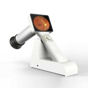
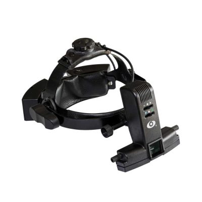
| Portable Fundus Camera is a portable medical camera for fundus imaging, diagnosis, and especially for fundus disease screening. It's compact, easy to obtain high definition fundus image. It can be conveniently applied to rapid screening, out diagnosis, bedside diagnosis and remote medical treatment, etc.
Features of Portable Fundus Camera:
- Images real-time display, one-button magnifying
- Non-mydriatic and high definition imaging.
- Micro SD memory card up to 32GB for 80,000 images
- USB and WiFi connection
- Low weight 450g, movable and portable
- A rechargeable battery provides up to more than 4 hours of continuous operation.
- Optional imaging modules for examination: posterior ophthalmoscope, anterior segment ophthalmoscope, otoscope, rhinoscope, laryngoscope and dermatoscope
- High definition and stable image
- Easy one-handed operation and excellent portability
- Large data service and a variety of image acquisition mode
Technical Specifications of Portable Fundus Camera:
| Model |
MC-600 |
| FOV |
45° |
| Minimum pupil |
2.5mm |
| Refractive compensation |
-20D~+20D |
| Light source |
White LED/IR |
| Image resolution |
1920×1080 |
| Focus Mode |
Manual |
| Screen |
3.5" color |
| Power |
≤6VA |
| Storage |
8GB Micro SD card |
| Power supply |
3.7VLithium Battery |
| Interface |
Mini USB/Wifi |
| N. Weight |
450g(Typical) |
| G. Weight |
2.5kg |
| Size |
160mm*90mm*190mm |
| Packing size |
360mm*310mm*160mm |
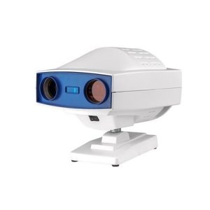
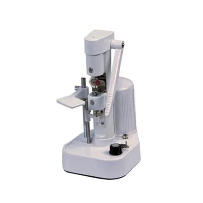
| Digital Chart Projector-Features:
- Easy installation and maintenance: It is quite easy to replace light bulks, and unnecessary to adjust its focus and position.
- Customer Programmable: Icons can be displayed in predefined sequence by programming.
- Display Single Icon: Display single icon by multi-function mask plate.
- Tilted Placement: Perfect image position can be obtained by adjusting the horizontal axis of projector.
- Standards parts: Metal plate with polarized coating (410x 410mm)
Technical Specifications of Digital Chart Projector:
|
Projection Distance |
1.5m~6m |
|
Projection Magnification |
30x (at 5m) |
|
Projection Size |
330x270mm (at 5 m) |
|
Chart Switching Speed |
One chart per 0.03s |
|
Mask Switching Speed |
1 open, 5 horizontal different lines, 8 vertical lines, 21 single letters, 1 red/green |
|
Program |
2 sets programs, each program contains up to 30 steps |
|
Speed of mask conversion |
One mask per 0.03s |
|
Lamp |
LED lamp |
|
Auto-off function |
After 10 minutes idle time |
|
Power Source |
AC 220V, 50Hz or 110V, 60Hz |
|
Power consumption |
40W |
|
Accessories |
Remote control, polarized metal screen, halogen lamp, polarized glasses, fuses (2), batteries (2) |
| Features:
- 7.0-inch color LCD touch panel.
- Hartman sensor with 108 multiple measure points.
- Green measurement light beam.
- Dyeing lens, sunglasses measured easily.
Technical Specifications:
- Sphere lenses: 0~±25D
- Cylinder lenses: 0~±10D
- Cylinder Axis Angle: 0°~180°
- Add.:0~10D
- Power supply: 100~240V, 50/60HZ, 30W
- Weight: 5kg
|


Reviews
There are no reviews yet.