


DRGEM DR System
$0.00
Ship from abroad
ACQUIDR is the digital imaging system composed of a Flat Panel Detector(FPD) and an imaging workstation with software. The digital FPD and full-feature imaging software with excellent digital image processing, designed for DRGEM X-ray machine.
Delivery & Availability:
Typically 21 working days – excluding furniture and heavy/bulky equipment. Please contact us for further information.

Description
DRGEM ACQUIDR (DRGEM DR System) is the digital imaging system composed of a Flat Panel Detector(FPD) and an imaging workstation with software. The digital FPD and full-feature imaging software with excellent digital image processing will meet all your needs in the diagnostic digital radiographic field.
Features of DRGEM DR System:
- Adaptable with most existing analog x-ray systems
- AED(Auto Exposure Detection) function available
- Simple and convenient installation
- Intuitive graphic user interface
- Over 1300 pre-loaded exam profiles with DRGEM’s generator
- Configurable automatic study
- PMS / EMR /HIS / RIS work list support
- Multiple printing formats
- DICOM 3.0 compliant
- Brightness and contrast adjustment
- Rotation
- Horizontal and vertical flip
- Zoom and pan
- Left / right makers
- Optimized work flow
- Automatic shutters
- Support DAP interface
- Exposure index (EI)
- DR upgrade solution by retrofit.
- Portable and Wireless FPD DR solution for maximum flexibility in virtually all general radiographic specialties.
- Portable and wireless detector fits into most existing analogue x-ray system.
- Faster workflow after DR upgrade.
- With AED(Auto Exposure Detection) function, there is no DR trigger cable between detector and generator providing very easy DR upgrade.
- Easy to interface with any kind of x-ray generator.
- Radmax acquisition workstation
- Turns any analogue X-Ray system into a fully DR system
- Optional image stitching program
- Vet software available
- Detector format: 17×17” / 17×14″, wired/wireless
Quick Comparison
| DRGEM DR System remove | Sonoscape P15 Ultrasound Machine With Four Probes remove | Genoray Full Field Digital Mammography DMX-600 remove | Sonoscape S8 Exp Portable Ultrasound remove | Genoray Analogue Mammography MX-600 remove | ASPEL AsPEKT 712 Holter Monitor and Software remove | |
|---|---|---|---|---|---|---|
| Name | DRGEM DR System remove | Sonoscape P15 Ultrasound Machine With Four Probes remove | Genoray Full Field Digital Mammography DMX-600 remove | Sonoscape S8 Exp Portable Ultrasound remove | Genoray Analogue Mammography MX-600 remove | ASPEL AsPEKT 712 Holter Monitor and Software remove |
| Image | 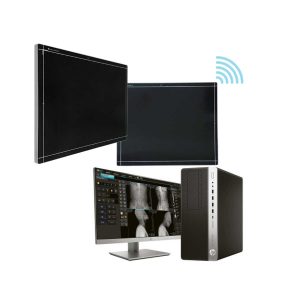 |  |  |  | 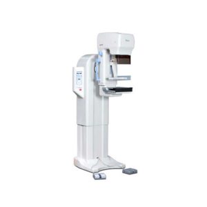 |  |
| SKU | SF1033560074-8 | SF1033560012-8 | SF1033560097-7 | SF1033560012-15 | SF1033560097-6 | SF1033560075-4 |
| Rating | ||||||
| Price |
| $13,900.00 |
| $9,350.00 |
| $1,991.00 |
| Stock | ||||||
| Availability | ||||||
| Add to cart | ||||||
| Description | Ship from abroad ACQUIDR is the digital imaging system composed of a Flat Panel Detector(FPD) and an imaging workstation with software. The digital FPD and full-feature imaging software with excellent digital image processing, designed for DRGEM X-ray machine. Delivery & Availability: Typically 21 working days – excluding furniture and heavy/bulky equipment. Please contact us for further information. | In Stock A feature-rich system inheriting the Wi-Sono high-end platform, the P15 uses an array of advanced tools to help enhance the image quality. It's a cost-effective, simplified console with an intuitive user interface and multiple intelligent functions. Delivery & Availability: Typically 2 working days – excluding furniture and heavy/bulky equipment. Please contact us for further information. | Shipped from Abroad
The DMX-600 is a full-field digital mammography system with smart C-arm rotation and fully motorized movements for fast and accurate positioning and efficient examinations. It features crystalline Silicon CMOS active pixel detector for higher contrast and higher resolution images and dual target X-ray tube for dose reduction.
| Shipped from Abroad With ultra-modern innovative design and the clinically-proven technologies, S8 Exp is portable ultrasound scanner well equipped as a low-physical-effort and enhanced-image-quality ultrasound scanner, which not only provides optimized solutions for versatile applications, but does help to improve the user-experience for both routine and non-traditional challenges. Delivery & Availability: Typically 5-7 working days – excluding furniture and heavy/bulky equipment. Please contact us for further information. | Shipped from Abroad
MX-600 is designed compactly for easy install and operation.
| Shipped from Abroad The Holta Monitor allows quick analysis of ECG examination and detection, reviewing and editing capability in the qualitative assessment of VE, VT, Single SVE, PSVT, Pauses, Irregular Rhythm, VT, IVR, Brady - and Tachycardia, Couplets, ST-segment elevation and depression, Maximum, Minimum and averaged Heart Rates, artifacts Delivery & Availability: Typically 10 working days – excluding furniture and heavy/bulky equipment. Please contact us for further information. |
| Content | DRGEM ACQUIDR (DRGEM DR System) is the digital imaging system composed of a Flat Panel Detector(FPD) and an imaging workstation with software. The digital FPD and full-feature imaging software with excellent digital image processing will meet all your needs in the diagnostic digital radiographic field.
Features of DRGEM DR System:
| DETAILS
Super Wide-bandwidth Platform
Inheriting Wi-sono's ultra-wide system platform and with the advanced probe technology, high-resolution and deep penetration images are provided for precision medicine.
Spatial Compound Imaging
Spatial Compound Imaging utilizes several lines of sight for optimal contrast resolution, speckle reduction and border detection, with which P15 is ideal for superficial and abdominal imaging with better clarity and improved continuity of structures.
μ-Scan+
The new generation μ-Scan imaging technology gives you better image quality by reducing noise, improving signal strength and improving visualization.
Dynamic Color
Dynamic color improves upon already existing color Doppler technologies for a clearer capture of color flow and detailed visualization of even tiny veins with lower velocities.
Real-time Panoramic
With real-time panoramic, you can acquire an extended field of view for large organs or long vessels for easy measurement and diagnostic efficiency. Accomplished in real-time for the convenience of the sonographers, any mistake can also be easily back tracked and corrected without interrupting the scan.
3D/4D
Outstanding volume performance with speed and convenience makes P15 outshine others on volume imaging.
Tissue Doppler Imaging
Tissue Doppler Imaging allows clinical doctors to quantitatively evaluate local myocardial movements and functions, facilitating them with the ability to analyze and compare the motions of the different parts of the patient's heart.
Auto IMT
Quick measurement of intra-media vessel thickness ensures good reproducibility and high diagnostic efficiency.
Click Here To Download Catalogue | The DMX-600 is a full-field digital mammography system with smart C-arm rotation and fully motorized movements for fast and accurate positioning and efficient examinations. It features crystalline Silicon CMOS active pixel detector for higher contrast and higher resolution images and dual target X-ray tube for dose reduction.
Features:
Excellent & High resolution Image quality:
- Innovative and accurate technology of Crystalline Silicon CMOS active pixel detector with higher contrast, higher resolution, brilliant images and economic maintenance cost.
- High performance of dual target X-ray tube for dose reduction.
- High output power of HF Generator with stability and efficiency.
- Stable hardware with the best reproducibility.
Large F.O.V. (Field Of View) by Multi-format technology:
- Optimally suited for screening and diagnosis with the financial advantages.
- Patent Application No. : 10-2011-0139676
- Available large F.O.V. to 23x23cm for almost patient.
User friendly Versatile functions:
- Smart C-arm rotation and fully motorized movements for fast & accurate positioning and efficiency examination by ASP & ASE (CC, RMLO, LMLO)
- Smart Automatic collimation with easy operation.
- Smart compression System by Microprocessor and Intelligent AEC control.
- Ergonomic design for quick and easy set-up.
Smart Software user alike:
- Customized and dedicated acquisition workstation.
- Perfect PACS accessibility with full DICOM capability.
- Extremely fast saving and transferring images for quick access and optimized workflow.
Click Here To Download Catalogue | Sonoscape S8 Exp Portable Ultrasound scannerDETAILS Agile and Versatile With ultra-modern innovative design and the clinically-proven technologies, S8 Exp Portable Ultrasound scanner is well equipped as a low-physical-effort and enhanced-image-quality ultrasound scanner, which not only provides optimized solutions for versatile applications but does help to improve the user experience for both routine and non-traditional challenges. Working with S8 Exp, it will trigger your unlimited reverie and endow you with endless charm. Carrying forward the classical design of SonoScape's portable ultrasound products, S8 Exp successfully combines the best ergonomics, attractive design and ease of use. This charismatic identity is also enhanced by a sophisticated color palette—with sedate grey as its interior paint color and pearl white as exterior cover, S8 Exp reveals a style of aristocrat and strong character among SonoScape's ultrasound systems. Workflow The S8 Exp is a portable ultrasound scanner that adapts to your workflow, whether you are in the consulting room, at the bedside, or at a remote location. With easy-to-use new platform designed for sonographers' needs and full connection interfaces for easy connectivity and data sharing, S8 Exp leads to improved user comfort and clinical outcome as well as patient throughput and working efficiency. Powerful Platform Embedded with SonoScape's core imaging technologies such as μ-scan, PHI and Spatial Compound, S8 Exp boasts exceptional 2D image, sensitive spectral, Color and Power Doppler, displaying well-defined anatomy and pathology and facilitating a highly optimized diagnostic user environment for conclusive diagnoses. Besides, S8 Exp offers a comprehensive selection of electronic probes to maximally extend its capabilities to meet a wide range of applications including the abdomen, pediatric, OB/GYN, cardiovascular, musculoskeletal, etc. The advanced probe technologies also effectively enhance the image quality and confidence in reaching clinical diagnoses even in difficult patients.Click Here To Download Catalogue | MX-600 is designed compactly for easy install and operation.
Features:
Generator
Tube
Radiographic support
Bucky device
Automatic exposure control (AEC)
Collimator: Automatic. Accessories
Options
Click Here To Download Catalogue | The Holter Monitor allows quick analysis of ECG examination (arrhythmias and ST segment).
Technical specifications:
HolCARD 24W Software:
Click Here To Download Catalogue |
| Weight | N/A | N/A | N/A | N/A | N/A | N/A |
| Dimensions | N/A | N/A | N/A | N/A | N/A | N/A |
| Additional information |


Reviews
There are no reviews yet.