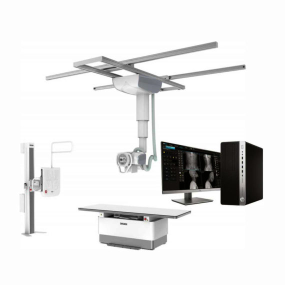
DrGem Ceiling Mounted Digital X-ray
$0.00
In Stock
The GXR-SD is a diagnostic digital radiography system that provides reliable high quality digital radiographic images with a reduced dose. The GXR-SD DR systems offer comprehensive digital solutions to all radiography needs, featuring ACQUIDR digital imaging system with stationary or portable digital flat-panel detectors as well as reliable high-frequency x-ray generators that are known worldwide for their excellent performance, lifetime and stability. Patient tables and wall stands are also offered.
Delivery & Availability:
Typically 21 working days – excluding furniture and heavy/bulky equipment. Please contact us for further information.

Description
DrGem Ceiling Mounted Digital X-ray is a diagnostic digital radiography system that provides reliable high quality digital radiographic images with a reduced dose. The GXR-SD DR systems offer comprehensive digital solutions to all radiography needs, featuring ACQUIDR digital imaging system with stationary or portable digital flat-panel detectors as well as reliable high-frequency x-ray generators that are known worldwide for their excellent performance, lifetime and stability. Patient tables and wall stands are also offered.
Features:
- TS-CSA-A (Vertical movement, 1.6m stroke, rail length 3x4meter) including HV cable 15m
- WBS-TA: Vertical movement
- V Stroke:1,450mm in Uprigh Bucky Position,
- 1,526mm in Horizontal Bucky position.
- PBT-4 is a 4 way Floating Tabletop. A large tabletop with extended travel enables all radiography studies with minimal patient movement. Fully fat tabletop without a frame on the edge makes cleanliness and odors free
- Digital Flat Panel Detector (FPD) – Wireless 17X14 (Csl, 4336W) with Auto Exposure Detection (AED) function, there is no DR trigger cable between detector and generator.
- Full Featured Imaging Software & Excellent Digital Image Processing:
- Provides convenient user interface and easy operation
- Anatomical view-based digital image processing automatically optimizes and enhances the quality of the captured image for the pictured anatomy.
- Radiographic stand & automatic collimator control function
- DICOM 3.0 networking interface includes Worklist, Print, Store, Query for integration with any PACS or RIS
- Included – Software, HP Laptop Computer
- CPU≥3.2GHz
- Memory capacity:≥4GB
- Hard drive capacity :≥500 GB
- Resolution: 1280 x 1024
- Display size: 21 inch color LCD screen
- 64 bit Windows 10 operation system
- Core: i5
Technical Specification:
- Power Rating – 32KW
- Generator – GXR-32S
- Rotor – Dual Speed Starter(DSS)
- Input Power – 400/480VAC, Three phase
- Line Frequency – 50/60Hz
- X-ray tube – DXT-12M, (0.6/1.2mm, 300kHU)
- Tube Voltage – 40 to 150kV, 1kV Step
- Tube Current – 10 to 640mA
- Output – 640mA@81kV, 500mA@104kV, 400mA@130kV, 320mA@150kV
- Time Range – 1ms to 10s
- mAs Range – 0.1 to 800mAs
- Reproducibility – Coecient of Variation : kV < 0.005, Time < 0.005,mAs < 0.01
- Accuracy – kV < ±(1%+1kV), mA < ±(3%+1mA), Time <±(1%+0.5ms), mAs < ±(3%+0.1mAs)
- Linearity – Coecient of Linearity < 0.01 : CL = (X1-X2)/(X1+X2), where X is mR/mAs
- Mechanical Parts:
-TS-CSA-A (Vertical movement, 1.6m, stroke rail length 3x4meter) including HV cable 15m
– PBT-4: 4 way Floating Tabletop.
– WBS-TA: a. Vertical movement
- V Stroke:1,450mm in Upright Bucky
- Position, 1,526mm in Horizontal Bucky position.
– HVC-15: 15M HV cable
– Auto Collimator
Click Here To Download Catalogue
Review(1)
Quick Comparison
| DrGem Ceiling Mounted Digital X-ray remove | Genoray Full Field Digital Mammography DMX-600 remove | ASPEL Stress ECG with Treadmill and Software remove | SIGNERS SUPiA Dry Thermal X-ray Film Printer remove | Sonoscape P50 Ultrasound Machine remove | Sonoscape P20 Ultrasound Machine remove | |
|---|---|---|---|---|---|---|
| Name | DrGem Ceiling Mounted Digital X-ray remove | Genoray Full Field Digital Mammography DMX-600 remove | ASPEL Stress ECG with Treadmill and Software remove | SIGNERS SUPiA Dry Thermal X-ray Film Printer remove | Sonoscape P50 Ultrasound Machine remove | Sonoscape P20 Ultrasound Machine remove |
| Image |  |  |  | 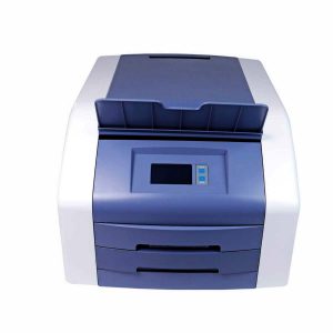 |  |  |
| SKU | SF1033560074-4 | SF1033560097-7 | SF1033560075-2 | SF1033560050-02 | SF1033560012-11 | SF1033560012-9 |
| Rating | ||||||
| Price |
|
| $6,542.00 | $3,520.00 |
|
|
| Stock | ||||||
| Availability | ||||||
| Add to cart | ||||||
| Description | In Stock The GXR-SD is a diagnostic digital radiography system that provides reliable high quality digital radiographic images with a reduced dose. The GXR-SD DR systems offer comprehensive digital solutions to all radiography needs, featuring ACQUIDR digital imaging system with stationary or portable digital flat-panel detectors as well as reliable high-frequency x-ray generators that are known worldwide for their excellent performance, lifetime and stability. Patient tables and wall stands are also offered. Delivery & Availability: Typically 21 working days – excluding furniture and heavy/bulky equipment. Please contact us for further information. | Shipped from Abroad
The DMX-600 is a full-field digital mammography system with smart C-arm rotation and fully motorized movements for fast and accurate positioning and efficient examinations. It features crystalline Silicon CMOS active pixel detector for higher contrast and higher resolution images and dual target X-ray tube for dose reduction.
| Shipped from Abroad It is a system with professional tool dedicated to exercise and resting ECG examination. Treadmill has 12 lead ECG modules. With ECG Analyzing Software. Delivery & Availability: Typically 21 working days – excluding furniture and heavy/bulky equipment. Please contact us for further information. | Shipped from Abroad
Signers Digital Dry Thermal X-ray Film Printer - Is a dry imager designed to output information processed through DICOM network protocol, supports 4 films sizes. 8”x10”, 10”x12”, 11”x14” and 14”x17” Achieve the convenience of multiple sizes.
Delivery & Availability: Typically 14 working days – excluding furniture and heavy/bulky equipment. Please contact us for further information. | Shipped from Abroad Easily accomplish more with SonoScape’s new P50 ultrasound system. Incorporating single crystal clarity, automatic corrections and calculation, and user defined flexibility promises a confident diagnostic experience as well as opening new doors of opportunity for ultrasound use. Delivery & Availability: Typically 7-14 working days – excluding furniture and heavy/bulky equipment. Please contact us for further information. | Shipped from Abroad Incorporating innovative technologies, P20’s user-friendly design with a simple operation panel, intuitive user interface and a variety of intelligent auxiliary scanning tools, will significantly improve your daily examination experience. Besides general imaging applications, P20 has entitled with diagnostic 4D technology which has an extraordinary performance in obstetrics and gynecology applications. Delivery & Availability: Typically 5-7 working days – excluding furniture and heavy/bulky equipment. Please contact us for further information. |
| Content | DrGem Ceiling Mounted Digital X-ray is a diagnostic digital radiography system that provides reliable high quality digital radiographic images with a reduced dose. The GXR-SD DR systems offer comprehensive digital solutions to all radiography needs, featuring ACQUIDR digital imaging system with stationary or portable digital flat-panel detectors as well as reliable high-frequency x-ray generators that are known worldwide for their excellent performance, lifetime and stability. Patient tables and wall stands are also offered.
Features:
Click Here To Download Catalogue | The DMX-600 is a full-field digital mammography system with smart C-arm rotation and fully motorized movements for fast and accurate positioning and efficient examinations. It features crystalline Silicon CMOS active pixel detector for higher contrast and higher resolution images and dual target X-ray tube for dose reduction.
Features:
Excellent & High resolution Image quality:
- Innovative and accurate technology of Crystalline Silicon CMOS active pixel detector with higher contrast, higher resolution, brilliant images and economic maintenance cost.
- High performance of dual target X-ray tube for dose reduction.
- High output power of HF Generator with stability and efficiency.
- Stable hardware with the best reproducibility.
Large F.O.V. (Field Of View) by Multi-format technology:
- Optimally suited for screening and diagnosis with the financial advantages.
- Patent Application No. : 10-2011-0139676
- Available large F.O.V. to 23x23cm for almost patient.
User friendly Versatile functions:
- Smart C-arm rotation and fully motorized movements for fast & accurate positioning and efficiency examination by ASP & ASE (CC, RMLO, LMLO)
- Smart Automatic collimation with easy operation.
- Smart compression System by Microprocessor and Intelligent AEC control.
- Ergonomic design for quick and easy set-up.
Smart Software user alike:
- Customized and dedicated acquisition workstation.
- Perfect PACS accessibility with full DICOM capability.
- Extremely fast saving and transferring images for quick access and optimized workflow.
Click Here To Download Catalogue | It is a system with professional tool dedicated to exercise and resting ECG examination. Treadmill has 12 lead ECG modules. With ECG Analyzing Software.
Technical Specification:
Click Here To Download Catalogue | Signers Digital Dry Thermal X-ray Film Printer - Is a dry imager designed to output information processed through DICOM network protocol, supports 4 films sizes. 8”x10”, 10”x12”, 11”x14” and 14”x17” Achieve the convenience of multiple sizes.
Technical Specifications:
Click Here To Download Catalogue | DETAILS
Powerful Compact Precision
Taking into consideration the evolving expectations and needs for ultrasound, the P50 is a slim and unobtrusive trolley system that is comfortable in tight, congested spaces with little room to work in. Providing everything you need for a comfortable examination in a small space for both you and your patient.
Single Crystal Transducer
Wideband single crystal probes greatly improve the signal ratio, acquire stunning images and provide superior sensitivity and resolution for both the near and far-fields.
μ-Scan+
The new generation μ-Scan imaging technologies give you better image quality by reducing noise, improving signal strength and improving visualization.
Dynamic Color
Dynamic colour improves upon already existing colour Doppler technologies for clear capture of colour flow and detail visualization of even tiny veins with lower velocities.
Solution for Radiology
P50, is a leading-edge ultrasound system that can meet the demands of any clinical setting. You can experience a superior performance in multi-dimensional imaging for a full range of clinical applications – abdominal, breast and cardiovascular.
C-xlasto Imaging
By understanding that tissue stiffness varies depending on the type of tissue, we can use C-xlasto Imaging to easily find abnormalities and tumours within soft tissue. The differences in tissue responses are detected and visualized in real-time by the elastography algorithms through different representations, which can be particularly helpful in analyzing breast, thyroid and musculoskeletal structures. Predominately used only in linear probes, SonoScape’s new transvaginal and bi-plane probe for gynaecology and urology are breaking the mould and expanding elastography applications.
Real-time Color Panoramic
With the combination of colour flow and real-time panoramic, visualizing the blood flow of an entire vein or artery is now an easy task. Accomplished in real-time for the convenience of the sonographers, any mistakes can also be easily backtracked and corrected without interrupting the scan.
Contrast Imaging
Contrast Imaging on P50 makes full use of the infra harmonic signal and second harmonic signal to improve the image resolution and deep penetration. What’s more, the Dynamic Acoustic Control technology effectively controls the acoustic pressure for the contrast agent, decreasing the required agent dose and assures uniform image quality, guaranteeing longer contrast agent duration and better lesion perfusion of delayed phase observation.
Solution for OB/GYN
P50 has superior image quality, automated measurement tools, and a variety of volume technologies to provide ideal solutions for clinical examinations such as pregnancy examinations, and gynecologic disease diagnosis. With a new 4D transvaginal probe, P50 helps you to see and detect fetal abnormalities and significantly improves your diagnostic confidence during your examinations.
S-Live Silhouette
A unique transparent 3D anatomical image of the fetus for improved initial anatomical review. By using this new application, the system can create completely different fetal images from conventional ultrasound images, which can depict the fetal's intracorporeal anatomical structure.
Pelvic Floor 4D
Working in conjunction with SonoScape’s latest transvaginal probes, trans-perineal 4D pelvic floor ultrasound provides a useful clinical assessment of the impact of vaginal delivery on the female anterior compartment. Allowing doctors to judge whether the pelvic organs prolapsed or not, the extent of prolapse, and determining whether the pelvic muscles tore correctly.
S-Guide
S-Guide gives the user an extensive list of example obstetric ultrasound images as reference guides and a convenient checklist system to keep track of their progress during their obstetrics examination.
Auto Face
Automatically removes masking layers in front of the fetus’s face for a clearer vision of the fetus’s face.
AVC Follicle
AVC Follicle automatically identifies how many follicles are present and calculates their individual volumes.
Solution for Cardiology
P50 provides clear 2D clinical images and Doppler sensitivity to assess critical cardiac performance. Compatible with SonoScape’s single crystal probes, the P50 can provide images with better resolution and penetration in Cardiac diagnosis.
Tissue Doppler Imaging
Tissue Doppler Imaging allows clinical doctors to quantitatively evaluate local myocardial movements and functions, facilitating them with the ability to analyze and compare the motions of the different parts of the patient’s heart.
Stress Echo
Stress echocardiography is the combination of 2D echocardiography with physical, pharmacological or electrical stress of the patient. It also then provides users with report management tools such as configurable template editor, multiple loops to select one for storage, wall motion scoring, stress echo report, etc
Auto IMT
Auto IMT is used when determining the level of vascular sclerosis present in the patient by automatically tracing and calculating the thickness of the carotid vessels. What distinguishes the P50 is that it provides an instant and accurate Mean and Max index at the touch of a single button.
Auto EF
Automated 2D Cardiac Quantification is a fully intelligent trace function for endocardium with 19 easily-adjustable points providing rapid access to proven 2D EF and volumes.
Click Here To Download Catalogue | DETAILS
Upgraded Images with More Clarity
SonoScape never stops making progress in improving the image quality of its ultrasound products to enhance the confidence of diagnosis for doctors. With extraordinary images provided by P20, the anatomy structures are clearer than ever.
C-Xlasto Imaging
With C-xlasto Imaging, P20 enables comprehensive quantitative elastic analysis. Meanwhile, C-xlasto on P20 is supported by linear, convex and transvaginal probes, to ensure good reproducibility and highly consistent quantitative elastic results.
S-Live
S-Live allows for detailed visualization of subtle anatomical features, thereby enabling intuitive diagnosis with real-time 3D images and enriching patient communication.
Pelvic Floor 4D
Transperineal 4D pelvic floor ultrasound can provide useful clinical values in assessing the vaginal delivery impact on the female anterior compartment, judging whether the pelvic organs are prolapsed or not and the extent, determining if the pelvic muscles were torn accurately.
Anatomic M Mode
Anatomic M Mode helps you observe the myocardial motion at different phases by freely placing sample lines. It accurately measures the myocardial thickness and the heart size of even difficult patients and supports the myocardial function and LV wall-motion assessment.
Tissue Doppler Imaging
P20 is endowed with Tissue Doppler Imaging which provides velocities and other clinical information on myocardial functions, facilitating clinical doctors with the ability to analyze and compare the motions of different parts of the patient's heart.
Click Here To Download Catalogue |
| Weight | N/A | N/A | N/A | N/A | N/A | N/A |
| Dimensions | N/A | N/A | N/A | N/A | N/A | N/A |
| Additional information |


xqgkyhiwcj
Muchas gracias. ?Como puedo iniciar sesion?