DrGem Ceiling Analogue X-ray Machine
$0.00
Shipped from abroad
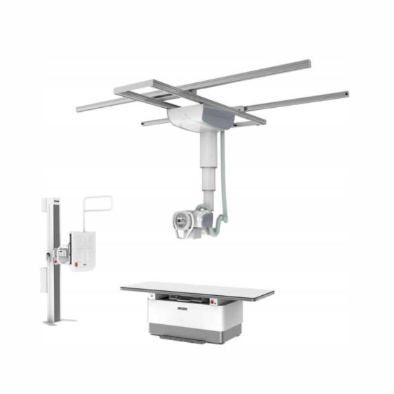
The DrGem Ceiling Analogue X-ray Machine is a diagnostic radiography system that provides reliable high quality radiographic images with a reduced dose. The reliable high-frequency x-ray generators that are known worldwide for their excellent performance, lifetime and stability. Patient tables and wall stands are also offered.
Delivery & Availability:
Typically 21 working days – excluding furniture and heavy/bulky equipment. Please contact us for further information.

Description
DrGem Ceiling Analogue X-ray Machine is a diagnostic radiography system X-ray Machine that provides reliable high quality radiographic images with a reduced dose. The reliable high-frequency x-ray generators that are known worldwide for their excellent performance, lifetime and stability. Patient tables and wall stands are also offered.
Features of DrGem Ceiling Analogue X-ray Machine
- TS-CSA-A (Vertical movement, 1.6m stroke, rail length 3x4meter) including HV cable 15m
- WBS-TA: Vertical movement
- V Stroke:1,450mm in Uprigh Bucky Position,
- 1,526mm in Horizontal Bucky position.
- PBT-4 is a 4 way Floating Tabletop with. A large tabletop with extended travel enables all radiography studies with minimal patient movement. Fully fat tabletop without a frame on the edge makes cleanliness and odors free
Technical Specifications of DrGem Ceiling Analogue X-ray Machine
- Power Rating – 32KW
- Generator – GXR-32S
- Rotor – Dual Speed Starter(DSS)
- Input Power – 400/480VAC, Three phase
- Line Frequency – 50/60Hz
- X-ray tube – DXT-12M, (0.6/1.2mm, 300kHU)
- Tube Voltage – 40 to 150kV, 1kV Step
- Tube Current – 10 to 640mA
- Output – 640mA@81kV, 500mA@104kV, 400mA@130kV, 320mA@150kV
- Time Range – 1ms to 10s
- mAs Range – 0.1 to 800mAs
- Reproducibility – Coecient of Variation : kV < 0.005, Time < 0.005,mAs < 0.01
- Accuracy – kV < ±(1%+1kV), mA < ±(3%+1mA), Time <±(1%+0.5ms), mAs < ±(3%+0.1mAs)
- Linearity – Coecient of Linearity < 0.01 : CL = (X1-X2)/(X1+X2), where X is mR/mAs
- Mechanical Parts:
-TS-CSA-A (Vertical movement, 1.6m, stroke rail length 3x4meter) including HV cable 15m
– PBT-4: 4 way Floating Tabletop with Elevating Feature (66cm).
– WBS-TA: a. Vertical movement
- V Stroke:1,450mm in Upright Bucky
- Position, 1,526mm in Horizontal Bucky position.
– HVC-15: 15M HV cable
– Auto Collimator
Click Here To Download Catalogue
Review(1)
Quick Comparison
| DrGem Ceiling Analogue X-ray Machine remove | ASPEL Stress ECG with Ergometer and Software remove | ASPEL AsPEKT 712 Holter Monitor and Software remove | SIGNERS SUPiA Dry Thermal X-ray Film Printer remove | Genoray Full Field Digital Mammography DMX-600 remove | DrGem Diamond All-In-One Digital X-ray Machine remove | |
|---|---|---|---|---|---|---|
| Name | DrGem Ceiling Analogue X-ray Machine remove | ASPEL Stress ECG with Ergometer and Software remove | ASPEL AsPEKT 712 Holter Monitor and Software remove | SIGNERS SUPiA Dry Thermal X-ray Film Printer remove | Genoray Full Field Digital Mammography DMX-600 remove | DrGem Diamond All-In-One Digital X-ray Machine remove |
| Image |  | 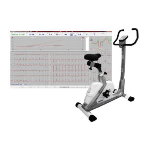 |  | 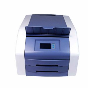 | 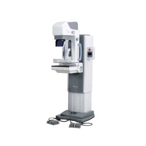 |  |
| SKU | SF1033560074-7 | SF1033560075-1 | SF1033560075-4 | SF1033560050-02 | SF1033560097-7 | SF1033560074-3 |
| Rating | ||||||
| Price |
| $4,202.00 | $1,991.00 | $3,520.00 |
|
|
| Stock | ||||||
| Availability | ||||||
| Add to cart | ||||||
| Description | Shipped from abroad The DrGem Ceiling Analogue X-ray Machine is a diagnostic radiography system that provides reliable high quality radiographic images with a reduced dose. The reliable high-frequency x-ray generators that are known worldwide for their excellent performance, lifetime and stability. Patient tables and wall stands are also offered. Delivery & Availability: Typically 21 working days – excluding furniture and heavy/bulky equipment. Please contact us for further information. | Shipped from Abroad Ergometer CRG 200 is dedicated for Exercise Stress Tests System CardioTEST. The Ergometer has been designed according to modern technologies. It is controlled from PC equipped in CardioTEST software. Load level is controlled by a microprocessor, therefore it does not depend on speed, which in turn can be adjusted according to patient’s individual needs. Ergometer is equipped with ECG mode recording 12 standard leads. Delivery & Availability: Typically 21 working days – excluding furniture and heavy/bulky equipment. Please contact us for further information. | Shipped from Abroad The Holta Monitor allows quick analysis of ECG examination and detection, reviewing and editing capability in the qualitative assessment of VE, VT, Single SVE, PSVT, Pauses, Irregular Rhythm, VT, IVR, Brady - and Tachycardia, Couplets, ST-segment elevation and depression, Maximum, Minimum and averaged Heart Rates, artifacts Delivery & Availability: Typically 10 working days – excluding furniture and heavy/bulky equipment. Please contact us for further information. | Shipped from Abroad
Signers Digital Dry Thermal X-ray Film Printer - Is a dry imager designed to output information processed through DICOM network protocol, supports 4 films sizes. 8”x10”, 10”x12”, 11”x14” and 14”x17” Achieve the convenience of multiple sizes.
Delivery & Availability: Typically 14 working days – excluding furniture and heavy/bulky equipment. Please contact us for further information. | Shipped from Abroad
The DMX-600 is a full-field digital mammography system with smart C-arm rotation and fully motorized movements for fast and accurate positioning and efficient examinations. It features crystalline Silicon CMOS active pixel detector for higher contrast and higher resolution images and dual target X-ray tube for dose reduction.
| Shipped from Abroad DrGem Diamond All-In-One Digital X-ray Machine is a fully automatic digital radiography system providing state-of-the-art image quality, image processing and user interface. With a wide selection of anatomical studies on the imaging software, DIAMOND automatically sets up the x-ray generator’s preprogrammed exposure technique settings, motorized radiographic stand positioning, x-ray collimation and post-image processing for the selected study. Specifically designed to increase workflow, this fully digital system offers convenient auto-positioning and advanced image processing to achieve big performance with little effort. Delivery & Availability: Typically 21 working days – excluding furniture and heavy/bulky equipment. Please contact us for further information. |
| Content | DrGem Ceiling Analogue X-ray Machine is a diagnostic radiography system X-ray Machine that provides reliable high quality radiographic images with a reduced dose. The reliable high-frequency x-ray generators that are known worldwide for their excellent performance, lifetime and stability. Patient tables and wall stands are also offered.
Features of DrGem Ceiling Analogue X-ray Machine
Click Here To Download Catalogue | Ergometer CRG 200 is dedicated for Exercise Stress Tests System CardioTEST. The Ergometer has been designed according to modern technologies. It is controlled from PC equipped in CardioTEST software. Load level is controlled by a microprocessor, therefore it does not depend on speed, which in turn can be adjusted according to patient’s individual needs. Ergometer is equipped with ECG mode recording 12 standard leads.
Technical Specifications:
Click Here To Download Catalogue | The Holter Monitor allows quick analysis of ECG examination (arrhythmias and ST segment).
Technical specifications:
HolCARD 24W Software:
Click Here To Download Catalogue | Signers Digital Dry Thermal X-ray Film Printer - Is a dry imager designed to output information processed through DICOM network protocol, supports 4 films sizes. 8”x10”, 10”x12”, 11”x14” and 14”x17” Achieve the convenience of multiple sizes.
Technical Specifications:
Click Here To Download Catalogue | The DMX-600 is a full-field digital mammography system with smart C-arm rotation and fully motorized movements for fast and accurate positioning and efficient examinations. It features crystalline Silicon CMOS active pixel detector for higher contrast and higher resolution images and dual target X-ray tube for dose reduction.
Features:
Excellent & High resolution Image quality:
- Innovative and accurate technology of Crystalline Silicon CMOS active pixel detector with higher contrast, higher resolution, brilliant images and economic maintenance cost.
- High performance of dual target X-ray tube for dose reduction.
- High output power of HF Generator with stability and efficiency.
- Stable hardware with the best reproducibility.
Large F.O.V. (Field Of View) by Multi-format technology:
- Optimally suited for screening and diagnosis with the financial advantages.
- Patent Application No. : 10-2011-0139676
- Available large F.O.V. to 23x23cm for almost patient.
User friendly Versatile functions:
- Smart C-arm rotation and fully motorized movements for fast & accurate positioning and efficiency examination by ASP & ASE (CC, RMLO, LMLO)
- Smart Automatic collimation with easy operation.
- Smart compression System by Microprocessor and Intelligent AEC control.
- Ergonomic design for quick and easy set-up.
Smart Software user alike:
- Customized and dedicated acquisition workstation.
- Perfect PACS accessibility with full DICOM capability.
- Extremely fast saving and transferring images for quick access and optimized workflow.
Click Here To Download Catalogue | DrGem Diamond All-In-One Digital X-ray Machine is a fully automatic digital radiography system providing state-of-the-art image quality, image processing and user interface. With a wide selection of anatomical studies on the imaging software, DIAMOND automatically sets up the x-ray generator’s pre-programmed exposure technique settings, motorized radiographic stand positioning, x-ray collimation and post-image processing for the selected study. Specifically designed to increase workflow, this fully digital system offers convenient auto-positioning and advanced image processing to achieve big performance with little effort.
Features of DrGem Diamond All-In-One Digital X-ray Machine:
Outstanding Image Quality -
Digital radiography via at panel detector improves your workflow, exam speed and comfort with efficiency. Digital at panel detector with Csl screen provides excellent spatial resolution, MTF, DQE and stability based on ne pixel pitch. A 3-field ion-chamber is provided for AEC function.
Automatic Collimation –
Automatic x-ray eld size control of the motorized collimator corresponds to dierent SIDs. Includes user adjustable lamp timer with on/oswitch.
Automatic Positioning –
Click Here To Download Catalogue |
| Weight | N/A | N/A | N/A | N/A | N/A | N/A |
| Dimensions | N/A | N/A | N/A | N/A | N/A | N/A |
| Additional information |


vorbelutrioperbir
Only wanna input that you have a very nice website , I enjoy the design it really stands out.