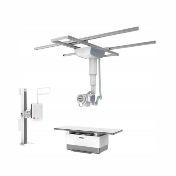
DrGem Ceiling Analogue X-ray Machine
$0.00
Shipped from abroad
The DrGem Ceiling Analogue X-ray Machine is a diagnostic radiography system that provides reliable high quality radiographic images with a reduced dose. The reliable high-frequency x-ray generators that are known worldwide for their excellent performance, lifetime and stability. Patient tables and wall stands are also offered.
Delivery & Availability:
Typically 21 working days – excluding furniture and heavy/bulky equipment. Please contact us for further information.

Description
DrGem Ceiling Analogue X-ray Machine is a diagnostic radiography system X-ray Machine that provides reliable high quality radiographic images with a reduced dose. The reliable high-frequency x-ray generators that are known worldwide for their excellent performance, lifetime and stability. Patient tables and wall stands are also offered.
Features of DrGem Ceiling Analogue X-ray Machine
- TS-CSA-A (Vertical movement, 1.6m stroke, rail length 3x4meter) including HV cable 15m
- WBS-TA: Vertical movement
- V Stroke:1,450mm in Uprigh Bucky Position,
- 1,526mm in Horizontal Bucky position.
- PBT-4 is a 4 way Floating Tabletop with. A large tabletop with extended travel enables all radiography studies with minimal patient movement. Fully fat tabletop without a frame on the edge makes cleanliness and odors free
Technical Specifications of DrGem Ceiling Analogue X-ray Machine
- Power Rating – 32KW
- Generator – GXR-32S
- Rotor – Dual Speed Starter(DSS)
- Input Power – 400/480VAC, Three phase
- Line Frequency – 50/60Hz
- X-ray tube – DXT-12M, (0.6/1.2mm, 300kHU)
- Tube Voltage – 40 to 150kV, 1kV Step
- Tube Current – 10 to 640mA
- Output – 640mA@81kV, 500mA@104kV, 400mA@130kV, 320mA@150kV
- Time Range – 1ms to 10s
- mAs Range – 0.1 to 800mAs
- Reproducibility – Coecient of Variation : kV < 0.005, Time < 0.005,mAs < 0.01
- Accuracy – kV < ±(1%+1kV), mA < ±(3%+1mA), Time <±(1%+0.5ms), mAs < ±(3%+0.1mAs)
- Linearity – Coecient of Linearity < 0.01 : CL = (X1-X2)/(X1+X2), where X is mR/mAs
- Mechanical Parts:
-TS-CSA-A (Vertical movement, 1.6m, stroke rail length 3x4meter) including HV cable 15m
– PBT-4: 4 way Floating Tabletop with Elevating Feature (66cm).
– WBS-TA: a. Vertical movement
- V Stroke:1,450mm in Upright Bucky
- Position, 1,526mm in Horizontal Bucky position.
– HVC-15: 15M HV cable
– Auto Collimator
Click Here To Download Catalogue
Review(1)
Quick Comparison
| DrGem Ceiling Analogue X-ray Machine remove | Sonoscape P10 Ultrasound Machine remove | ASPEL Stress ECG with Ergometer and Software remove | Sonoscape P15 Ultrasound Machine With Four Probes remove | ASPEL AsPEKT 712 Holter Monitor and Software remove | ASPEL Stress ECG with Treadmill and Software remove | |
|---|---|---|---|---|---|---|
| Name | DrGem Ceiling Analogue X-ray Machine remove | Sonoscape P10 Ultrasound Machine remove | ASPEL Stress ECG with Ergometer and Software remove | Sonoscape P15 Ultrasound Machine With Four Probes remove | ASPEL AsPEKT 712 Holter Monitor and Software remove | ASPEL Stress ECG with Treadmill and Software remove |
| Image |  |  |  |  |  |  |
| SKU | SF1033560074-7 | SF1033560012-7 | SF1033560075-1 | SF1033560012-8 | SF1033560075-4 | SF1033560075-2 |
| Rating | ||||||
| Price |
| $9,350.00 | $4,202.00 | $13,900.00 | $1,991.00 | $6,542.00 |
| Stock | ||||||
| Availability | ||||||
| Add to cart | ||||||
| Description | Shipped from abroad The DrGem Ceiling Analogue X-ray Machine is a diagnostic radiography system that provides reliable high quality radiographic images with a reduced dose. The reliable high-frequency x-ray generators that are known worldwide for their excellent performance, lifetime and stability. Patient tables and wall stands are also offered. Delivery & Availability: Typically 21 working days – excluding furniture and heavy/bulky equipment. Please contact us for further information. | Shipped from Abroad The P10 color Doppler ultrasound system is a new generation product from SonoScape. It is designed to give high quality images, rich probe configurations, various clinical tools and automatic analysis software to provide you with comprehensive solutions for your growing demand for clinical applications. Delivery & Availability: Typically 5-7 working days – excluding furniture and heavy/bulky equipment. Please contact us for further information. | Shipped from Abroad Ergometer CRG 200 is dedicated for Exercise Stress Tests System CardioTEST. The Ergometer has been designed according to modern technologies. It is controlled from PC equipped in CardioTEST software. Load level is controlled by a microprocessor, therefore it does not depend on speed, which in turn can be adjusted according to patient’s individual needs. Ergometer is equipped with ECG mode recording 12 standard leads. Delivery & Availability: Typically 21 working days – excluding furniture and heavy/bulky equipment. Please contact us for further information. | In Stock A feature-rich system inheriting the Wi-Sono high-end platform, the P15 uses an array of advanced tools to help enhance the image quality. It's a cost-effective, simplified console with an intuitive user interface and multiple intelligent functions. Delivery & Availability: Typically 2 working days – excluding furniture and heavy/bulky equipment. Please contact us for further information. | Shipped from Abroad The Holta Monitor allows quick analysis of ECG examination and detection, reviewing and editing capability in the qualitative assessment of VE, VT, Single SVE, PSVT, Pauses, Irregular Rhythm, VT, IVR, Brady - and Tachycardia, Couplets, ST-segment elevation and depression, Maximum, Minimum and averaged Heart Rates, artifacts Delivery & Availability: Typically 10 working days – excluding furniture and heavy/bulky equipment. Please contact us for further information. | Shipped from Abroad It is a system with professional tool dedicated to exercise and resting ECG examination. Treadmill has 12 lead ECG modules. With ECG Analyzing Software. Delivery & Availability: Typically 21 working days – excluding furniture and heavy/bulky equipment. Please contact us for further information. |
| Content | DrGem Ceiling Analogue X-ray Machine is a diagnostic radiography system X-ray Machine that provides reliable high quality radiographic images with a reduced dose. The reliable high-frequency x-ray generators that are known worldwide for their excellent performance, lifetime and stability. Patient tables and wall stands are also offered.
Features of DrGem Ceiling Analogue X-ray Machine
Click Here To Download Catalogue | DETAILS
B + Compound
B + Compound utilizes several lines of sight for optimal contrast resolution, speckle reduction and border detection, with which P10 is ideal for superficial and abdominal imaging with better clarity and improved continuity of structures.
μ-Scan
The new generation μ-Scan imaging technology gives you better image quality by reducing noise, improving signal strength and improving visualization.
P10 offers a comprehensive selection of electronic probes to maximize its capabilities to meet a wide range of applications including abdomen, pediatric, OB/GYN, cardiovascular, musculoskeletal, etc. The advanced probe technologies also effectively enhance the image quality and confidence in reaching clinical diagnoses, even in difficult patients.
Convex Probe 3C-A
Ideal for an abundant of application such as abdomen, gynecology, obstetrics, urology and even abdomen biopsy.
Linear Probe L741
This linear probe is designed to satisfy vascular, breast, thyroid, and other small parts diagnosis, and its adjustable parameters could also present users a clear view of MSK and deep vessels.
Phase Array Probe 3P-A
For the purpose of adult and pediatric cardiology and emergency, the phase array probe provides elaborate presets for different exam modes, even for difficult patients.
Intracavitary Probe 6V1
Intracavitary probe could face application of gynecology, urology, prostate, and its temperature detection technology not only protects the patient but also extends the service life.
Click Here To Download Catalogue | Ergometer CRG 200 is dedicated for Exercise Stress Tests System CardioTEST. The Ergometer has been designed according to modern technologies. It is controlled from PC equipped in CardioTEST software. Load level is controlled by a microprocessor, therefore it does not depend on speed, which in turn can be adjusted according to patient’s individual needs. Ergometer is equipped with ECG mode recording 12 standard leads.
Technical Specifications:
Click Here To Download Catalogue | DETAILS
Super Wide-bandwidth Platform
Inheriting Wi-sono's ultra-wide system platform and with the advanced probe technology, high-resolution and deep penetration images are provided for precision medicine.
Spatial Compound Imaging
Spatial Compound Imaging utilizes several lines of sight for optimal contrast resolution, speckle reduction and border detection, with which P15 is ideal for superficial and abdominal imaging with better clarity and improved continuity of structures.
μ-Scan+
The new generation μ-Scan imaging technology gives you better image quality by reducing noise, improving signal strength and improving visualization.
Dynamic Color
Dynamic color improves upon already existing color Doppler technologies for a clearer capture of color flow and detailed visualization of even tiny veins with lower velocities.
Real-time Panoramic
With real-time panoramic, you can acquire an extended field of view for large organs or long vessels for easy measurement and diagnostic efficiency. Accomplished in real-time for the convenience of the sonographers, any mistake can also be easily back tracked and corrected without interrupting the scan.
3D/4D
Outstanding volume performance with speed and convenience makes P15 outshine others on volume imaging.
Tissue Doppler Imaging
Tissue Doppler Imaging allows clinical doctors to quantitatively evaluate local myocardial movements and functions, facilitating them with the ability to analyze and compare the motions of the different parts of the patient's heart.
Auto IMT
Quick measurement of intra-media vessel thickness ensures good reproducibility and high diagnostic efficiency.
Click Here To Download Catalogue | The Holter Monitor allows quick analysis of ECG examination (arrhythmias and ST segment).
Technical specifications:
HolCARD 24W Software:
Click Here To Download Catalogue | It is a system with professional tool dedicated to exercise and resting ECG examination. Treadmill has 12 lead ECG modules. With ECG Analyzing Software.
Technical Specification:
Click Here To Download Catalogue |
| Weight | N/A | N/A | N/A | N/A | N/A | N/A |
| Dimensions | N/A | N/A | N/A | N/A | N/A | N/A |
| Additional information |


vorbelutrioperbir
Only wanna input that you have a very nice website , I enjoy the design it really stands out.