DRGEM JADE Analogue Mobile X-ray Machine with DR System

$16,928.00

In Stock

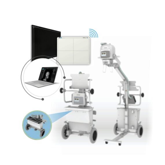
DRGEM JADE Analogue Mobile X-ray Machine with Flat Panel Detector (MAN04336W), 17×14 Inch, Portable, Wireless, Csl Workstation (laptop) including Software – Is one of the lightest portable X-ray systems on the market, allowing it to be used in any imaginable way including bedside, operating rooms, intensive care units and in veterinary ­fields. With a simple, easy-to-use operator console, three-way control, two-step foldable stand and auto lock system, JADE is a user-friendly portable X-ray system.

Delivery & Availability:
Typically 14-21 working days – excluding furniture and heavy/bulky equipment. Please contact us for further information.

SF1033560070
DrGem Corporation

Description

DRGEM JADE Mobile X-ray Machine with Flat Panel Detector (MAN04336W), 17×14 Inch, Portable, Wireless, Csl Workstation (laptop) including Software – Is one of the lightest portable X-ray systems on the market, allowing it to be used in any imaginable way including bedside, operating rooms, intensive care units and in veterinary ­fields. With a simple, easy-to-use operator console, three-way control, two-step foldable stand and auto lock system, JADE is a user-friendly portable X-ray system.


Features of DRGEM JADE Analogue Mobile X-ray Machine:

  • Vehicle loadable
  • Wheel lock
  • Automatic tube arm lock at any angle
  • Storage space for cassettes or detectors
  • User Programmable APR, save up to 9 APR settings
  • Three way X-ray exposure
  • USB interface & Bluetooth
  • Remote control (Option)
  • 5kg including X-ray unit, collimator and stand
  • Maximum hight of 228.6cm
  • Exposure Hand Switch
  • Foldable, two-step stand
  • In-Built Battery


Technical Specifications of DRGEM JADE Analogue Mobile X-ray Machine:

  • Power Rating – 4kW,
  • 100kHz, high frequency X-ray Generator
  • kVp Range – Maximum 140kVp
  • mAs Range – 0.1-250mAs
  • mA Range – 10 to 100mA
  • 330~2000mm FD

Collimator with 30 seconds LED lamp timer

Click Here To Download Catalogue

Reviews

There are no reviews yet.

Be the first to review “DRGEM JADE Analogue Mobile X-ray Machine with DR System”

Your email address will not be published. Required fields are marked *

Quick Comparison

SettingsDRGEM JADE Analogue Mobile X-ray Machine with DR System removeSingle X-Ray Viewing Box removeSonoscape S22 Ultrasound Machine removeAnke Supermark 1.5T MRI Machine removeSonoscape P20 Ultrasound Machine removeSonoscape P50 Ultrasound Machine remove
NameDRGEM JADE Analogue Mobile X-ray Machine with DR System removeSingle X-Ray Viewing Box removeSonoscape S22 Ultrasound Machine removeAnke Supermark 1.5T MRI Machine removeSonoscape P20 Ultrasound Machine removeSonoscape P50 Ultrasound Machine remove
ImageMRI SuperMart
SKUSF1033560070SF1033560084-203SF1033560012-3SF1033560092-4SF1033560012-9SF1033560012-11
Rating
Price

$16,928.00

$95.20

Ask for Price$0.00

Ask for Price$0.00

Ask for Price$0.00

Ask for Price$0.00

Stock
Availability
Add to cart

DescriptionIn Stock
DRGEM JADE Analogue Mobile X-ray Machine with Flat Panel Detector (MAN04336W), 17x14 Inch, Portable, Wireless, Csl Workstation (laptop) including Software - Is one of the lightest portable X-ray systems on the market, allowing it to be used in any imaginable way including bedside, operating rooms, intensive care units and in veterinary ­fields. With a simple, easy-to-use operator console, three-way control, two-step foldable stand and auto lock system, JADE is a user-friendly portable X-ray system. Delivery & Availability: Typically 14-21 working days – excluding furniture and heavy/bulky equipment. Please contact us for further information.
In stock
  • Length: 355 mm
  • Height: 560 mm
  • Diameter: 150 mm
  • Power: 30 w
  • Scattering diffusion: Greater than 0.9
Delivery & Availability: Typically 5-7 working days – excluding furniture and heavy/bulky equipment. Please contact us for further information.
Shipped from Abroad As SonoScape steps forward to add value and efficiency to ultrasound, the latest S22 was designed in a user-friendly platform to address current and future demanding needs. It represents an excellent mix in performance and price. Delivery & Availability: Typically 5-7 working days – excluding furniture and heavy/bulky equipment. Please contact us for further information.Shipped from Abroad SuperMark 1.5T is a new generation superconducting MRI system based on years of experience in production and research. It's applicable to whole body scan, such as, nervous system, spine, joint soft tissue, pelvic and abdominal cavity, etc

Delivery & Availability:

Typically 90 working days – excluding furniture and heavy/bulky equipment. Please contact us for further information.
Shipped from Abroad Incorporating innovative technologies, P20’s user-friendly design with a simple operation panel, intuitive user interface and a variety of intelligent auxiliary scanning tools, will significantly improve your daily examination experience. Besides general imaging applications, P20 has entitled with diagnostic 4D technology which has an extraordinary performance in obstetrics and gynecology applications. Delivery & Availability: Typically 5-7 working days – excluding furniture and heavy/bulky equipment. Please contact us for further information.Shipped from Abroad Easily accomplish more with SonoScape’s new P50 ultrasound system. Incorporating single crystal clarity, automatic corrections and calculation, and user defined flexibility promises a confident diagnostic experience as well as opening new doors of opportunity for ultrasound use. Delivery & Availability: Typically 7-14 working days – excluding furniture and heavy/bulky equipment. Please contact us for further information.
ContentDRGEM JADE Mobile X-ray Machine with Flat Panel Detector (MAN04336W), 17x14 Inch, Portable, Wireless, Csl Workstation (laptop) including Software - Is one of the lightest portable X-ray systems on the market, allowing it to be used in any imaginable way including bedside, operating rooms, intensive care units and in veterinary ­fields. With a simple, easy-to-use operator console, three-way control, two-step foldable stand and auto lock system, JADE is a user-friendly portable X-ray system.   Features of DRGEM JADE Analogue Mobile X-ray Machine:
  • Vehicle loadable
  • Wheel lock
  • Automatic tube arm lock at any angle
  • Storage space for cassettes or detectors
  • User Programmable APR, save up to 9 APR settings
  • Three way X-ray exposure
  • USB interface & Bluetooth
  • Remote control (Option)
  • 5kg including X-ray unit, collimator and stand
  • Maximum hight of 228.6cm
  • Exposure Hand Switch
  • Foldable, two-step stand
  • In-Built Battery
  Technical Specifications of DRGEM JADE Analogue Mobile X-ray Machine:
  • Power Rating - 4kW,
  • 100kHz, high frequency X-ray Generator
  • kVp Range - Maximum 140kVp
  • mAs Range - 0.1-250mAs
  • mA Range – 10 to 100mA
  • 330~2000mm FD
Collimator with 30 seconds LED lamp timer
Click Here To Download Catalogue
  • Length: 355 mm
  • Height: 560 mm
  • Diameter: 150 mm
  • Power: 30 w
  • Scattering diffusion: Greater than 0.9
DETAILS As SonoScape steps forward to add value and efficiency to ultrasound, the latest S22 was designed in a user-friendly platform to address current and future demanding needs. It represents an excellent mix in performance and price. S22, is a shared service ultrasound system with a slim and elegant package that has combined mobility with utility to fit in specific clinical situations including emergency department, ICU, operating room and so on. Furthermore, its ergonomic design, easy operating and flexible data management will give you a memorable experience. SPECIFICATION • Large high-resolution widescreen LED • Sensitive touch screen • Four transducer sockets plus one socket for pencil probe • A comprehensive selection of probes: linear, Convex, Micro-convex, Volumetric, Endocavity, Bi-plane, Phased Array, TEE, Intraoperative, Pencil • Premium application technology: 4D, μ-scan speckle reduction, compound imaging, Pulse Inversion Harmonic Imaging, Color M-Mode, Steer M-Mode, PDI, TDI, Real-time Panoramic Imaging, Trapezoid Imaging, Auto-IMT… • Full patient database and image management solutions: DICOM 3.0, AVI/JPG, USB 2.0, HDD, DVD, PDF report • Multi-Language Input Keyboard • Built-in battery  
Click Here To Download Catalogue
SuperMark 1.5T is a new generation superconducting MRI system based on years of experience in production and research. It's applicable to whole body scan, such as, nervous system, spine, joint soft tissue, pelvic and abdominal cavity, etc. SuperMark 1.5T provides not only conventional pulse sequences and clinical diagnosis functions, but also provides advanced functional applications, for instance, 3D angiography and water imaging. It adopts brand new ANKE APEX operating system which ensures easy operation and fast diagnosis. Technical Advantages:
  • Reliable short cavity superconducting magnet system with zero liquid helium consumption
  • New generation fully digitalized and extensible multichannel spectrometer
  • Powerful high efficiency and high fidelity gradient system; Multi-channel PA RF receiving coil with intelligent identification
  • English operating system and high extensible computer system
  • High resolution conventional clinical images; Practical advanced functional imaging
Superconducting MRI System:
  • Highly open and humanization design -> Streamlined design
  • Rich sequences and technology satisfy clinical needs -> Efficient service
Low Investment:
  • High cost performance superconducting MRI system
  • Zero liquid helium consumption, low running and maintenance cost
  • Core technology by independent R & D supports full upgrade
  • Low electric consumption
  • Compact magnet design, minimum installation space: 35 square meters
High Return:
  • High resolution thin slice images improve diagnosis
  • Short cavity magnet design makes patients comfortable
  • Fast scan speed improves work efficiency
  Technical Specifications:
No. Technique Description Parameter
1 Magnet System
 1.1  Magnet Type Permanent Magnet Automatic constant temperature system
1.2 Field Strength 0.51T
1.3 Magnet Shape Dual-pillar shape
1.4 Homogeneity(40cm,DSV,VRMS) ≤1.6ppm
1.5 Shim Method Active/Passive
1.6 Magnet Vertical Gap (Cover) 40cm
1.7 Magnetic Pole Dia. (Exclude Cover) 145cm
1.8 Accessibility(Horizontal Opening Angle, 280°
1.9 5 Gauss fringe field X-axis:horizontal ≤2.5m Y-axis:Vertical ≤2.5m Z-axis:horizontal ≤2.5m
2 Patient Couch and Communication
2.1 Patient Couch Driven mode Motor-driven
2.2 Max. Patient Weight ≥200kg(440lbs)
2.3 Patient Positioning Tools Laser Light Localizer for positioning of center slice Motor-driven transfer to center of imaging volume
2.4 Position accuracy ±1mm
2.5 Emergency Call Key Yes
2.6 Intercom System Yes
3 Gradient System
3.1 Gradient Field Strength(Single Axis) ≥30mT/m
3.2 Gradient Slew Rate (Single Axis) ≥100mT/m/ms
3.3 Rise Time ≤0.3ms
3.4 Gradient Cooling System ( Gradient coils and Power electronics) Air Cooling
4 RF System
4.1 RF System Type Digital Transmit and Receive signal
4.2 Number of RF Channels 4
4.3 Transmitter Amplifier Peak Power 6kW
4.4 RF Bandwidth of Receiver ≥1.25MHz
4.5 Head Coil Yes
4.6 Neck Coil Yes
4.7 Body/Spine Coil (17 inch) Yes
4.8 Body/Spine Coil (21 inch) Yes
4.9 Knee Coil Yes
4.10 Shoulder Coil Yes
4.11 Flexible Coil Optional
4.12 Breast Coil Optional
5 Computer System
5.1 Host Computer DELL Computer (for MR)
5.2 System Software Windows XP
5.3 Operation Software APEX
5.4 CPU Clock rate 3.0GHz
5.5 Main Memory 4GB
5.6 Color LCD Monitor 19”
5.7 Keyboard and Mouse Standard
5.8 Image Reconstruction Speed(256 x 256 Matrix) 200 frame/Sec.
5.9 Hard Disk 500GB
5.10 Image Storage Capacity(256 x 256 Matrix) 500,000
5.11 Media Driver DVD RW
5.12 DICOM 3.0 Yes
5.13 Ethernet Yes
5.14 Operation Console Yes
5.15 Operation Chair Yes
6 Scanning Parameter
6.1 Max. FOV 410mm
6.2 Min. FOV 5mm
6.3 Min. TE(SE) 5ms
6.4 Min. TR(SE) 11ms
6.5 Min. TE(GR) 1ms
6.6 Min. TR(GR) 3ms
6.7 Min. 2D Thickness 1.0mm
6.8 Min. 3D Thickness 0.1mm
6.9 Max. Image Matrix 512x512
7 Scanning Sequence & Imaging Technique
7.1 Spin Echo 2D/3D (SE 2D/3D) Yes
7.2 DE/QE Yes
7.3 Fast Spin Echo 2D/3D(FSE 2D/3D) Yes
7.4 Single Shot FSE 2D/3D Yes
7.5 Inversion Recovery(IR) Yes
7.6 Fast Inversion Recovery(FIR) Yes
7.7 Gradient Echo 2D/3D(GR 2D/3D) Yes
7.8 Fast GR 2D/3D Yes
7.9 SPGR Yes
7.10 FLAIR Yes
7.11 Fat Imaging Yes
7.12 Fat Suppression imaging Yes
7.13 Water-Fat Separation imaging Yes
7.14 TOF MRA(2D/3D) Yes
7.15 MRCP(2D/3D) Yes
7.16 MRU (2D/3D) Yes
7.17 MRM Yes
7.18 Fast Hydrograph Imaging Yes
7.19 Diffusion Weighted Imaging(DWI) Yes
7.20 Max. b Value 1000s/mm2
7.21 Breath Hold Technology Yes
7.22 Magnetization Transfer Contrast(MTC) Yes
7.23 Multi-slice and Angle-free Presaturation Yes
7.24 Saturation Tracking Yes
7.25 Maximum Intensity Projection(MIP) Yes
7.26 Multi-Angle Projection(MAP) Yes
7.27 3D Reconstruction Yes
7.28 Multi-planar Reconstruction(MPR) Yes
7.29 Multi-Artifacts Eliminating technology Yes
7.30 Checking with Part Metal Implant Yes
7.31 Online Image Filtration Yes
7.32 Online Post Procession Yes
7.33 3D Scout Yes
7.34 Scanning Protocol Preset Yes
7.35 Scanning Protocol Queue Waiting Yes
7.36 Advanced Image Post Processing Yes
7.37 Image Fusion Technology of Vascular Yes
7.38 Image Fusion Technology of Spine Yes
 
Click Here To Download Catalogue
DETAILS Upgraded Images with More Clarity SonoScape never stops making progress in improving the image quality of its ultrasound products to enhance the confidence of diagnosis for doctors. With extraordinary images provided by P20, the anatomy structures are clearer than ever. C-Xlasto Imaging With C-xlasto Imaging, P20 enables comprehensive quantitative elastic analysis. Meanwhile, C-xlasto on P20 is supported by linear, convex and transvaginal probes, to ensure good reproducibility and highly consistent quantitative elastic results. S-Live S-Live allows for detailed visualization of subtle anatomical features, thereby enabling intuitive diagnosis with real-time 3D images and enriching patient communication. Pelvic Floor 4D Transperineal 4D pelvic floor ultrasound can provide useful clinical values in assessing the vaginal delivery impact on the female anterior compartment, judging whether the pelvic organs are prolapsed or not and the extent, determining if the pelvic muscles were torn accurately. Anatomic M Mode Anatomic M Mode helps you observe the myocardial motion at different phases by freely placing sample lines. It accurately measures the myocardial thickness and the heart size of even difficult patients and supports the myocardial function and LV wall-motion assessment. Tissue Doppler Imaging P20 is endowed with Tissue Doppler Imaging which provides velocities and other clinical information on myocardial functions, facilitating clinical doctors with the ability to analyze and compare the motions of different parts of the patient's heart.  
Click Here To Download Catalogue
DETAILS Powerful Compact Precision Taking into consideration the evolving expectations and needs for ultrasound, the P50 is a slim and unobtrusive trolley system that is comfortable in tight, congested spaces with little room to work in. Providing everything you need for a comfortable examination in a small space for both you and your patient. Single Crystal Transducer Wideband single crystal probes greatly improve the signal ratio, acquire stunning images and provide superior sensitivity and resolution for both the near and far-fields. μ-Scan+ The new generation μ-Scan imaging technologies give you better image quality by reducing noise, improving signal strength and improving visualization. Dynamic Color Dynamic colour improves upon already existing colour Doppler technologies for clear capture of colour flow and detail visualization of even tiny veins with lower velocities. Solution for Radiology P50, is a leading-edge ultrasound system that can meet the demands of any clinical setting. You can experience a superior performance in multi-dimensional imaging for a full range of clinical applications – abdominal, breast and cardiovascular. C-xlasto Imaging By understanding that tissue stiffness varies depending on the type of tissue, we can use C-xlasto Imaging to easily find abnormalities and tumours within soft tissue. The differences in tissue responses are detected and visualized in real-time by the elastography algorithms through different representations, which can be particularly helpful in analyzing breast, thyroid and musculoskeletal structures. Predominately used only in linear probes, SonoScape’s new transvaginal and bi-plane probe for gynaecology and urology are breaking the mould and expanding elastography applications. Real-time Color Panoramic With the combination of colour flow and real-time panoramic, visualizing the blood flow of an entire vein or artery is now an easy task. Accomplished in real-time for the convenience of the sonographers, any mistakes can also be easily backtracked and corrected without interrupting the scan. Contrast Imaging Contrast Imaging on P50 makes full use of the infra harmonic signal and second harmonic signal to improve the image resolution and deep penetration. What’s more, the Dynamic Acoustic Control technology effectively controls the acoustic pressure for the contrast agent, decreasing the required agent dose and assures uniform image quality, guaranteeing longer contrast agent duration and better lesion perfusion of delayed phase observation. Solution for OB/GYN P50 has superior image quality, automated measurement tools, and a variety of volume technologies to provide ideal solutions for clinical examinations such as pregnancy examinations, and gynecologic disease diagnosis. With a new 4D transvaginal probe, P50 helps you to see and detect fetal abnormalities and significantly improves your diagnostic confidence during your examinations. S-Live Silhouette A unique transparent 3D anatomical image of the fetus for improved initial anatomical review. By using this new application, the system can create completely different fetal images from conventional ultrasound images, which can depict the fetal's intracorporeal anatomical structure. Pelvic Floor 4D Working in conjunction with SonoScape’s latest transvaginal probes, trans-perineal 4D pelvic floor ultrasound provides a useful clinical assessment of the impact of vaginal delivery on the female anterior compartment. Allowing doctors to judge whether the pelvic organs prolapsed or not, the extent of prolapse, and determining whether the pelvic muscles tore correctly. S-Guide S-Guide gives the user an extensive list of example obstetric ultrasound images as reference guides and a convenient checklist system to keep track of their progress during their obstetrics examination. Auto Face Automatically removes masking layers in front of the fetus’s face for a clearer vision of the fetus’s face. AVC Follicle AVC Follicle automatically identifies how many follicles are present and calculates their individual volumes. Solution for Cardiology P50 provides clear 2D clinical images and Doppler sensitivity to assess critical cardiac performance. Compatible with SonoScape’s single crystal probes, the P50 can provide images with better resolution and penetration in Cardiac diagnosis. Tissue Doppler Imaging Tissue Doppler Imaging allows clinical doctors to quantitatively evaluate local myocardial movements and functions, facilitating them with the ability to analyze and compare the motions of the different parts of the patient’s heart. Stress Echo Stress echocardiography is the combination of 2D echocardiography with physical, pharmacological or electrical stress of the patient. It also then provides users with report management tools such as configurable template editor, multiple loops to select one for storage, wall motion scoring, stress echo report, etc Auto IMT Auto IMT is used when determining the level of vascular sclerosis present in the patient by automatically tracing and calculating the thickness of the carotid vessels. What distinguishes the P50 is that it provides an instant and accurate Mean and Max index at the touch of a single button. Auto EF Automated 2D Cardiac Quantification is a fully intelligent trace function for endocardium with 19 easily-adjustable points providing rapid access to proven 2D EF and volumes.
Click Here To Download Catalogue
WeightN/AN/AN/AN/AN/AN/A
DimensionsN/AN/AN/AN/AN/AN/A
Additional information
Model

Advanced, Advanced Plus, Basic, Smart

Select the fields to be shown. Others will be hidden. Drag and drop to rearrange the order.
  • Image
  • Rating
  • Price
  • Description
Click outside to hide the comparison bar
Compare